Rectal cancer - what is it?
This malignant cancer originates from the epithelium of the rectum. This tumor has the characteristic features of any malignant formation - rapid growth, invasion of neighboring tissues, metastasis.
Men and women are equally susceptible to the disease. An increase in the number of cases is observed from the age of 45, and the peak incidence occurs among 75-year-olds.
About the disease
The mucous membrane of the final portion of the colon is covered with columnar epithelium with a large number of glands. Their cells produce mucus. Under the influence of risk factors, pathological cells appear with uncontrolled division and loss of the apoptosis mechanism - programmed death. Gradually, a tumor of the rectum is formed from them.
Presumably the causes of rectal cancer have been established:
- Poor nutrition. The predominance of meat foods and animal fats in the diet with a simultaneous deficiency of plant fiber impairs the passage of feces, contributes to constipation and the development of colorectal tumors.
- The role of heredity has been noticed in the occurrence of certain forms of neoplasms.
- Precancerous pathologies - polyps, Crohn's disease, ulcerative colitis.
- Immunodeficiency conditions, carcinoma of the genital organs or breast in women.
How does an adenoma turn into carcinoma?
Rectal cancer.
Rectal cancer is one of the most studied malignant tumors. Typically, colorectal cancer develops from benign intestinal polyps (adenomas). This process may take more than one year. The change from polyp to cancer involves irreversible changes (mutations) in the genes of the cells lining the intestinal wall. This leads to the loss of natural mechanisms of cell division, which, in turn, causes their uncontrolled growth, that is, transformation into cancer cells. They ignore the normal boundaries of surrounding tissues and grow into nearby structures. This process is called invasive tumor growth. These cells penetrate the thickness of the intestinal wall, pushing aside and destroying the natural cell mass. Having reached the blood and lymphatic vessels, they are transferred to other parts of the human body, forming daughter tumors. These colonies are called metastases. The process of carcinoma formation from a benign intestinal polyp can take approximately 5-10 years. The risk of cancer increases with age. Most patients with colorectal cancer are over 50 years old. Gene mutations can also be hereditary, that is, transmitted by inheritance. In such cases, the process of tumor formation proceeds faster and cancer may occur at a younger age.
Types of rectal cancer
The type of tumor is determined by its location:
- Anorectal location is typical for 5-8% of cases.
- Ampullary occurs more often than others, up to 80%. This is a pathology in the widest part of the intestine.
- Supraampullary localization in up to 12% of patients.
The nature of tumor growth can be:
- exophytic – grows into the intestinal lumen;
- endophytic - grows through the thickness of the wall, infiltrates it, and can cover it circularly;
- mixed growth.
According to the histological picture, the neoplasm can be:
- glandular cancer (adenocarcinoma);
- solid;
- signet ring cell;
- skirr;
- squamous.
Symptoms of colorectal cancer, first signs
Discussing rectal problems is something shameful for many people. Therefore, the first signs of pathology often remain without due attention. The initial symptoms of rectal cancer are changes in stool character. Frequent changes in constipation and diarrhea appear, and the shape of stool changes. It becomes much thinner than before. The feeling of incomplete defecation, discomfort, and increased formation of gases are disturbing.
The development of the tumor process causes long-term signs of rectal cancer:
- mucus and blood in stool;
- purulent discharge;
- feeling of fullness in the intestines;
- bloating;
- pain in the rectal area;
- chronic fatigue, fatigue;
- anemia;
- weight loss.
In the initial stages, there may be no manifestations of the disease. If anemia of unknown origin is detected, it is worth looking for the cause of hidden bleeding. Perhaps it's cancer.
Late stages are characterized by damage to other organs:
- germination into neighboring organs, formation of interorgan fistulas;
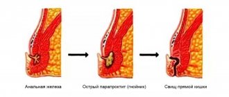
- purulent-inflammatory processes – paraproctitis, pelvic phlegmon;
- perforation of a tumor of supramullary localization with the development of peritonitis;
- bleeding.
Colonoscopy.
Rectal cancer.
This is the most informative method of examining the colon. It involves inserting a flexible instrument (endoscope) through the anus and is accompanied by illumination and examination of the inner surface of the colon and rectum. To maximize visualization of the intestinal wall, it must be adequately cleansed using special laxatives or an enema. Colonoscopy is the only method to confirm colorectal cancer because tissue samples (biopsy) can be obtained. In addition, intestinal polyps can not only be detected, but also removed during this procedure.
Diagnostics
After contacting a doctor, they determine the symptoms that bother the patient and suggest a disease of the rectal region.
Screening study
A stool occult blood test is ordered
Laboratory research
A general analysis of urine and blood reflects the condition of the body, the presence of inflammation, anemia, kidney and bladder function
Mandatory digital rectal examination
This method allows you to identify tumors of anorectal localization. The condition of the sphincter and mucous membrane is determined.
Endoscopic methods
To examine the inner surface of the rectum in more distant areas: - sigmoidoscopy - examination of the intestine using a rigid tube with an optical device at the end; - colonoscopy - examination using a flexible endoscope, allows you to examine the entire intestine.
Biopsy
Parts of tissue are taken from suspicious and altered areas and examined for cancer cells or dysplastic precancerous processes.
Parallel examination of the large intestine
For differential diagnosis of cancer, detection at an early stage of changes that can lead to cancer.
Ultrasound of the intestines
To determine the prevalence of a malignant process. It can be performed abdominally - through the abdominal wall, and transrectally - through the anus.
Gynecological examination (for women)
Often there is a combination of intestinal tumors with gynecological carcinomas - cancer of the uterus, ovaries, and breast.
X-ray methods for diagnosing rectal cancer
Using a contrast agent. The photographs show defects in intestinal filling, pathological tissue growths, and stenoses caused by neoplasms.
MRI diagnostics
It detects even small-sized pathological foci, metastases and tumor invasion into neighboring organs, and its topographic position relative to other structures. The method is safe and does not create radiation exposure, unlike CT.
Radioisotope methods
Tumor metastases are detected.
Diagnostic laparoscopy
carried out to clarify the diagnosis. It can be used to detect metastases in the peritoneum.
Risk factors
Factors contributing to the development of colorectal cancer include:
- Poor diet with an emphasis on heavy fatty meats and minimal fiber.
- Avitaminosis. It is especially bad if the body lacks vitamins A, C and E.
- Obesity. This type of rectal cancer is most common among overweight people.
- Lack of regular activity, sedentary lifestyle.
- Smoking and alcohol abuse.
- Contact with certain chemicals - for example, skatole, indole.
- Heredity.
- Some benign formations that can eventually become malignant are the same as polyps.
- HPV in the anal area. Papillomaviruses can cause the development of cancer in various organs, including the rectum.
All these factors are contributing factors - this does not mean that they will lead to colorectal cancer. But eliminating them significantly increases the chance of better health.
Stages of rectal cancer
The course of the cancer process progresses in the absence of proper treatment. The stage is determined by the degree of damage to the intestine itself, its growth through the wall, the presence of metastases in the lymph nodes, and distant lesions of other organs.
In this regard, tumors are divided into 4 stages. This distribution is universal for any malignant tumors.
Stage 1 – the tumor is small in size, grows on the mucous layer, does not affect neighboring organs and lymph nodes.
Stage 2 is divided into A and B. 2A - this is a lesion from a third to half the circumference of the intestinal tube, but grows strictly in the wall or lumen, there are no metastases. 2B – the size of the lesion is the same, but there are metastases in the peri-intestinal lymph nodes.
3A – the tumor occupies more than half the circumference of the intestine, grows through all layers and peri-intestinal tissue. There may be single metastases in nearby lymph nodes.
3B – any tumor size, metastases in distant lymph nodes receiving lymph from the rectal area.
Stage 4 – metastases spread to internal organs and distant lymph nodes. The primary tumor can be of any size.
What methods are used to prevent and early diagnose cancer?
Digital rectal examination.
Digital rectal examination is the main and simplest diagnostic method in the arsenal of a coloproctologist, which does not require the use of special equipment. During this manipulation, the specialist examines the rectum by inserting the index finger into the intestine. When performing a digital rectal examination, it is possible to determine the presence of intra- and extraintestinal formations at a height of up to 10 cm from the edge of the anus, assess the nature of the formation (benign or malignant), and the involvement of neighboring organs and tissues in the tumor process. In the presence of intraluminal formations, patients must undergo a biopsy (a piece is taken) in order to determine the histological structure and make an accurate diagnosis.
Treatment methods for colorectal cancer
The small size of the tumor and its growth only through the mucous and submucosal layer of the rectum, without affecting the muscular and serous layer, allows surgical removal of the tumor itself. Sometimes it is possible to perform surgery through the colon using a colonoscope.
If it has grown into the muscle layer, then rectal resection or extirpation (complete removal of the organ) is indicated. The perirectal tissue and lymph nodes are also removed, in which metastases are already detected in 20% of cases. To perform the operation, two approaches are used - laparotomy (dissection of the abdominal wall) and laparoscopy (operation using video equipment through several punctures in the abdomen).
The type of surgery is selected based on the location of the tumor. The high position makes it possible to remove the tumor and temporarily bring the end of the intestine to the abdominal wall - to form a colostomy for defecation. Such manipulation is necessary if it is not yet possible to sew the ends of the intestine together. The second stage, after some time, restores the integrity of the intestine.
If the tumor process is located low, if there is no healthy tissue left below it, the affected area and anus are removed, and a colostomy is applied to the abdominal wall.
Survival prognosis
After radical surgery, survival rate for 5 years ranges from 34-68%. The outcome of treatment is influenced by the stage at which the tumor was diagnosed, the condition of the patient himself, his age, and concomitant diseases.
Depending on the stage of the tumor process, five-year survival rate is determined by the following figures:
- Stage 1 – up to 77%;
- Stage 2 – up to 73%;
- stage 3 – 46%;
- Stage 3b – 43%.
Stage 4 is not considered in these statistics. Radical operations are often impossible to carry out, because tumor metastases are disseminated throughout the body. The lethal outcome depends on the general condition of the patient.
Prognosis for bowel cancer
When predicting the success of cancer treatment, oncologists use the concept of five-year survival. Such forecasts are based on statistical data, which is formed from the results of treatment of a large number of patients. Experts take into account the number of patients who lived after successful treatment for more than 5 years. The study takes into account the stage of the tumor for which treatment was carried out:
- stage 1 – 95% five-year survival rate;
- stage 2 – 75%;
- stage 3 – 50%;
- stage 4 – 5%.
The figures given are averages, so they are considered approximate. For each patient, a more accurate prognosis is made, which additionally takes into account all related factors:
- age group;
- functionality of the immune system;
- accompanying illnesses.
It must be remembered that the chances of a complete cure for intestinal cancer depend not only on the timeliness of diagnosis of the disease, but also on the chosen clinic. When visiting a clinic abroad, the likelihood of successful treatment will be high even in the later stages. If cancer cannot be cured completely, then doctors take all measures to prevent the disease from progressing, eliminate painful symptoms, and significantly prolong the life of the patient.
Contraindications
The operation is contraindicated under the following conditions:
- severe chronic diseases of the patient - arterial hypertension, coronary heart disease, when it is impossible to give anesthesia;
- advanced age of the patient;
- advanced stages of cancer.
In case of a widespread process with metastasis to many organs, palliative resections are used, aimed at alleviating the patient’s condition. Symptomatic operations - the application of bypass anastomoses to relieve the intestines and avoid complications in the last stages of cancer.
Treatment before and after surgery
Chemotherapy and radiation therapy are indicated for patients with stage 2 or higher tumors.
If before the operation metastases were detected in several lymph nodes, and the tumor has grown into the muscle layer, then at the stage of preparation for the operation a short course of radiation therapy is carried out for 5 days. This allows you to destroy early metastases and reduce the size of the formation itself.
Treatment of rectal cancer after surgery is carried out after obtaining pathomorphological data on the removed tissues. The issue of radiation or its combination with chemotherapy is being decided. Radiation therapy after surgery destroys the remaining cells in the area of the primary tumor and prevents its recurrence. In inoperable patients, it alleviates the condition.
Sensitivity to chemotherapy is detected in 30% of patients. It is prescribed for therapeutic purposes to destroy metastases.
Chemotherapy is also carried out adjuvantly - to prevent the spread of carcinoma if damage to several lymph nodes is detected. This method of therapy improves the quality and life expectancy of patients with metastases. Platinum preparations, 5-fluorouracil, leucovarin, and calcium folinate are used. Medicines are administered intravenously in courses of several days. Chemotherapy is also used in combination with radiation before surgery for locally advanced cancer. This combined treatment is carried out for 1-1.5 months, and after the end of irradiation, surgery is performed 6 months later.
Positron emission tomography (PET).
Rectal cancer.
During their growth, cancer cells spend more energy and absorb more glucose than healthy cells. These characteristics can be used to diagnose a tumor and its metastases. During positron emission tomography (PET), radioactively labeled glucose is injected intravenously and taken up by rapidly growing metabolically active cells (mostly cancer cells), making them visible during the study. Considering the high cost of the method, PET is used to perform specific tasks, such as searching for metastases and diagnosing possible relapse of the disease.