Synonyms cutaneous dyslipoidosis, Oppenheim-Urbach disease, diabetic necrobiosis lipoidica, nerobiosis lipoidica.
A rare chronic dermatosis of a vascular-metabolic nature, manifested by multi-colored (red, yellow, brown) plaques with clear boundaries on the anterior and lateral surfaces of the legs. The etiology of the disease is unknown. The pathogenesis is based on angtopathy of various origins.
In the dermatogastroenterology section you can learn about other diseases
Clinic
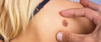
The disease usually begins with the appearance of small bluish-pink or reddish-brown spots or nodules of round or irregular shape, which later merge into plaques. The central part of the plaque is atrophic, waxy-yellow in color with telangiectasia, and glossy. The peripheral part is represented by a narrow brownish rim. Plaques can ulcerate over time, leaving scars after the ulcers heal. There are no subjective sensations. Pain occurs only with ulceration.
Localization: 80% of the lower leg, often symmetrical damage.
Less commonly – feet, torso, arms, face, scalp.
Introduction
Necrobiosis lipoidica (NL) is a rare inflammatory granulomatous skin disease resulting from collagen degeneration (1, 2). Incidence rates are higher in women than in men and in adults than in children (3). NL usually appears between the ages of 30 and 40 years (4). There appears to be a significant association between NL and diabetes mellitus (DM) in numerous studies, starting with the study by Cohen et al., who are of the opinion that more than fifty percent of patients with NL have comorbid diabetes; however, less than one percent of diabetic patients also have NL (5). Two other studies, on the other hand, estimate that 80 and 60% of patients with NL are diabetic, respectively (1, 6). Regarding the significant association between NL and DM and the unknown etiology of this disease, microangiopathy appears to play a prominent role in the pathogenesis of NL (7). It is characterized by well-circumscribed erythematous plaques, mainly affecting the tibial region of the lower extremities (8, 9). However, involvement of other body sites has also been reported (7). NL is a chronic relapsing-remitting disease in which rashes are accompanied by ulceration due to trauma (1) and cannot always be distinguished from those of other inflammatory skin diseases (10). We reviewed approximately seventy articles and intend to discuss all proposed and tested treatments for NL in the literature as follows (summarized in Supplementary Table S1).
Corticosteroids
Systemic corticosteroids
Taniguchi et al. extensively studied the effectiveness of oral corticosteroids for NL eruptions, leading to their clinical cure (11). In a study of 6 patients with NL, administration of systemic corticosteroids resulted in complete healing of all ulcers except atrophic ones (12). Plus, treatment with prednisolone at a dose of 6 mg/day provoked improvement (13). Two other cases in the same category, one of which was described by Tan et al. and another described by Dwyer and Dick (14, 15) have also been published. In addition, Bukhanik et al. presented a case report in which combination therapy with topical corticosteroids and hyperbaric oxygen resulted in remission of NL (16). Thus, corticosteroids may be considered an effective therapeutic option for NL, but their use in diabetics remains controversial given the potential effect of destabilizing diabetes control (14).
Intralesional corticosteroid injection
The effectiveness of triamcinolone acetonide and saline injections was studied in five patients with NL, three of whom achieved complete resolution of the lesions and one who experienced only partial improvement. Despite the high relapse rate, reintroduction was advisable (17). Asteroid bodies were found in a case of NL in which dipyridamole combined with intralesional triamcinolone for two months resulted in complete regression of the lesions, which persisted even after cessation of therapy (18).
Topical corticosteroids
Topical clobetasol propionate was effective in two case studies (19).
Intravenous administration of immunoglobulin and methylprednisolone
In one study, intravenous immunoglobulin (IVIG) was effective in treating refractory NL ulcers in a patient, but repeated administration of IVIG was less effective. The use of intravenous methylprednisolone also caused improvement in the lesions (20).
Pentoxifylline
The role of Pentoxifylline as a treatment for NL was studied in a 20-year-old diabetic patient with NL who was administered the drug 3 times daily at a dose of 400 mg, with improvement after one month of therapy. A 2-year follow-up did not reveal relapses; in addition, an improvement in the patient’s psychosomatic status was observed. No treatment side effects were noted (21). Noz et al. also presented a case of NL ulceration that did not respond to topical and oral ASA therapy but resolved fairly quickly with pentoxifylline (22). The diagnosis of NL was made by identifying a rash on the glans penis of a nondiabetic patient who responded well to pentoxifylline (23). Therapy with Trental (pentoxifylline) in 17 patients with NL demonstrated a positive effect both in terms of correction of hematological parameters (for example, blood viscosity, erythrocyte aggregation and stability of erythrocyte aggregates) and in terms of the dynamics of skin status (24). Thus, pentoxifylline may be a good treatment option for NL. The recommended dosage is 400 mg three times daily for at least 6 months (21).
Anti-TNF alpha therapy
Infliximab
A study evaluating the effect of intralesional infliximab in 3 NL patients showed complete regression of NL lesions, but follow-up after therapy revealed disease recurrence. No noticeable side effects were noted except for painful injections (25). Additionally, an 84-year-old type 1 diabetic woman with intractable NL demonstrated a robust response to the first intravenous infusion of infliximab, resulting in complete healing of the ulcers after three infusions (26).
Etanercept
A marked improvement was noted after subcutaneous etanercept was administered in the treatment of NL (27–29). However, subcutaneous administration of adalimumab had no success (29).
Based on the above-mentioned studies, anti-TNF-alpha drugs (etanercept/infliximab) may be effective in the treatment of refractory ulcerative lesions in NL through both intralesional and intravenous routes of administration; however, more research is needed to elucidate further details regarding this treatment (dosage and duration of treatment) (27).
Skin graft
Several case reports have presented skin grafting as an effective surgical intervention for severe ulcerative NL, which was performed using split-thickness skin grafts and combined with the use of a xenograft in one case (30–33). Additionally, a study including 7 cases of NL at Stanford University Medical Center found that split-thickness skin grafting was effective and did not cause subsequent recurrence (34).
UVA1 phototherapy
Several studies have reported that ultraviolet A1 (UVA1) phototherapy or topical psoralen therapy combined with ultraviolet A (PUVA) therapy is effective for treatment-resistant forms of NL that have not responded to other treatments, such as corticosteroids or surgery (2, 35). -38). Additionally, a 68-year-old diabetic patient with NL demonstrated the same results when undergoing topical PUVA therapy (39).
Cyclosporine
The administration of systemic cyclosporine A has shown encouraging results in two different clinical trials (1, 40). Another study examining the effectiveness of cyclosporine in 2 patients with NL over a 4-month period demonstrated complete healing of ulcers, which also persisted after discontinuation of cyclosporine therapy (41). In addition, the regimen of cyclosporine at a dose of 2.5 mg/kg/day for treatment-resistant ulcerative forms of NL was very effective; however, a relapse was detected 3 months after discontinuation of therapy, which regressed after resumption of cyclosporine (42). Smith also suggested cyclosporine A as an effective treatment for NL (43).
Aspirin and dipyridamole (antiplatelet therapy)
The double-blind, randomized, placebo-controlled study included 14 patients with NL (2 groups) who received either aspirin and dipyridamole or placebo; neither treatment showed any significant improvement (44). In contrast, administration of acetylsalicylic acid and dipyridamole in 7 patients with NL caused improvement of lesions by reducing thromboxane levels (which, according to the author, were elevated in all patients before therapy) (45). In addition, Quimby et al. low platelet survival time in patients with NL has been reported, returning to normal in response to antiplatelet therapy; however, the response of NL lesions to treatment ranged from resolution to no noticeable improvement (46). Beck and Bjerring correlated low-dose acetylsalicylic acid (ASA) therapy with a marked reduction in cutaneous blood flow at the center of NL lesions, measured by laser Doppler (47). Császár et al. demonstrated that combination therapy with ASA and dipyridamole is effective against manifestations of NL and also reduces platelet aggregation (48). Last but not least, a double-blind controlled trial showed an increase in the number of NL lesions after both low-dose ASA and placebo therapy (49). Other findings from these studies suggest that the effectiveness of antiplatelet therapy remains to be determined.
Topical tacrolimus
Barth et al. used topical tacrolimus ointment in the treatment of a patient with diabetes and NL, also with protein S deficiency and antiphospholipid syndrome, which led to significant improvement (50). Another study also found significant healing of NL lesions after topical tocrolimus 0.1%, with continued therapy after 1 year of follow-up (51).
Hyperbaric O2 Therapy
Bruengger studied exposure to 100% oxygen in 9 nondiabetic NL patients, which resulted in a significant increase in oxygen pressure (pCO2) at the lesion, although markedly lower than in normal skin, and a sharp increase in PcCO2 levels within the lesion ( 52). In another study, hyperbaric oxygen therapy resulted in complete clearance of all ulcers after 98 sessions (53).
Pancreas transplant
A retrospective study of NL patients with diabetes assessed the effectiveness of pancreas transplantation compared with kidney transplantation, determining whether regression of diabetes could lead to resolution of NL. Pancreas transplantation with or without kidney transplantation contributed to the resolution of NL lesions, whereas kidney transplantation alone had no success (54). Two case reports also described that simultaneous pancreas transplantation after renal transplantation was effective, and a corresponding effect on the skin process was observed (8, 55).
Fumaric acid esters
A prospective study of 18 patients with NL evaluating the effectiveness of fumaric acid esters (FAEs) on NL demonstrated marked regression of the disease with increased skin density assessed by 20 MHz ultrasound. After a follow-up period of 6 months, no relapses were found, suggesting that FAE is an effective drug for the treatment of NL (56). In addition, another study reported regression of NL lesions following FAE therapy (57).
Pulsed dye laser
Pulsed dye laser was investigated as a treatment approach for lesions up to 4 cm NL on the anterior leg of a nondiabetic patient who received 3 treatment sessions. The results were inconclusive, as the left half of the lesion showed signs of resolution, while the right upper quadrant of the lesion showed little improvement (58). Additionally, a study of a 23-year-old diabetic patient with NL receiving pulsed dye laser found that this treatment is ineffective at both high and low intensities, as it may be harmful to the skin and therapeutically insignificant, respectively (59) . As can be seen, pulsed dye laser is not a safe or effective treatment for NL.
Antimalarials
Nguyen et al. demonstrated that oral chloroquine is useful in the treatment of NL (60). Improvement was also noted after administration of antimalarial drugs in 7 of 8 patients (61).
Topical tretinoin
Heymann confirms the effectiveness of topical tretinoin in the treatment of atrophic manifestations of NL (62). In addition, favorable results were achieved with combination therapy of topical glucocorticoids and topical tretinoin in a 58-year-old nondiabetic woman with a 7-year history of NL (63).
Photodynamic therapy
Photodynamic therapy using 632 nm red light and methyl aminolevulinate as a topical photosensitizer showed good results in the treatment of a 60-year-old woman with advanced NL (64). However, it was effective in only 40% of clinical trials (65).
Local granulocyte-macrophage colony-stimulating factor
Remes and Rennemaa studied the effectiveness of GM-CSF in two patients with diabetes mellitus suffering from chronic refractory NL and concluded that this treatment method is beneficial, since improvement was noted even after 1 episode of GM-CSF use ending in complete regression lesions without recurrence after 3 years of follow-up (66).
Clofazimine
A clinical study involving 10 patients suffering from NL evaluated the effectiveness of clofazimine administered at a daily dose of 200 mg PO. The study was inconclusive because clinical response to clofazimine varied among patients from no improvement to complete remission. Side effects ranged from minor to severe (67).
PROMOGRAN
A study of the clinical effectiveness of PROMOGRAN (a new protease modulating matrix complex) revealed complete resolution of NL ulcers after 8 weeks of therapy (68).
Benzoyl peroxide
Application of 20% topical benzoyl peroxide to NL ulcers caused rapid improvement of the lesions (69).
Nicotinamide
Treatment with high doses of nicotinamide has proven effective in controlling NL (70).
Bovine collagen
Treatment with topical bovine collagen resulted in significant improvement of NL lesions (71).
Thalidomide
Thalidomide treatment of a 51-year-old woman with NL resulted in regression of the lesions after 4 months of therapy (72).
Discussion and conclusions
Because NL is a rare disease, there are not enough cases to conduct effective prospective clinical trials, so most studies conducted to date are case reports and small-scale clinical trials. In addition, there are several comparative studies between different NL treatments, which lead us to conclude that despite the introduction of all these NL treatments, the therapy of choice remains an ongoing debate requiring further large-scale studies. But to summarize, we can call the use of corticosteroids by various routes of administration (systemic, intralesional and local) a priority in the treatment of NL, despite the possibility of destabilizing blood sugar control in patients with diabetes.
Acrochordons
Acrochordons (filamentous warts) are growths of normal skin tissue on a narrow stalk, usually localized in the neck, axilla, eyelids and groin area [4]. They consist of hyperplastic soft growths of the dermis and epidermis and are skin-colored or brownish in color (Fig. 1A) [8]. Acrochordons are a fairly common phenomenon, occurring in approximately 25-46% of the adult population; their number and prevalence also increase with age and during pregnancy [9]. The association between acrochordons and metabolic syndrome (obesity, dyslipidemia, hypertension, insulin resistance and elevated C-reactive protein levels) is well established [10,11].
Picture 1.
(A) Acrochordons: pink, thick, pedunculated papules. (B) Acanthosis - acanthosis nigricans: hyperpigmented, velvety plaques on the neck. Images courtesy of DermnetNZ. Published online at: https://www.dermnetnz.org
The diagnosis of acrochordons is based on the clinical picture and localization of lesions [4,8]. In rare cases, they may raise suspicion of malignancy and should be sent for histological examination [4]. The presence of acrochordons on the body should be an indicator for the local doctor to exclude possible diabetes, if such a diagnosis has not previously been made. These lesions are often asymptomatic and should not be removed unless desired by the patient for cosmetic reasons or in cases of irritation [4]. Treatment of acrochordons involves excision and can be performed with tweezers, small scissors, cryosurgery with liquid nitrogen, or electrical excision [4,8].
Eruptive xanthomatosis
Eruptive xanthomatosis is a dermatosis characterized by the sudden appearance of yellow papules with an erythematous base on the buttocks, shoulders and extensor surfaces of the extremities (Fig. 2) [17,18]. The sudden appearance of such formations can cause anxiety in patients and serve as a reason to visit their local doctor [4]. Eruptive xanthomatosis is associated with markedly elevated serum triglyceride levels, as occurs in hyperlipidemia syndromes (eg, Fredrickson-Levy hyperlipidemia types I, IV, and V) with poorly controlled type 2 diabetes mellitus, hypothyroidism, obesity, pancreatitis, nephrotic syndrome, cholestatic liver diseases, dysglobulinemia, and may also be a side effect of certain medications (for example, estrogens, corticosteroids, systemic retinoids) [19-22]. Eruptive xanthomatosis may be the first sign of diabetes mellitus [4].
Figure 2.
Eruptive xanthomatosis: yellow to erythematous papules on the extensor surfaces of the hands. Figure reproduced with permission from DermnetNZ. Published online at: https://www.dermnetnz.org.
The diagnosis of eruptive xanthomatosis can be made clinically and confirmed by biopsy from the lesions [4]. Following diagnosis, fasting lipid levels should be determined [4], as patients with eruptive xanthomatosis are at increased risk for early onset coronary artery disease and pancreatitis caused by hypertriglyceridemia [23]. Treatment of eruptive xanthomatosis involves treatment of the underlying disease, and the treating physician should aim to reduce triglyceride levels through dietary changes and systemic medications [20,21,24,25]. Eruptive xanthomatosis usually resolves within 6–8 weeks with appropriate treatment [20,21].
Diagnostics
People who have symptoms of necrobiosis lipoidica should see a doctor as soon as possible. Although the disease itself is harmless, complications such as infection or scarring can sometimes occur.
The doctor will examine the patient and ask about underlying health conditions to diagnose necrobiosis lipoidica.
If there is any doubt, the doctor may take a small sample (biopsy) of the affected tissue for testing. He will do this using local anesthesia.
Xerosis
The term xerosis, or xeroderma, refers to dry skin. It is the second most common cutaneous manifestation of diabetes mellitus [4]. One prospective study found xerosis in 26.4% of patients with diabetes mellitus [3]. Patients with diabetes often suffer from autonomic neuropathy, the complication of which increases the risk of xerosis [58,59]. The presence of xerosis increases the risk of complications, including infection and ulceration.
Clinically, xerosis appears gradually with a tendency to worsen while the disease persists. When the skin is too dry, it initially turns red and then cracks appear [60]. If dryness persists, the skin may begin to peel or flake. Winter is the peak time for dry skin due to low ambient humidity and heating devices that heat and dry the air in the home or workplace [61]. Air conditioning can also cause xerosis because it removes most of the moisture from the air [61, 62].
The diagnosis of xerosis is made based on clinical signs and history. Referral to a dermatologist is not necessary [4]. Local doctors should point out to patients the importance of maintaining skin hygiene and the possibility of using fragrance-free creams or lotions within 3 minutes after bathing to increase skin hydration [4].
Causes of necrobiosis lipoidica
It is generally accepted that this pathological condition occurs more often against the background of diabetes mellitus or conditions close to it (impaired glucose tolerance). As a result of a significant increase in sugar levels, as well as “jumps” in glucose levels, all metabolic processes in the patient’s body are disrupted.
First of all, this concerns fat and carbon metabolism. Metabolic disorders lead to insufficient nutrition of all organs and tissues of the human body, the skin is no exception. In patients suffering from diabetes, the microcirculation function of certain areas of the skin often fails, as a result of which degenerative changes occur. They occur at the cellular level and lead to fatty degeneration of skin cells.
To summarize, we can say that necrobiosis lipoidica of the skin is a consequence of metabolic disorders in the patient’s body, which leads to vascular pathologies, insufficient nutrition of skin cells and, accordingly, to their final death.