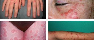
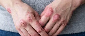
Erythema nodosum (ICD-10 code: L52) is a fairly common disease that most often affects pregnant women. This pathology is characterized by the formation of medium-sized nodules with a diameter of one to three centimeters in the thighs, on the front surface of the legs and on the buttocks. As a rule, the appearance of these nodules is symmetrical - they appear on both the right and left legs. When pressing on the nodules, the patient experiences pain. There is thinning, thickening, and shine of the skin above them.
Erythema nodosum occurs not only in pregnant women, but also in people using hormonal contraception or suffering from tuberculosis and diseases caused by the introduction of streptococcal flora.
Causes
The exact causes of the development of erythema nodosum have not been thoroughly studied to date. However, it is known that the occurrence of this disease can be provoked by the following factors:
- hereditary predisposition;
- infectious diseases of various origins;
- sarcoidosis;
- the use of medications belonging to different groups: bromides, oral contraceptives, sulfonamides;
- Crohn's disease;
- allergic reactions and general intoxication of the body;
- ulcerative colitis.
An important role in the clinical picture of erythema nodosum is given to the condition of the blood vessels of the lower extremities. The likelihood of relapse of erythema increases significantly in patients suffering from thrombophlebitis and varicose veins.
Due to increased stress on the vessels of the lower extremities during pregnancy, during this period of a woman’s life the risk of developing erythema nodosum is also considered increased.
However, there are cases where erythema nodosum occurred in the absence of visible causes. According to doctors, this may occur due to an allergic reaction, the development of which was caused by an unknown allergen.
Why does inflammation of the skin vessels develop?
There are several factors that provoke the onset of the inflammatory process in the vessels of the epidermis:
- bacterial infectious diseases caused by streptococci - scarlet fever, tonsillitis, pharyngitis, otitis media, etc., after which erythema nodosum of the lower extremities develops as a complication of the disease;
- long-term systemic disease - tuberculosis, lymphogranulomatosis, trichophytosis, yersiniosis;
- long-term course of treatment with antibiotics, bromides, salicylates, iodides or sulfonamides;
- vaccination;
- ulcerative colitis;
- cancer;
- Behçet's disease;
- pregnancy and the associated hormonal changes in the female body, and the risk of developing erythema nodosum increases significantly if the pregnant woman has a history of chronic infectious foci;
- hereditary predisposition to the disease: it is often diagnosed in several generations of the same family or in close relatives;
- allergies, which contribute to the development of a chronic form of pathology.
Erythema nodosum most often affects young people 20-30 years of age. Before the onset of puberty, the incidence is the same in boys and girls, but starting from adolescence, the disease occurs mainly in women, among whom the incidence is 3-6 times higher than in men. There is a certain seasonality: in winter and spring, the number of visits to the doctor about this increases significantly due to decreased immunity and an increase in the number of infectious diseases, including streptococcal infections.
Types of disease, symptoms
The clinical picture of erythema nodosum depends on its form, which can be acute or chronic.
Acute erythema at the initial stage is manifested by the following symptoms:
- the appearance of a feverish state;
- increased body temperature;
- headaches;
- swelling of the legs;
- formation of bright red nodules on the lower extremities.
At the same time, patients experience an enlargement of the inguinal lymph nodes, accompanied by the appearance of pain when walking or with increased stress on the legs.
After two to three weeks, a gradual regression of the nodules is observed, they change color: from purple-bluish to greenish, then yellowish (as with a resolving hematoma).
Erythema nodules can be localized at different depths under the skin, affecting the middle and deep layers of the dermal layer, as well as subcutaneous fat - in these cases allergic vasculitis can be diagnosed. An increase in the size of the nodes occurs within several hours, then, as a rule, their growth stops.
The chronic form of erythema nodosum is called angiitis nodosum. The occurrence of this pathology is most often observed in female, elderly patients suffering from concomitant diseases caused by relapses of chronic infectious processes.
An exacerbation of the second wave of erythema nodosum (relapse of the inflammatory process) is observed in the autumn or spring months - the climatic transition period.
Under the skin in the shin area, dense, painful, bluish-colored lumps the size of a walnut form. The localization of dermatological manifestations is most often observed on the legs along large vessels, but in some cases they can also be located in the thighs. Severe swelling forms in the legs, and the patient has difficulty walking.
A characteristic symptom of the subacute form of the disease, called Vilanova-Piñol migratory hypodermitis, is the acute onset of the inflammatory process and the appearance on the legs of several single nodules located in the subcutaneous fatty tissue.
After some time, the nodes become flattened and flat structures with different structures form. The diameter of the infiltrate surface can reach up to 20 centimeters. The spots do not have clear boundaries; the intensity of inflammation of the skin surface increases closer to the center.
This form of dermatological formation is typical only for adults. The duration of the disease can be more than six months, after which pigment spots remain on the surface of the skin, which gradually peel off. No traces of formations remain.
Erythema nodosum migrans is characterized by the formation of asymmetrical nodular formations on the legs and feet. Palpation most often reveals the pain of these rashes. At the initial stage, a small number of compacted formations appear, which subsequently migrate around the periphery, which is accompanied by an increase in body temperature. Relapses occur quite often, especially in the absence of timely treatment of the underlying infection.
In case of repeated development of the disease, the patient requires a thorough examination of the respiratory system with a detailed X-ray of the lungs.
Often, a similar dermatological process occurs in patients with sarcoid pathologies, which are characterized by the localization of nodular formations in the lungs.
An X-ray examination is carried out to exclude a malignant process affecting the paratracheal lymph nodes, which provides the doctor with the opportunity to select an appropriate therapeutic regimen.
Therapy of the inflammatory process
Treatment of erythema nodosum is aimed, first of all, at getting rid of foci of chronic infection or other causative pathology. It includes:
- antibiotic therapy if the inflammatory process is infectious;
- desensitizing therapy if the cause of the disease is an allergic reaction of the body;
- taking anti-inflammatory drugs to relieve inflammation and relieve pain;
- plasmapheresis, cryopheresis or hemosorption to quickly eliminate intoxication;
- corticosteroid and anti-inflammatory ointments for inflamed joints and erythema nodes;
- physiotherapeutic treatment - ultraviolet irradiation, laser therapy, background cutter, magnetic therapy, etc.
For pregnant patients, clinical recommendations for erythema nodosum are significantly limited, since many drugs are undesirable to use. Previously, this disease served as a justification for artificial termination of pregnancy, but it has now been proven that it does not affect the development of the fetus.
Diagnostics
When making a preliminary diagnosis of “erythema nodosum”, doctors at the Yusupov Hospital take into account the patient’s complaints, medical history, and the results of an objective examination. To confirm or refute it, additional instrumental and laboratory studies are prescribed:
- clinical blood test - to determine signs of the inflammatory process;
- blood test for rheumatoid tests - to identify rheumatoid factor;
- bacterial culture from the nasopharynx - to detect streptococcal infection;
- tuberculin diagnostics – if tuberculosis is suspected;
- stool culture - if there is suspicion of yersiniosis;
- biopsy of nodular formations and subsequent microscopic examination of the biomaterial;
- rhinoscopy and pharyngoscopy - to identify chronic foci of infection;
- chest radiography;
- computed tomography of the chest;
- ultrasound examination of the veins and rheovasography of the lower extremities, allowing to determine their patency and the severity of the inflammatory process;
- consultations with doctors of related specialties - infectious disease specialists, otorhinolaryngologists, pulmonologists, phlebologists, etc.
The diagnostic regimen is selected by highly qualified specialists at the Yusupov Hospital individually for each patient, taking into account the clinical picture of the disease and other important data.
Prognosis and prevention
Typically, erythema nodosum (photos and treatment are presented in the article) does not pose a threat to human life and health; it is only necessary to promptly treat concomitant diseases that can provoke the development of negative consequences. The prognosis for this pathology is favorable. In the chronic course of the disease, relapses may occur, but they do not pose a danger to human health.
Prevention of the disease should be aimed at timely treatment of infectious and inflammatory diseases, and also consists of mandatory sanitation of the body from foci of infection. Doctors recommend monitoring the condition of the vascular system; at the first signs of varicose veins, you should consult a specialist. It is also important not to come into contact with allergens, periodically undergo routine medical examinations and treat chronic pathologies. By following these recommendations and instructions, you can avoid the development of the disease or begin timely treatment if it is present.
Treatment, reviews
The main goal of treatment is to eliminate the identified disease that caused the development of this nonspecific immunoinflammatory syndrome.
To treat the underlying disease of infectious etiology, the use of antibacterial, antifungal and antiviral agents is prescribed.
Primary erythema nodosum requires treatment using drugs from the following groups:
- nonsteroidal anti-inflammatory drugs (Movalis, Nimesulide, Celecoxib, Diclofenac);
- corticosteroids (Prednisolone, Methylprednisolone) – used in the absence of sufficient effectiveness of NSAIDs;
- aminoquinoline drugs (Delagil, Plaquenil) - prescribed to patients with frequently recurrent and protracted forms of the disease;
- antihistamines (Suprastin, Loratadine, Cetirizine).
To quickly regress the symptoms of the disease, extracorporeal methods can also be used - plasmapheresis, hemosorption and laser irradiation of blood.
In addition, local treatment is effective: anti-inflammatory ointments, in particular hormonal ointments, as well as compresses based on dimexide, can be applied to the skin.
Positive results in the treatment of erythema nodosum are also achieved by using physiotherapeutic methods: magnetic and laser therapy, ultraviolet irradiation in erythema doses, phonophoresis with hydrocortisone on the affected areas.
Treatment of erythema nodosum at home can have serious negative consequences, since the drugs used have side effects that can cause significant damage to the patient’s health.
ethnoscience
Doctors do not recommend using traditional medicine to treat the disease, as they will not give a positive result and the person will waste time. But despite this, many patients prefer to use traditional recipes to combat the disease. Traditional therapy is possible only in the case of an additional method and only after consultation with the attending physician, since some medicinal herbs can provoke the development of side effects.
Symptoms
Erythema nodosum is characterized by the following clinical features:
- nodes that are dense to the touch, sometimes without clear boundaries;
- hemispherical shape;
- painful;
- no itching;
- up to 5 cm in size;
- the skin above them is hyperemic and smooth (then the color successively changes to brown, bluish and yellowish);
- with unclear boundaries;
- rising above the level of unchanged skin;
- localized mainly on the front of the legs;
- often symmetrical.
The nodes resolve without disintegration with peeling and hyperpigmentation.
