Causes of mycoses
Mycosis is an infectious dermatological disease caused by filamentous fungi. The source of infection is a sick person; in addition, fungal spores can be found on personal items, the use of which leads to infection.
The main methods of transmission of mycoses are:
- Close contact with an infected person.
- Penetration of microbes through microcracks, abrasions, abrasions and wounds on the body.
- The use of towels, manicure items, shoes, and combs contaminated with fungal microorganisms.
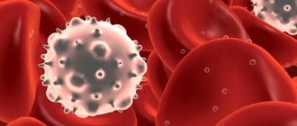
Contact with the skin and subcutaneous layers of pathogenic fungi does not always lead to the development of mycosis. If a person has a strong immune system, then the body itself destroys pathogens. The likelihood of developing the disease increases under the influence of provoking factors, these include:
- Violation of the normal functioning of the immune system.
- Long-term drug treatment.
- Lack of personal hygiene.
- Poor nutrition.
- Chronic diseases.
- Increased body sweating.
Mycoses most often affect the area of the feet and large folds. Sometimes the cause of the disease is one’s own fungi; when immunity decreases, their normal number may increase, which disrupts the skin microflora and leads to the formation of foci of infection.
Treatment
Therapy for systemic fungal infections depends on the general condition of the patient, the type of pathogen, the form and severity of the disease, concomitant diseases, and drug tolerance.
Treatment of deep mycoses is based on the use of fungicidal drugs.
For systemic candidiasis, amphotericin B is used. Sometimes treatment is supplemented with flucytosine. With simultaneous fungal skin infections, local forms of ketoconazole are prescribed. One of the effective treatments for skin manifestations of candida infection is Clotrimazole lotion for skin and nails. This drug for the treatment of mycoses is highly effective, safe and has an affordable price. You can order it in our online store fitosila.ru.
Ketoconazole can be used orally and in the form of lotion or other external forms to prevent candidiasis in patients with immunodeficiency. For candidal endocarditis, surgery may be necessary to replace the affected valve with an artificial one.
Aspergillosis is treated using amphotericin B and itraconazole. In the allergic variant of the disease, accompanied by attacks of bronchial asthma, glucocorticoid hormones are additionally prescribed. In case of severe lung damage, accompanied by hemoptysis, an operation is performed - removal of a lobe or the entire lung.
Amphotericin B or ketoconazole is used to treat histoplasmosis, cryptococcosis, coccidioidomycosis, paracoccidioidomycosis, blastomycosis. Only amphotericin B is used in the treatment of mucormycosis and sporotrichosis.
Additionally, pathogenetic and symptomatic therapy is carried out, aimed at relieving intoxication and maintaining the vital functions of the body:
- infusions of saline solutions, hemodez, plasma;
- vitamins and restoratives;
- bronchodilators;
- painkillers and others.
In cases of severe respiratory failure caused by fungal pneumonia, oxygen therapy or artificial ventilation is indicated.
Classification of mycoses
Mycoses are usually divided according to the degree of involvement of the skin, mucous membranes and appendages of the skin in the pathological process:
- Keratomycosis
. Fungal microorganisms multiply only in the stratum corneum of the skin; keratomycosis does not cause extensive inflammatory changes on the body. - Dermatophytes
. The changes affect the dermis and epidermis, as well as nails and hair follicles. - Candidiasis
. In addition to the skin, mucous membranes may be involved in the pathological process. - Deep mycoses
. Not only the skin is affected, but also a number of internal organs.
conclusions
Superficial fungal infections are most often caused by dermatophytes from the genera Trichophyton, Epidermophyton and Microsporum. These organisms metabolize keratin and cause a number of pathological clinical manifestations, including tinea pedis, dermatophytal dermatitis, dermatophytosis, etc. Based on clinical findings, the diagnosis of cutaneous dermatophyte infection can be strongly suspected. To confirm the diagnosis, potassium hydroxide should be used. Most dermatophyte infections can be managed with topical treatment. Examples of effective topical antifungal agents include azoles, allylamines, ciclopirox, butenafine, and tolnaftate. Oral antifungal therapy is used for widespread infections or infections that are resistant to topical therapy. Nystatin is not effective against dermatophyte infections.
List of literature / References
- Havlickova B, Czaika VA, Friedrich M. Epidemiological trends in skin mycoses worldwide // Mycoses. 2008 Sep;51 Suppl 4:2-15. doi: 10.1111/j.1439-0507.2008.01606.x. Erratum in: Mycoses. 2009 Jan;52(1):95. PMID: 18783559.
- Havlickova B, Czaika VA, Friedrich M. Epidemiological trends in skin mycoses worldwide // Mycoses 2008; 51 Suppl 4:2.
- Seebacher C, Bouchara JP, Mignon B. Updates on the epidemiology of dermatophyte infections // Mycopathologia 2008; 166:335.
- Ameen M. Epidemiology of superficial fungal infections // Clin Dermatol 2010; 28:197.
- El-Gohary M, van Zuuren EJ, Fedorowicz Z, et al. Topical antifungal treatments for tinea cruris and tinea corporis // Cochrane Database Syst Rev 2014; :CD009992.
- Alston SJ, Cohen BA, Braun M. Persistent and recurrent tinea corporis in children treated with combination antifungal/ corticosteroid agents // Pediatrics 2003; 111:201.
- Greenberg HL, Shwayder TA, Bieszk N, Fivenson DP. Clotrimazole/betamethasone diproprionate: a review of costs and complications in the treatment of common cutaneous fungal infections // Pediatr Dermatol 2002; 19:78.
- Rosen T, Elewski BE. Failure of clotrimazole-betamethasone dipropionate cream in treatment of Microsporum canis infections // J Am Acad Dermatol 1995; 32:1050.
- Hawkins DM, Smidt AC. Superficial fungal infections in children // Pediatr Clin North Am 2014; 61:443.
- Crawford F, Hollis S. Topical treatments for fungal infections of the skin and nails of the foot // Cochrane Database Syst Rev 2007; :CD001434.
- Korting HC, Tietz HJ, Brautigam M, et al. One week terbinafine 1% cream (Lamisil) once daily is effective in the treatment of interdigital tinea pedis: a vehicle controlled study. LAS-INT-06 Study Group // Med Mycol 2001; 39:335.
- Gupta AK, Cooper EA. Update in antifungal therapy of dermatophytosis // Mycopathologia 2008; 166:353.
- Bell-Syer SE, Khan SM, Torgerson DJ. Oral treatments for fungal infections of the skin of the foot // Cochrane Database Syst Rev 2012; 10:CD003584.
- Adams BB. Tinea corporis gladiatorum // J Am Acad Dermatol 2002; 47:286.
- van Zuuren EJ, Fedorowicz Z, El-Gohary M. Evidence-based topical treatments for tinea cruris and tinea corporis: a summary of a Cochrane systematic review // Br J Dermatol 2015; 172:616.6.
- Bourlond A, Lachapelle JM, Aussems J, et al. Double-blind comparison of itraconazole with griseofulvin in the treatment of tinea corporis and tinea cruris // Int J Dermatol 1989; 28:410.
- Cole GW, Stricklin G. A comparison of a new oral antifungal, terbinafine, with griseofulvin as therapy for tinea corporis // Arch Dermatol 1989; 125:1537.
- Panagiotidou D, Kousidou T, Chaidemenos G, et al. A comparison of itraconazole and griseofulvin in the treatment of tinea corporis and tinea cruris: a double-blind study // J Int Med Res 1992; 20:392.
- Faergemann J, Mörk NJ, Haglund A, Odegård T. A multicentre (double-blind) comparative study to assess the safety and efficacy of fluconazole and griseofulvin in the treatment of tinea corporis and tinea cruris // Br J Dermatol 1997; 136:575.
- Elewski BE, Hughey LC, Sobera JO. Fungal diseases. In: Dermatology, 3rd ed, Bolognia JL, Jorizzo JL, Schaffer JV (Eds), Elsevier Limited, Philadelphia; London 2012. Vol 2, p.1251.
- Voravutinon V. Oral treatment of tinea corporis and tinea cruris with terbinafine and griseofulvin: a randomized double blind comparative study // J Med Assoc Thai 1993; 76:388.
- Farag A, Taha M, Halim S. One-week therapy with oral terbinafine in cases of tinea cruris/corporis // Br J Dermatol 1994; 131:684.
- Smith KJ, Neafie RC, Skelton HG 3rd, et al. Majocchi's granuloma // J Cutan Pathol 1991; 18:28.
- Gill M, Sachdeva B, Gill PS, et al. Majocchi's granuloma of the face in an immunocompetent patient // J Dermatol 2007; 34:702.
- Cho HR, Lee MH, Haw CR. Majocchi's granuloma of the scrotum // Mycoses 2007; 50:520.
- Tse KC, Yeung CK, Tang S, et al. Majocchi's granuloma and posttransplant lymphoproliferative disease in a renal transplant recipient // Am J Kidney Dis 2001; 38:E38.
- Kim ST, Baek JW, Kim TK, et al. Majocchi's granuloma in a woman with iatrogenic Cushing's syndrome // J Dermatol 2008; 35:789.
- Akiba H, Motoki Y, Satoh M, et al. Recalcitrant trichophytic granuloma associated with NK-cell deficiency in a SLE patient treated with corticosteroid // Eur J Dermatol 2001; 11:58.
- Ilkit M, Durdu M, Karakaş M. Majocchi's granuloma: a symptom complex caused by fungal pathogens // Med Mycol 2012; 50:449.
- Novick NL, Tapia L, Bottone EJ. Invasive trichophyton rubrum infection in an immunocompromised host. Case report and review of the literature // Am J Med 1987; 82:321.
- Feng WW, Chen HC, Chen HC. Majocchi's granuloma in a 3-year-old boy // Pediatr Infect Dis J 2006; 25:658.
- Gupta AK, Prussick R, Sibbald RG, Knowles SR. Terbinafine in the treatment of Majocchi's granuloma // Int J Dermatol 1995; 34:489.
- McMichael A, Sanchez DG, Kelly P. Folliculitis and the follicular occlusion tetrad. In: Dermatology, 2nd ed, Bolognia JL, Jorizzo JL, Rapini RP (Eds), Elsevier Limited, St. Louis 2008.
- Gupta AK, Groen K, Woestenborghs R, De Doncker P. Itraconazole pulse therapy is effective in the treatment of Majocchi's granuloma: a clinical and pharmacokinetic evaluation and implications for possible effectiveness in tinea capitis // Clin Exp Dermatol 1998; 23:103.
- Burg M, Jaekel D, Kiss E, Kliem V. Majocchi's granuloma after kidney transplantation // Exp Clin Transplant 2006; 4:518.
- Liao YH, Chu SH, Hsiao GH, et al. Majocchi's granuloma caused by Trichophyton tonsurans in a cardiac transplant recipient // Br J Dermatol 1999; 140:1194.
- Bonifaz A, Vázquez-González D. Tinea imbricata in the Americas // Curr Opin Infect Dis 2011; 24:106.
- Cheng N, Rucker Wright D, Cohen BA. Dermatophytid in tinea capitis: rarely reported common phenomenon with clinical implications // Pediatrics 2011; 128:e453.
- Romano C, Rubegni P, Ghilardi A, Fimiani M. A case of bullous tinea pedis with dermatophytid reaction caused by Trichophyton violaceum // Mycoses 2006; 49:249.
- Al Aboud K, Al Hawsawi K, Alfadley A. Tinea incognito on the hand causing a facial dermatophytid reaction // Acta Derm Venereol 2003; 83:59.
- Veien NK, Hattel T, Laurberg G. Plantar Trichophyton rubrum infections may cause dermatophytids on the hands // Acta Derm Venereol 1994; 74:403.
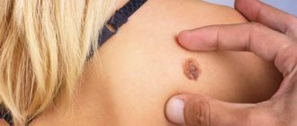
General symptoms of mycoses
During the incubation period, that is, during the proliferation of fungi, nothing bothers the infected person. The disease begins to manifest itself after the fungi begin to negatively affect the skin.
Depending on the type of fungal microorganism and the location of the infection, mycoses can manifest themselves in different ways, but there are several common symptoms:
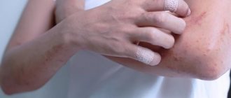
- Itching of the skin.
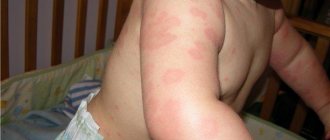
- The appearance of red spots, rashes, blisters.
- Peeling of individual areas of the skin.
When localized on the feet and hands, peeling of the dermis between the fingers is possible.
Mycosis involving the nail plates is called onychomycosis. Nail fungus damage is indicated by a change in their color, delamination, the appearance of dark or yellowish spots, and thickening of the plate.
Diagnosis of fungal diseases
To diagnose skin fungus, use:
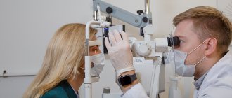
- Examination, dermatoscopy. After a dermatologist examines a patient's skin, he may suspect the presence of some kind of fungal infection.
- Examination under Wood's lamp. Wood's lamp is a special ultraviolet lamp. When examining skin elements under its rays, the latter acquire a specific color, or other effects are noted.
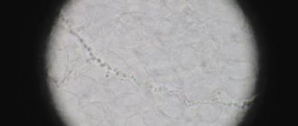
- Microscopy. To study under a microscope, they usually take hair, pieces of nails, and scrapings from the skin.
- Sowing the material on a nutrient medium.
Modern diagnostic methods used in the multidisciplinary CELT clinic make it possible to diagnose various mycoses with a high degree of accuracy.
Photos before and after
You can evaluate how mycosis of smooth skin is eliminated in our clinic from the photos taken before and after treatment. In 90% of cases, it is possible to achieve results without traces of the disease. If treatment is not done in a timely manner, complications may result, leaving barely noticeable scars and other changes in the epidermis. Therefore, to achieve maximum effectiveness of treatment, we recommend contacting a specialist when the first symptoms appear.
We are waiting for your questions by phone in Moscow. To make an appointment with a dermatologist, leave a request on the website.