Classification
The upper limit of normal platelet count varies from 350,000 to 400,000 per microliter depending on the reference intervals of the specific laboratory performing the analysis. According to the degree of increase, the following types of thrombocytosis are distinguished:
- Soft
: from 350-400 to 700 thousand. - Moderate
: from 700 to 900 thousand. - Heavy
: from 900 to 1000 thousand. - Extreme
: over 1,000,000.
The cause of extreme and severe thrombocytosis is oncohematological pathologies. According to the origin of thrombocytosis there are:
- Primary
(tumor, clonal). They account for approximately 10-15% of all cases of thrombocytosis. The cause is tumor diseases of the blood system. - Secondary
(reactive). The most common variety (about 85%). The cause is infectious, systemic inflammatory processes, and anemia. - False
(pseudothrombocytosis). The reason is an error in the hematology analyzer, which mistakes tumor cell fragments during treatment with chemotherapy drugs, small red blood cells, or red blood cells that have undergone hemolysis as platelets. Pseudothrombocytosis is also observed with cryoglobulinemia. - Hereditary
(family). This is a rare genetic disease, the cause of which lies in a mutation of the genes encoding the synthesis of thrombopoietin and its receptors (THPO, MPL).
Causes of thrombocytosis
Physiological conditions
An elevated platelet level does not always indicate pathology. There is physiological (short-term, transient) thrombocytosis, caused by various circumstances, for example, stress, intense physical activity. The reason is the mobilization of blood platelets, or rather their transition from the marginal position to the central blood flow in the vessels of the spleen and lungs.
In addition, minor physiological thrombocytosis is observed in children from the neonatal period to 11 years. There is also a so-called hemoconcentration thrombocytosis, which is caused by dehydration. This phenomenon is due to a decrease in the volume of the liquid part of the blood (plasma) and a relative increase in formed elements (platelets, erythrocytes, leukocytes). In this situation, it is necessary to focus on the hematocrit - with dehydration it is increased.
Infections
This is the most common cause of thrombocytosis (about 40%). An increase in the level of blood platelets develops when:
- Bacterial infections
. In adults, thrombocytosis occurs mainly with local (pneumonia, pyelonephritis, meningitis, endocarditis) and systemic (sepsis, tuberculosis) bacterial infections. - Viral and fungal infections
. Thrombocytosis occurs less frequently during infection with viruses (hepatitis B, C) and pathogenic fungi (aspergillosis). - Helminthiasis.
In children, parasitic infestations (toxocariasis, ascariasis) are recognized as a common cause of an increase in the number of blood platelets.
There are two pathogenetic mechanisms for the development of thrombocytosis in response to an infectious disease. First, during the fight against pathogens, leukocytes produce a large number of inflammatory mediators, including interleukin-6, which stimulates bone marrow megacaricytopoiesis (the formation of platelet precursors). Secondly, platelets themselves are part of the anti-infective immunity - they are able to produce bactericidal substances, capture, neutralize, and even phagocytose certain types of bacteria, viruses, and foreign particles.
Platelets facilitate the migration of leukocytes to the site of infectious inflammation by interacting with endothelial cells of the vascular wall. Thrombocytosis during infections occurs abruptly, correlates with the severity of the disease, and quickly resolves after the pathogen is eliminated from the body and the inflammatory process subsides. Thrombocytosis is usually mild or moderate; in septic conditions it can reach a severe degree; in children it is somewhat more pronounced than in adults.
Autoimmune diseases
Another common cause of thrombocytosis is considered to be chronic rheumatological pathologies that occur with autoimmune inflammation. The mechanism for increasing the content of blood platelets is the overproduction of substances such as interleukin-6, colony-stimulating factors, which activate bone marrow platelet formation. The degree of thrombocytosis corresponds to the activity of inflammation (minimal during remission, maximum during relapse).
In diseases such as rheumatoid arthritis, systemic lupus erythematosus (SLE), inflammatory bowel diseases (Crohn's disease, ulcerative colitis), a mild or moderate degree is observed. In systemic vasculitis with necrotizing destruction of vessel walls, severe thrombocytosis occurs, since in vasculitis, in addition to other inflammatory mediators, tumor necrosis factor is synthesized in large quantities, which also has a stimulating effect on platelet formation. High platelet counts are especially common in children.
Anemia
Iron deficiency anemia is often the cause of thrombocytosis, especially in children. The exact mechanisms of this phenomenon have not yet been clarified, but an inverse relationship has been clearly established between reduced levels of iron metabolism (ferritin, iron-binding capacity of serum) and an increased level of blood platelets. Iron is thought to have an inhibitory effect on the maturation of megakaryocytes (precursor cells).
In addition, some pluripotent stem cells in the bone marrow are unable to transform into red blood cells under conditions of iron deficiency. As a result, a kind of “bypass” occurs and a larger percentage of stem cells begin to mature along the megakaryocyte pathway. Thrombocytosis in iron deficiency anemia is mild, sometimes moderate. Platelets quickly return to normal after correction of iron deficiency and an increase in hemoglobin.
However, as anemia worsens, platelet levels drop to a state of thrombocytopenia. The cause of iron deficiency can be a lack of iron in food, increased iron consumption (growth period in children, pregnancy, lactation) or chronic blood loss (long menstruation, bleeding from the gastrointestinal tract due to gastric ulcer).
Malignant blood diseases
The cause of approximately 15% of all thrombocytosis is hemoblastosis - chronic myeloid leukemia, Ph-negative myeloproliferative pathologies (essential thrombocythemia, polycythemia vera, and primary myelofibrosis). The increase in the number of blood platelets in these diseases is due to clonal (tumor) transformation of the megakaryocyte lineage of the bone marrow due to various mutations, which leads to hyperproduction of platelets.
These diseases are more common in adults and older people, in children - only in exceptional cases. Initially, thrombocytosis is moderate, as it progresses it increases, reaching a severe or extreme degree, which is why microcirculation disorders, arterial and venous thrombosis of various localizations often occur. The concentration of blood platelets normalizes very slowly, only after courses of specific myelosuppressive treatment.
Splenectomy
The spleen, being an organ that deposits blood, retains a large number of formed elements, including platelets. The spleen is also directly involved in thrombocytopoiesis, secreting the hormones thrombocytopenin and splenin, which suppress bone marrow maturation of megakaryocytes. Therefore, thrombocytosis after splenectomy is caused by two mechanisms: the release of platelets, normally located in the splenic depot, into the circulating blood, and the phenomenon of “disinhibition of the bone marrow,” i.e. increased platelet production.
An increase in the number of blood platelets does not occur immediately, but approximately a week after splenectomy, reaches a maximum by the 13-14th day (up to 700-800 thousand), often becoming the cause of venous thrombosis of the portal vein, and then slowly returns to normal over several weeks or months .
Injuries and surgeries
Massive tissue damage (wound during abdominal surgery, fracture, extensive burns) causes activation of the blood coagulation system, namely the vascular-platelet unit, which is the first stage of hemostasis. It involves vasospasm, as well as adhesion and aggregation of platelets at the site of damage to the vascular wall. The consumption of platelets stimulates their active release from the depot and a compensatory increase in their bone marrow production. The extent of damage correlates with the degree of thrombocytosis. This type of thrombocytosis usually does not require treatment.
Oncological diseases
The cause of thrombocytosis in solid (non-hematopoietic) tumors is the ability of cancer cells to produce interleukin-6, which stimulates thrombocytopoiesis. This feature was found in small cell lung cancer, colon adenocarcinoma, and malignant mesothelioma. In addition, tumor disintegration often causes bleeding, leading to iron deficiency anemia. The degree of thrombocytosis is usually moderate, in children it can be severe, and regresses after long-term treatment with chemotherapy.
Rare causes
- Functional asplenia
: sickle cell anemia, chronic alcoholism, celiac disease. - Use of medications
: vincristine, adrenaline. - Rebound phenomenon
: development of thrombocytosis 1-2 weeks after treatment of thrombocytopenia or discontinuation of medications that cause thrombocytopenia (methotrexate, vitamin B12, prednisolone).
Increased platelet levels
Elevated platelets are a pathological condition. It is called thrombocytosis. The main danger of the pathology is the increased risk of blood clots.
The cause of an increase in the level of platelets in the blood can be various diseases. Most often thrombocytosis occurs against the background of:
- Malignant neoplasms.
- Infectious diseases.
- Helminthic infestations.
- Surgical operations.
- Autoimmune pathologies.
- Kidney failure.
High levels of platelets in the blood are observed in older people. Temporarily, indicators may increase after heavy physical exertion, for example, after playing sports.
The symptoms of thrombocytosis are characteristic, but mild. It is imperative to conduct a general blood test if the following symptoms are noted:
- Pain in the fingers and toes.
- Itching of skin surfaces.
- Unreasonable weakness, which leads to decreased performance.
- Lack of appetite.
Diagnostics
Thrombocytosis is detected in a clinical blood test. Although very high platelet counts are more common in hematologic diseases, platelet levels alone cannot determine the cause of thrombocytosis. Therefore, if it is detected, you should visit a therapist. The doctor carefully asks about the patient’s complaints, how long ago the symptoms occurred, and conducts a general examination of the patient. Then, based on the data obtained, an additional examination is prescribed, including:
- Blood tests
. In a general blood test, the content of other formed elements (erythrocytes, leukocytes) is determined, and the leukocyte formula is calculated. The concentration of inflammatory markers (ESR, CRP) is measured. The indicators of serum iron, TBC, ferritin are assessed. The presence of autoantibodies (RF, ACCP, antibodies to the cytoplasm of neutrophils) is checked. In case of endocarditis and sepsis, an analysis for procalcitonin and presepsin is performed. - Pathogen identification
. To identify the pathogen, microscopy, bacterial culture of urine and sputum are performed. If tuberculosis is suspected, an intradermal test with tuberculin is prescribed. Using an enzyme immunoassay, antibodies to viruses, parasites, and fungi are detected, and using the polymerase chain reaction method, their DNA and RNA are detected. To diagnose meningitis, a cerebrospinal fluid analysis is informative. - Genetic research
. In patients with myeloproliferative pathologies, mutations of Janus kinase (JAK2V617F), thrombopoietin receptors (MPL), and erythropoietin are determined using fluorescent hybridization (FISH) and PCR. Sometimes chromosomal abnormalities are detected - trisomies, deletions. In chronic myeloid leukemia, cytogenetic analysis reveals the Philadelphia chromosome (Ph). - X-ray
. On an X-ray of the lungs, in case of pneumonia, foci of darkening and infiltrates are noted, in case of tuberculosis - enlargement of the mediastinal lymph nodes, expansion of the roots of the lungs, rounded shadows (cavities) of the upper lobes of the lungs. In patients with arthritis, x-rays of the joints show a narrowing of the joint space, areas of erosion, and marginal osteoporosis. - Ultrasound
. Ultrasound of the abdominal organs in case of pyelonephritis determines compaction and expansion of the pyelocaliceal system, and in case of blood diseases - splenomegaly. In bacterial endocarditis, cardiac echocardiography reveals vegetations of the valves and sometimes effusion into the pericardial cavity. - Endoscopy
. In patients with inflammatory bowel pathologies, fibrocolonoscopy is performed, which reveals hyperemia of the mucous membrane, lack of vascular pattern, erosion, and ulcerative defects. Crohn's disease is characterized by the "cobblestone pavement" symptom - alternating deep ulcers with unchanged mucous membrane. - Histological studies
. In bone marrow aspirate for malignant hematological pathologies, hyperplasia of the megakaryocyte lineage of hematopoiesis is noted (in polycythemia vera - all three lineages), a large number of blast cells (in myeloid leukemia), proliferation of reticulin and collagen fibers (fibrosis). In case of vasculitis, a biopsy of a vessel reveals pronounced perivascular infiltration with lymphocytes and plasma cells.
Platelet count according to Fonio
How to lower platelets in the blood in women and men?
An increased level of platelets in the blood of women is corrected by the attending physician after a complete diagnosis. The prognosis of the outcome directly depends on the literacy and timeliness of the selected correction methods. During the course of therapy, emotional and physical stress is contraindicated for the patient.
When treating the primary form of high platelet count, it is important to take into account the age of the patient and the presence of associated complications. Treatment methods for reactive thrombocytosis directly depend on the root cause that caused this deviation.
How to reduce platelets in the blood with diet?
Diet for thrombocytosis is of particular importance for successful recovery. The patient will have to remove spicy and fatty foods, alcohol, as well as any types of sauces and ketchups from his diet. You will also have to give up bananas, nuts, mangoes, pomegranates and rosehips, since these foods increase the number of platelets.
Products that reduce platelets in the blood include:
- green apples;
- garlic;
- seafood;
- almond;
- buckwheat;
- fresh herbs;
- lingonberries; grape.
Fish oil and Omega-3® may also be prescribed.
By decision of the doctor, medications that promote the growth of the cells in question are discontinued. Particular attention should be paid to antidepressants and antibacterial drugs.
It is useful to take complexes of vitamins A, B12 and C. However, before taking them, it is advisable to determine the level of vitamins in the blood using a laboratory test. Since not only a deficiency, but also an excess of the substances in question negatively affects the process of hematopoiesis in the human body.
It is important to maintain the correct drinking regime. For an adult, the minimum amount of water per day should not be less than two liters. The result of drinking less water is a narrowing of blood vessels and thickening of the blood. Subsequently, this condition can cause false thrombocytosis.
How to reduce platelets in the blood with the help of drugs?
The question of the need to prescribe drugs, determining the required dosage and duration of administration is decided exclusively by the attending physician. Medicines are selected that thin the blood and also prevent platelet aggregation and the formation of blood clots.
The most popular medicine is aspirin®. Taking aspirin should be agreed with the attending physician and is carried out under the control of laboratory blood parameters.
Important: if surgical intervention is necessary, the patient must notify the attending physician about taking aspirin. Since the operation is accompanied by the risk of bleeding, which can become fatal if blood clotting slows down.
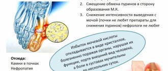
It should be noted that while taking aspirin, a person is less likely to remove uric acid from tissues. This can lead to the development of gout. The combined use of alcohol and aspirin is strictly contraindicated due to the high risk of bleeding of the digestive organs.
The administration of hydroxyurea is advisable for malignant neoplasms. The mechanism of action of the drug is based on the suppression of an enzyme necessary for the synthesis of DNA in mutant cancer cells after exposure to radiation or chemotherapy. Combined use with other methods of therapy significantly increases the inhibitory effect on the work of the bone marrow to produce blood cells.
The drug Bisulfan® has a similar effect. However, the mechanism of its destructive effect on tumor cells is the formation of cross-links between DNA strands. This leads to the impossibility of full functioning of the nucleic acid and cell death.
Anagrelide® is a drug that effectively reduces the number of platelets. However, the mechanism of its effect still remains questionable. The effectiveness and safety of anagrelide® has been proven in clinical and laboratory tests. It is assumed that the drug directly affects megakaryocytes, preventing their further differentiation into platelets. Megakaryocytes decrease in size and produce fewer blood cells.
Correction
Conservative therapy
In most cases, to correct thrombocytosis, it is enough to eradicate the cause, i.e. treatment of the underlying disease. Short-term thrombocytosis that develops due to stress or drug administration does not require intervention. In case of persistent long-term thrombocytosis, consultation with a hematologist is necessary to identify the cause and prescribe appropriate treatment. Therapy for thrombocytosis has several areas, including:
- Fighting infection
. To eliminate the infectious agent, antibacterial (amoxicillin), antifungal (fluconazole), and antiparasitic agents (mebendazole) are used. Treatment of viral hepatitis requires long-term use of peligated interferon in combination with antiviral drugs. - Treatment of iron deficiency anemia
. Correction of iron deficiency is carried out with tablet preparations (iron sulfate). For children, there are forms of syrup and drops for oral administration. The addition of ascorbic acid promotes better absorption. - Therapy of autoimmune diseases
. Treatment of autoimmune diseases is carried out using medications that suppress inflammation - glucocorticosteroids (prednisolone), immunosuppressants (cyclophosphamide). - Targeted therapy
. For myeloproliferative diseases, specific targeted treatment is prescribed to slow down the progressive growth of the malignant tumor. These drugs include Janus kinase inhibitors (ruxolitinib), tyrosine kinase inhibitors (imatinib, dasatinib). - Symptomatic treatment
. To relieve high thrombocytosis, medications are used that suppress the activity of the megakaryocyte lineage, and, consequently, the production of platelets - anagrelide, interferon-alpha, hydroxyurea. For polycythemia, regular bloodletting is successfully used as a treatment method to remove excess formed elements. - Blood thinning
. In case of high thrombocytosis, antiplatelet agents (acetylsalicylic acid) are prescribed to prevent thrombosis. In case of contraindications (peptic ulcer of the stomach, duodenum), platelet receptor blockers (clopidogrel, ticagrelor) are used. In people at high risk of thrombosis (elderly, patients with diabetes mellitus or atrial fibrillation), anticoagulants (warfarin, dabigatran) are used.
Specialized treatment
The only method that allows achieving complete recovery from a malignant hematological disease is allogeneic bone marrow transplantation. This requires HLA typing to select a compatible donor. However, due to the high risk of developing life-threatening complications, this method is used only if conservative treatment is ineffective.
Decreased platelet count
This condition is called thrombocytopenia ( platelet count 100 ) and often develops after uncontrolled medication use. Leukocytopenia can accompany pathologies such as cirrhosis, hepatitis, bone marrow damage, thyroid diseases, hematological diseases, alcoholism and other pathologies. As a result of these diseases, the fragility of blood vessels increases. Long periods of menstruation, cuts, tooth extraction, bleeding gums, and frequent nosebleeds can also contribute to low platelet counts.
Thrombocytosis rate table
First of all, medications are used to treat a low platelet count. If necessary, doctors prescribe platelet transfusions.
Forecast
The outcome depends on both the underlying pathology and the degree of thrombocytosis. For example, acute viral infection and iron deficiency anemia are characterized by a benign course. Patients with essential thrombocythemia, with proper selection of pathogenetic and symptomatic treatment, can live more than 80 years. People with chronic myeloid leukemia, on the other hand, live about 5-10 years from diagnosis.
Since reactive thrombocytosis almost always occurs in children, their prognosis is favorable. Thrombosis is not typical for mild to moderate thrombocytosis. In extreme or severe cases, there is a very high probability of fatal thrombosis leading to myocardial infarction, pulmonary infarction, and ischemic stroke.
If platelets are elevated in an adult, what does this mean?
At present, the definitive causes of such failures in the hematopoietic system have not been established. It is believed that increased megakaryocyte formation occurs due to mutations in the V617F gene.
Consider the main causes of reactive thrombocytosis:
- malignant neoplasms of the digestive, genitourinary or respiratory systems. Tumor cells produce substances that enhance the process of platelet formation;
- infectious infections of bacterial etiology. Thrombocytosis is rare in fungal or viral infections;
- damage to large bones, such as the femur or humerus;
- extensive surgery;
- splenectomy. Part of the platelets that should be deposited in the spleen enters directly into the bloodstream;
- dehydration;
- myeloproliferative pathologies;
- significant physical stress;
- bleeding in acute or chronic form. The acute form occurs abruptly against the background of mechanical trauma or surgery, the chronic form lasts for a long time, accompanies ulcers and oncopathologies of the digestive organs. The result is a decrease in the amount of iron ions in the blood against the background of an increased level of platelets. The mechanism of the phenomenon remains unclear;
- inflammation of organs and tissues, such as the walls of blood vessels, the large intestine, or joints. During inflammation, a specific substance is produced - thrombopoietin, which has a direct effect on the division of megakaryocytes, stimulating this process;
- taking synthetic analogues of hormones that are secreted by the adrenal glands, as well as chemotherapy drugs.
After the patient recovers, the criterion in question returns to within acceptable limits.
Read further: All the most important information about platelets in a blood test and their count according to Fonio