In this article you can not only evaluate your blood test, but also get answers to the following questions:
. | … |
Clinical (general) blood test
It is carried out with the aim of identifying the general picture of the state of blood in the human body, including helping in the diagnosis of various infectious and hematological diseases. Blood sampling from a person must be done on an empty stomach from a finger and does not require special preparation from the patient. During the analysis, the number of red blood cells, leukocytes, platelets, hemoglobin is counted, and the erythrocyte sedimentation rate is calculated. There are certain standards according to which any deviation from the required number of blood cells will be considered a sign of some kind of inflammatory process in the body or a serious disease, so the results of such an analysis will depend on whether the blood cell counts are normal or not.
In addition, you need to take into account that each laboratory has its own “standards” for a general (clinical) blood test, so it is better to ask your doctor all your questions.
A general blood test helps a doctor of any specialty. Based on the results of a blood test (hemogram), the doctor can competently assess the condition of the body, make a preliminary diagnosis and promptly prescribe appropriate treatment.
Norms of blood parameters
| Blood indicator | Normal values |
| Hemoglobin, g/l Men Women | 130,0-160,0 120,0-140,0 |
| Red blood cells (RBC), *1012/l Men Women | 4,0-5,0 3,9-4,7 |
| Hematocrit, % Men Women | 40-48 36-42 |
| Average hemoglobin content in erythrocytes (MCH), pg | 27,0-31,0 |
| Mean erythrocyte volume (MCV), fl, µm3 | 80,0-100,0 |
| Mean erythrocyte hemoglobin concentration (MCHC), g/dL | 30,0-38,0 |
| Red blood cell distribution width by volume (RDW-CV), % | 11,5-14,5 |
| Reticulocytes, ‰ (or%) | 2,0-12,0 (0,2-1,2) |
| Leukocytes, *109/l | 4,0-9,0 |
| Neutrophils, % (109/l) Bands Segmented | 1,0-6,0 (0,04-0,30) 47,0-72,0 (2,0-5,5) |
| Eosinophils | 0,5-5,0 (0,02-0,3) |
| Basophils | 0-1,0 (0-0,065) |
| Lymphocytes | 19,0-37,0 (1,2-3,0) |
| Monocytes | 3,0-11,0 (0,09-0,6) |
| Platelets, *109/l | 180,0-320,0 |
| Mean platelet volume (MPV), fl | 7,4-10,4 |
| Platelet distribution width by volume, (PDW), % | 10-20 |
| Thrombocrit (PCT), % | 0,15-0,40 |
| Erythrocyte sedimentation rate (ESR), mm/h | 2,0-20,0 |
How to take a general blood test and what is needed for this?
There are no complex, strict regulations regarding this testing, but there are some rules:
- For this examination, capillary blood is used, which is taken from a finger. Less often, according to the doctor's instructions, blood from a vein can be used.
- The analysis is carried out in the morning. The patient is prohibited from consuming food or water 4 hours before taking a blood sample.
- The main medical supplies used for drawing blood are a scarifier, cotton wool, and alcohol.
What does a general blood test show?
A general (clinical) blood test shows:
- number of red blood cells,
- erythrocyte sedimentation rate (ESR),
- hemoglobin content,
- number of leukocytes,
- leukocyte formula
- and other indicators, each of which we will dwell on in detail.
blood cells are also known as red blood cells. In humans, 1 mm³ of blood contains 4.5-5 million red blood cells. Red blood cells contain hemoglobin and carry oxygen and carbon dioxide. An increase in the number of red blood cells is a sign of diseases such as leukemia, chronic lung diseases, and congenital heart defects. Anemia (decreased number of red blood cells) can be caused by stress, increased physical activity, and fasting. If you cannot immediately determine the cause of the decrease in the number of red blood cells, then it is better to go to a hematologist and undergo additional examination.
A significant increase in the content of red blood cells may indicate erythremia (one of the blood diseases). In addition, an increase in the number of red blood cells (erythocytosis, polycythemia) is observed in acute poisoning, when due to severe vomiting and diarrhea there is a large deficiency of fluid in the body; with acidosis (due to metabolic disorders during exacerbation of certain diseases); when losing fluid for various reasons (heat, illness, heavy physical activity); with long-term cardiovascular or pulmonary diseases, when the body is not sufficiently supplied with oxygen and increases the number of red blood cells in an attempt to still deliver oxygen to the tissues; or when a person is in the highlands, when he no longer has enough oxygen.
Color index - its normal value for people of any age is 0.85-1.15. The blood color index is an indicator of the degree of saturation of red blood cells with hemoglobin and reflects the relationship between the number of red blood cells and hemoglobin in the blood. When its values differ from the norm, this generally indicates the presence of anemia. In this case, anemia is divided into: - hypochromic - color index less than 0.85; - hyperchromic - color index greater than 1.15.
However, anemia can also be normochromic - when the color indicator remains within the normal range.
Reticulocytes are young forms of red blood cells. Children have more of them, adults have less, because the formation and growth of the body has already been completed. An increase in the number of reticulocytes can be observed in anemia or malaria. A decrease in the number of reticulocytes or their absence is an unfavorable sign in anemia, indicating that the bone marrow has lost the ability to produce red blood cells.
The erythrocyte sedimentation rate (ESR) determines how quickly red blood cells settle in a test tube and separate from the blood plasma. In women, the ESR rate is slightly higher than in men; during pregnancy, the ESR increases. Normally, the ESR value in men does not exceed 10 mm/hour, and in women - 15 mm/hour . The ESR indicator may change depending on various factors, including due to various diseases.
An increase in ESR in a blood test is one of the indicators that makes the doctor assume that the patient has an acute or chronic inflammatory process (pneumonia, osteomyelitis, tuberculosis, syphilis), and an increase in ESR is characteristic of poisoning, myocardial infarction, trauma, bone fractures, anemia, kidney diseases, cancer. It is observed both after operations and as a result of taking certain medications. A decrease in ESR occurs during fasting, with a decrease in muscle mass, and when taking corticosteroids.
Hemoglobin is a complex iron-containing protein found in red blood cells - erythrocytes - of animals and humans, capable of reversibly binding with oxygen, ensuring its transfer to tissues. The normal content of hemoglobin in human blood is considered to be: for men 130-170 g/l, for women 120-150 g/l; in children - 120-140 g/l. Blood hemoglobin is involved in the transport of oxygen and carbon dioxide and maintains pH balance. Therefore, determining hemoglobin is one of the most important tasks of a general blood test.
Low hemoglobin (anemia) can be the result of large blood loss; a decrease in hemoglobin occurs when there is a lack of iron, a necessary material for the construction of hemoglobin. Also, low hemoglobin (anemia) is a consequence of blood diseases and many chronic diseases not associated with them.
A hemoglobin level higher than normal can be an indicator of many blood diseases, and a complete blood count will also show an increase in red blood cells. Increased hemoglobin is typical for people with congenital heart defects and pulmonary heart failure. An increase in hemoglobin can be caused by physiological reasons - in pilots after flights, mountain climbers, after significant physical activity, the hemoglobin level is higher than normal.
Leukocytes are the protectors of our body from foreign components. The blood of an adult contains an average of 4-9x10 9/l leukocytes . White blood cells fight viruses and bacteria and cleanse the blood of dying cells. There are several types of leukocytes (monocytes, lymphocytes, etc.). The leukocyte formula allows you to calculate the content of these forms of leukocytes in the blood.
If leukocytes are found in increased numbers in a blood test, this may mean the presence of viral, fungal or bacterial infections (pneumonia, tonsillitis, sepsis, meningitis, appendicitis, abscess, polyarthritis, pyelonephritis, peritonitis), and also be a sign of poisoning of the body (gout ). Previous burns and injuries, bleeding, postoperative condition of the body, myocardial infarction, lung, kidney or spleen, acute and chronic anemia, malignant tumors - all these “troubles” are accompanied by an increase in the number of blood leukocytes.
In women, a slight increase in leukocytes in the blood is also observed in the period before menstruation, in the second half of pregnancy and during childbirth.
A decrease in the number of white blood cells, which a blood test can show, may be evidence of viral and bacterial infections (influenza, typhoid fever, viral hepatitis, sepsis, measles, malaria, rubella, mumps, AIDS), rheumatoid arthritis, kidney failure, radiation sickness, some forms of leukemia, bone marrow diseases, anaphylactic shock, exhaustion, anemia. A decrease in the number of leukocytes can also be observed while taking certain medications (analgesics, anti-inflammatory drugs).
Platelets - these cells are also called blood plates. They are the smallest blood cells. The main role of platelets is participation in blood clotting processes. In blood vessels, platelets can be located near the walls and in the bloodstream. At rest, platelets have a disc-shaped shape. If necessary, they become like a sphere and form special outgrowths (pseudopodia). With their help, blood platelets can stick to each other or stick to a damaged vascular wall.
A decrease in the number of platelets is observed in women during menstruation and during normal pregnancy, and an increase occurs after physical activity. Also, the number of platelets in the blood has seasonal and daily fluctuations. Typically, platelet monitoring is prescribed when taking certain medications, when a person has burst capillaries for no reason, has frequent nosebleeds, or when being examined for various diseases.
An increase in the number of platelets in the blood (so-called thrombocytosis) occurs during: - inflammatory processes (acute rheumatism, tuberculosis, ulcerative colitis); - acute blood loss; - hemolytic anemia (when red blood cells are destroyed); — conditions after removal of the spleen; - observed during treatment with corticosteroids; - some rarer diseases.
A decrease in the number of platelets (thrombocytopenia) is observed in a number of hereditary diseases, but appears much more often in acquired diseases. The number of platelets decreases with: - severe iron deficiency anemia; - some bacterial and viral infections; - liver diseases; - diseases of the thyroid gland; - the use of a number of medications (vinblastine, chloramphenicol, sulfonamides, etc.); - systemic lupus erythematosus.
Hematocrit is the percentage (percentage) of the total blood volume that is made up of red blood cells. Normally, this figure is 40-48% for men, 36-42% for women.
The volume of red blood cells compared to plasma increases with: - dehydration (dehydration), which happens with toxicosis, diarrhea, vomiting; - congenital heart defects, accompanied by insufficient oxygen supply to the tissues; — a person being in high altitude conditions; - insufficiency of the adrenal cortex.
The volume of red blood cells relative to plasma decreases with blood thinning (hydremia) or with anemia.
Hydremia can be physiological if a person immediately drinks a lot of liquid. After significant blood loss, compensatory hydremia occurs when blood volume is restored. Pathological hydremia develops when water-salt metabolism is disturbed and occurs with glomerulonephritis, acute and chronic renal failure, and with heart failure during the period of swelling.
Blood formula . The study of the leukocyte formula has important diagnostic value, showing characteristic changes in a number of diseases. But these data should always be assessed together with other indicators of the blood system and the general condition of the patient.
For various diseases, a combination of the following signs is looked at: the total number of leukocytes; the presence of a nuclear shift of neutrophils (the so-called “shift according to the formula to the left”, that is, the appearance of young, immature forms of neutrophils in the blood); percentage of individual leukocytes; the presence or absence of degenerative changes in cells.
Blood chemistry
It is carried out to assess the functional state of the body, the functioning of its internal organs, as well as metabolism. This blood test can check your blood levels of protein, sugar, iron, cholesterol, bilirubin, triglycerides, various enzymes, calcium, magnesium, sodium, phosphorus and various gases. Any deviation from the norm may signal that some processes may be going on in the body that are invisible from the outside (parasitic infections, tumors, allergies). Patients with poor results of biochemical analysis may have diseases of the thyroid gland, liver, and possible diabetes mellitus and atherosclerosis. Depending on the type of biochemical analysis, preparation for blood sampling may vary, including following a diet for several days.
How to prepare for a biochemical blood test?
One day before taking blood for biochemistry, it is necessary to avoid drinking alcohol, and 1 hour before taking smoking. It is advisable to take blood samples on an empty stomach in the morning. There should be at least 12 hours between the last meal and the blood draw. Juice, tea, coffee, chewing gum are not allowed. You can drink water. It is necessary to exclude increased psycho-emotional and physical stress.
How are the results of a biochemical blood test evaluated?
The use of different diagnostic methods by different clinics leads to different results, and the units of measurement may also differ. Therefore, to correctly decipher the result of a biochemical blood test, a consultation with the attending physician is required.
What indicators are included in a standard biochemical blood test?
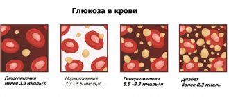
1) Glucose (in blood) is the main test in the diagnosis of diabetes mellitus. This analysis is very important when selecting therapy and assessing the effectiveness of diabetes treatment. A decrease in glucose levels is observed in some endocrine diseases and liver dysfunction.
Normal blood glucose levels:
| Age | Glucose level, mmol/l |
| < 14 years | 3,33 — 5,55 |
| 14 – 60 years | 3,89 — 5,83 |
| 60 - 70 years | 4,44 — 6,38 |
| > 70 years | 4,61 — 6,10 |
2) Common bilirubin is a yellow blood pigment that is formed as a result of the breakdown of hemoglobin, myoglobin and cytochromes. The main reasons for the increase in the amount of total bilirubin in the blood: damage to liver cells (hepatitis, cirrhosis), increased breakdown of red blood cells (hemolytic anemia), impaired outflow of bile (for example, cholelithiasis).
Normal values of total bilirubin: 3.4 - 17.1 µmol/l.
3) Direct bilirubin (conjugated, bound bilirubin) - a fraction of total blood bilirubin. Direct bilirubin increases with jaundice, which develops due to a violation of the outflow of bile from the liver.
Normal values of direct bilirubin: 0 - 7.9 µmol/l.
4) Indirect bilirubin (unconjugated, free bilirubin) - the difference between the indicators of total and direct bilirubin. This indicator increases with increased breakdown of red blood cells - with hemolytic anemia, malaria, massive hemorrhages in tissue, etc.
Normal values for indirect bilirubin: < 19 µmol/l.
5) AST (AST, aspartate aminotransferase) is one of the main enzymes synthesized in the liver. Normally, the content of this enzyme in the blood serum is low, since most of it is found in hepatocytes (liver cells). An increase is observed with liver and heart diseases, as well as with long-term use of aspirin and hormonal contraceptives.
Normal AST values:
- Women – up to 31 U/l;
- Men - up to 37 U/l.
6) ALT (ALT, alanine aminotransferase) is an enzyme synthesized in the liver. Most of it is located and works in liver cells, so normally the concentration of ALT in the blood is low. An increase is observed with massive death of liver cells (for example, with hepatitis, cirrhosis), severe heart failure and blood diseases.
Normal ALT values:
- women – up to 34 U/l;
- men – up to 45 U/l.
7) Gamma-GT (gamma-glutamyltransferase) is an enzyme found primarily in the cells of the liver and pancreas. An increase in its amount in the blood is observed with diseases of these organs, as well as with prolonged use of alcohol.
Normal gamma-GT values:
Men < 55 U/l
Women < 38 U/l
 Alkaline phosphatase is an enzyme widely distributed in human tissues. The hepatic and bone forms of alkaline phosphatase are of greatest clinical importance, the activity of which is determined in the blood serum.
Normal alkaline phosphatase values: 30-120 U/l.
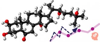
9) Cholesterol (total cholesterol) is the main blood lipid that enters the body with food and is also synthesized by liver cells.
Normal cholesterol levels: 3.2-5.6 mmol/l.
10) Low-density lipoproteins (LDL) are one of the most atherogenic, “harmful” lipid fractions. LDL is very rich in cholesterol and, transporting it to vascular cells, lingers in them, forming atherosclerotic plaques.
Normal LDL levels: 1.71-3.5 mmol/l.
11) Triglycerides are neutral fats found in the blood plasma, an important indicator of lipid metabolism.
Normal triglyceride levels: 0.41-1.8 mmol/l.
12) Total protein - an indicator reflecting the total amount of proteins in the blood. Its decrease is observed in some diseases of the liver and kidneys, accompanied by increased excretion of protein in the urine. Increased in blood diseases and infectious and inflammatory processes.
Normal values for total protein: 66-83 g/l.
13) Albumin is the most important blood protein, making up approximately half of all serum proteins. A decrease in albumin content can also be a manifestation of certain diseases of the kidneys, liver, and intestines. Elevated albumin is usually associated with dehydration.
Normal albumin values: 35-52 g/l
14) Potassium (K+) is an electrolyte found primarily inside cells. An increase in the level of potassium in the blood is most often observed in acute and chronic renal failure, a sharp decrease in the amount of urine excreted or its complete absence, most often associated with severe kidney disease.
Normal potassium values: 3.5-5.5 mmol/l.
15) Sodium (Na+) is an electrolyte found primarily in extracellular fluid and in smaller quantities inside cells. It is responsible for the functioning of nervous and muscle tissue, digestive enzymes, blood pressure, and water metabolism.
Normal sodium values: 136-145 mmol/l.
16) Chlorine (Cl-) is one of the main electrolytes, which is in the blood in an ionized state and plays an important role in maintaining water-electrolyte and acid-base balances in the body.
Normal chlorine values: 98-107 mmol/l.
17) Creatinine is a substance that plays an important role in the energy metabolism of muscle and other tissues. Creatinine is completely excreted by the kidneys, so determining its concentration in the blood is of greatest clinical importance for diagnosing kidney diseases.
Normal creatinine values:
- men - 62 – 115 µmol/l;
- women - 53 - 97 µmol/l.
18) Urea is a substance that is the end product of protein metabolism in the body. Urea is excreted by the kidneys, so determining its concentration in the blood gives an idea of the functional abilities of the kidneys and is most widely used for diagnosing renal pathology.
Normal urea values: 2.8-7.2 mmol/l
19) Uric acid is one of the end products of protein metabolism in the body. Uric acid is completely excreted by the kidneys. An increase in the concentration of uric acid occurs in kidney stones and other kidney diseases that occur with renal failure.
Normal uric acid values:
- men - 210 - 420 µmol/l;
- women - 150 - 350 µmol/l.
20) C-reactive protein (CRP) is a sensitive element in the blood that reacts faster than others to tissue damage. The presence of reactive protein in blood serum is a sign of an inflammatory process, injury, or penetration of foreign microorganisms into the body - bacteria, parasites, fungi. The more acute the inflammatory process, the more active the disease, the higher the C-reactive protein in the blood serum.
Normal values for C-reactive protein: 0 - 5 mg/l.
21) Iron (serum iron) is a vital microelement that is part of hemoglobin, is involved in the transport and deposition of oxygen and plays an important role in hematopoiesis.
Normal serum iron values:
- women - 8.95 - 30.43 µmol/l;
- men - 11.64 - 30.43 µmol/l.
Immunological blood test
Few people know what blood tests there are from the point of view of the human immunological system, since these tests are not so common. As a rule, such a blood test provides information about the immunodeficiency virus in the human body and is anonymous, since it is carried out at the request of the patient. For sampling, blood taken from a vein on an empty stomach is used, from which serum is obtained for testing by centrifugation. In addition, the study of blood serum can detect a number of sexually transmitted diseases (syphilis, herpes, chlamydia), as well as all types of hepatitis, measles, rubella, mumps and toxoplasmosis.
Basically, for ELISA analysis, the biological material being studied is blood, however, cerebrospinal fluid, vitreous contents, amniotic fluid, etc. can be examined.
What is immunoglobulin (antibody)?
Immunoglobulins are immune molecules that can bind to and neutralize most infectious pathogens and toxins in the body. In this case, the most important characteristic of immunoglobulins is their specificity, that is, the ability to bind to a specific antigen. It is this property that is used to conduct a blood test for immunoglobulin.
There are five types of immunoglobulins, but the most studied are immunoglobulins A, M, and G. Immunoglobulins M and G show their activity in the blood. Immunoglobulins A are a kind of barrier on the surface of the mucous membranes, as they are present there in large quantities.
An immunological blood test allows you to determine the type of immunoglobulins, thanks to which ELISA allows you not only to diagnose the disease, but also to determine the stage of the disease and monitor the dynamics of the disease:
- In the first 2 weeks of the disease, only immunoglobulins A are detected.
- From the 2nd to the 3rd week of the disease, immunoglobulins A and M are detected in the blood.
- From 3 to 4 weeks, a blood test for immunoglobulin determines all three types.
- During recovery, immunoglobulins M disappear from the blood, and the amount of A and G decreases by 2–4 times.
- In the presence of a chronic process, immunoglobulins G are necessarily present in the blood, immunoglobulins M are absent, immunoglobulins A may or may not be present.
Advantages of enzyme immunoassay (ELISA):
- Relatively high sensitivity (accuracy).
- Reasonable price.
- Allows for early diagnosis (due to the ability to determine immunoglobulin classes during analysis).
- Allows you to monitor the dynamics of the infectious process (thanks to the ability to determine immunoglobulin classes).
- Ease of use.
- An immunological blood test allows you to get a quick answer.
Disadvantages of ELISA blood test:
- Sometimes enzyme immunoassays give false positive or false negative results.
Scope of application of immunological blood test
- Diagnosis of viral diseases: hepatitis, herpes, Epstein-Barr virus, cytomegalovirus, etc.
- Sexually transmitted infections: chlamydia, gonorrhea, trichomonas, mycoplasma, ureaplasma.
- Syphilis.
- Endocrinology (determination of hormone levels).
- Tumor markers (diagnosis of cancer).
- Immunology (diagnosis of immunodeficiency).
- Allergology (diagnosis and treatment of allergies).
Serological blood test is a laboratory method of blood testing that is used to diagnose infectious diseases and determine the stage of the infectious process. The serological reaction is based on the interaction of antibodies and antigens.
Determination of antigens is used to determine the genus and species of microorganisms. This research method is used in urology and venereology. Blood for a serological blood test is taken in the morning on an empty stomach from a vein.
Why should you contact the Gynecology Clinic?
In our clinic, we offer to take a blood test for biochemistry and get a transcript of the tests quickly and without queues. We use modern equipment for carrying out biochemical reactions and accelerated methods for determining the genus and type of bacteria: photoelectrocolorimeter, a system of indicator papers, immunofluorescence express diagnostic method. The automated analyzers that our clinic has in its arsenal allow us to identify pathogens and also determine their sensitivity to antibiotics within a few hours.
All laboratory tests are carried out by highly qualified specialists. A competent approach to conducting a biochemical blood test and correct interpretation of the results obtained help the doctor understand the nature of the disease, make a diagnosis, prescribe treatment or take effective preventive measures. If you want to do blood biochemistry, call us, we guarantee comfortable service, efficiency and reliability of the research.
Clinic of Aesthetic Gynecology - 20 years of experience.
Do you want to undergo an examination, make an appointment with a gynecologist, take tests, do an ultrasound?
+7-343-385-72-88 Call by phone in Yekaterinburg
Qualified specialists of the Clinic of Aesthetic Gynecology - gynecologists, endocrinologists, urologists - will help in solving the problems that concern you.
You can also make an appointment with a specialist online. Fill out the application, indicate a convenient appointment time right now! For convenience, check the specialists' work schedule.
Don't put off taking care of your health!
Hormonal blood test
This type of study helps determine the general functioning of the organs of the human endocrine system, as well as identify diseases of the pituitary gland, thyroid gland, adrenal glands and other human endocrine organs. Blood is drawn on certain days depending on the production of the hormone that will be tested. This can be an analysis of thyroid hormones (T4, T3), pituitary gland (TSH, LH, FSH), sex glands (testosterone, estriol), adrenal glands (cortisol), as well as pregnancy hormone (hCG). The results will depend entirely on the age and gender of the person, as well as whether there are any diseases in his endocrine system.
Preparing for a hormone test
The amount of hormones in the blood depends on the time of day, since there is a daily rhythm of secretion (release of hormones). Blood for hormonal analysis should be taken in the morning, on an empty stomach.
In women, hormonal levels also depend on the stage of the menstrual cycle. The most favorable days for analysis are days 5-7 of the cycle, starting from the first day of menstruation.
On the eve of the test, you should not drink alcohol, and you should also avoid increased physical activity and stressful situations. It is advisable not to smoke for an hour before the test.
A week before the test, you must stop taking hormonal medications. If you are prescribed medication, discuss this with your doctor; the test may have to be postponed.
How to take a hormone test correctly?
The human hormonal system is connected to all organs and systems of the body. Therefore, the level of hormones in the blood may depend on various external influences and changes in the human body. General recommendations from doctors are as follows: you need to take a blood test for hormones in the morning on an empty stomach. Before donating blood for hormone testing, stop smoking, drinking alcohol and strenuous physical activity. Women need to have many hormones analyzed on certain days of the menstrual cycle. To ensure that the results of your hormone tests are as reliable as possible, consult your doctor and follow his recommendations.
When is a blood test for hormones prescribed?
Hormonal analysis is usually carried out if there is a suspicion of dysfunction of the endocrine glands or if an increase in the size of the glands is detected.
Indications for testing for female sex hormones (estrogens) are:
- menstrual irregularities;
- infertility;
- miscarriage;
- acne;
- overweight;
- fibrocystic mastopathy (breast disease).
Indications for testing for male sex hormones (androgens) are:
- suspicion of the development of tumor processes;
- ovarian dysfunction;
- kidney dysfunction;
- overweight (obesity);
- infertility;
- acne;
- in women – excessive growth of body hair.
Hormonal analysis is prescribed during pregnancy if pathological development of the fetus is suspected. An analysis for the hormone hCG (human chorionic gonadotropin), produced by the cells of the membrane of the embryo, can detect pregnancy already on the 6-10th day after fertilization.
Interpretation of blood test results for hormones
Blood from a vein is used to test hormones.
Depending on the clinical signs indicating a specific pathology, an analysis with tests for specific hormones is usually prescribed.
The most complete picture of your health can be obtained by taking a test for the following hormones.
Thyroid hormones:
- T3 (triiodothyronine) free – stimulates oxygen metabolism in tissues. Normal values: 2.6 - 5.7 pmol/l.
- T4 (thyroxine) – stimulates protein synthesis. Normal values: 0.7-1.48 ng/dl.
- Antibodies to thyroglobulin (AT-TG) are an important parameter for identifying a number of autoimmune diseases. Normal values: 0-4.11 U/ml.
- Some others.
Pituitary hormones:
- TSH (thyroid-stimulating hormone) – stimulates the production of thyroid hormones (T3 and T4). Normal values: 0.4-4.0 mU/l. An elevated TSH value usually indicates decreased thyroid function.
- FSH (follicle stimulating hormone) . Normal values: in women - depends on the phase of the menstrual cycle. Phase I – 3.35-21.63 mU/ml; Phase II – 1.11-13.99 mU/ml; postmenopause – 2.58-150.53 mU/ml; girls under 9 years old 0.2-4.2 mU/ml. In men – 1.37-13.58 mU/ml.
- LH (luteinizing hormone) . Normal values: in women - depends on the phase of the menstrual cycle. Phase I – 2.57-26.53 mU/ml; Phase II – 0.67-23.57 mU/ml; postmenopause - 11.3-40 mU/ml; girls under 9 years old - 0.03-3.9 mU/ml. In men – 1.26-10.05 mU/ml.
- Prolactin . The main function is to stimulate the development of mammary glands and lactation. Normal values: in women (from the first menstruation to menopause) - 1.2-29.93 ng/ml; in men - 2.58-18.12 ng/ml. Increased concentrations of prolactin are called hyperprolactinemia . There are physiological and pathological hyperprolactinemia. Physiological hyperprolactinemia can be caused by breastfeeding, pregnancy, severe physical activity, and stress. An increased concentration of prolactin in women leads to menstrual irregularities and may cause infertility. In men, hyperprolactinemia leads to decreased libido and impotence.
- ACTH (adrenocorticotropic hormone) – stimulates the synthesis and secretion of hormones from the adrenal cortex. Normal values: 9-52 pg/ml.
- Some others.
Sex hormones:
- Testosterone (male sex hormone) is produced by the adrenal glands and in the gonads (in men - in the testicles, in women - in the ovaries). Affects the development of the genital organs, the formation of secondary sexual characteristics, the growth of bones and muscles. Normal values: for men - 4.94-32.01 nmol/l, for women - 0.38-1.97 nmol/l.
- Estrogens (female sex hormones). The main estrogens - progesterone and estradiol - are produced by the adrenal glands and ovaries. Normal values of progesterone in women depend on the phase of the menstrual cycle: Phase I - 1.0-2.2 nM/l; Phase II - 23.0-30.0 nM/l; for postmenopause - 1.0-1.8 nM/l. Similarly for estradiol: phase I - 198-284 pM/l: phase II - 439-570 pM/l; for postmenopause - 51-133 pM/l. Elevated estrogen levels may indicate tumors of the ovaries and adrenal cortex, as well as cirrhosis of the liver. Reduced - for insufficient development and sclerosis of the ovaries.
Adrenal hormones:
- DEA-s (dehydroepiandrosterone sulfate) is necessary for the synthesis of testosterone and estrogens. The range of normal values for the concentration of this hormone: 3591 – 11907 nmol/l; in women - 810 - 8991 nmol/l. However, this is the general picture; when processing analysis data, the patient’s age must also be taken into account.
- Cortisol is involved in many metabolic processes and is actively produced as a result of the body’s response to hunger or stress. Normal values: for children under 16 years of age - 3-21 mcg/dl, for an adult - 3.7-19.4 mcg/dl.
- Aldosterone is responsible for regulating water-salt balance in the body. Normal values: 35 - 350 pg/ml.
Abnormal alkaline phosphatase - causes
The causes of increased alkaline phosphatase levels can be grouped as follows:
- Damage or destruction (destruction) of the liver, problems with the movement of bile:
- viral and autoimmune hepatitis;
- liver pathology caused by toxins and drugs;
- formation of bile in the ducts of stones;
- primary sclerosing cholangitis - manifested by inflammation and narrowing of the intrahepatic ducts;
- Infectious mononucleosis;
- stagnation of bile – cholestasis.
- Pathology of bone tissue:
- osteomalacia or softening of the bones - this systemic damage is characterized by a violation of mineral metabolism and loss of calcium salts, vitamins and phosphoric acid, as a result of which the bones soften and become deformed;
- increased metabolism in bone tissue that occurs during fracture healing;
- Paget's disease - characterized by significant destruction of bone tissue, deformation and weakening of bones, most often affecting men over 50 years of age;
- osteosarcoma and metastases penetrating into bone tissue.
III. Other pathologies:
- primary and secondary hyperparathyroidism are diseases of the endocrine system with severe disturbances in the metabolism of phosphorus and calcium;
- myocardial infarction;
- gastrointestinal pathology.
- Non-pathological reasons:
- alcohol poisoning;
- third trimester of pregnancy;
- lactation;
- the use of drugs with hepatotoxic effects - they can negatively affect the liver and cause structural and functional disorders;
- excessive physical activity;
- poor nutrition and vitamin deficiency;
- possible deviations during childbirth and after menopause vary from person to person.
Exceeding the enzyme norm does not always indicate the type of disease. Perhaps the person is healthy and the increase in alkaline phosphatase is due to physiological properties. Therefore, further tests need to be carried out to determine the exact causes.
Definitive blood tests
These types of blood tests are very different from those listed above in that they do not diagnose any disease or health condition of a person, but simply indicate the peculiarity of his circulatory system. These include tests to determine blood type, Rh factor and coagulation. As a rule, these studies are mandatory for people in special professions, as well as for all pregnant women. Blood is taken for analysis from a vein and does not require special preparation of the patient.
Preparing to donate blood for biochemistry
The quantitative and qualitative composition of blood normally does not undergo serious fluctuations. Diseases significantly affect hematological parameters. To reliably track them, before going for research, you must meet some basic conditions:
- food – the day before there should be a usual diet with the exception of fried, fatty, spicy, “exotic” dishes;
- medications - in agreement with the treating doctor, stop taking medications that affect biochemical blood parameters: hormones, statins, antibiotics (if this condition cannot be met, then you need to warn the doctor about this)
- bad habits - exclude alcohol and energy drinks for 2-3 days, and do not smoke immediately before the test;
- stressful situations - they need to be neutralized as much as possible, and before the manipulation, rest for a few minutes;
- on the day of donation - blood is drawn in the morning, always on an empty stomach.
The attending physician should check the norms and pathological abnormalities obtained in blood biochemistry, because independent interpretation of tests without knowledge of the physiology and pathophysiology of the body cannot be correct. You can simply miss or aggravate the course of the disease. Another important point is the study of blood over time, which is preferably done in the same laboratory.