Monocytes are the largest immune cells of all types of white blood cells and play an important role in protecting against bacteria and inflammation. What does an elevated monocyte count mean for your health? What factors can reduce their number? Find out about this in the article.
The article is based on the findings of 68 scientific studies
The article quotes authors such as:
- Rockefeller University, New York, USA
- Necker Hospital, Paris, France
- Department of Cell Biology and Genetics, Erasmus University Medical Center, Rotterdam, The Netherlands
Please note that the numbers in parentheses (1, 2, 3, etc.) are clickable links to peer-reviewed scientific studies. You can follow these links and read the original source of information for the article.
What are monocytes?
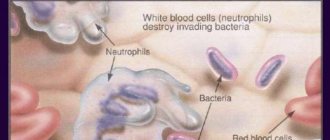
The front line of defense for your immune system
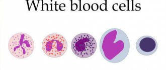
Monocytes are the largest type of white blood cell.
They help fight bacteria, viruses and other infections in your body. Along with other types of white blood cells, monocytes are a key part of your immune response. Only about 1% of your blood contains white blood cells, but they play a huge role in protecting against disease. Approximately 2 to 10% of all leukocytes are monocytes. (1) Your bone marrow produces monocytes and releases them into your bloodstream. Once they reach tissues in your body, they are called macrophages . There they isolate and devour germs and other harmful microorganisms. They also get rid of dead cells and promote the immune response. After leaving the bone marrow, monocytes circulate in the blood for several days before they enter tissues where they become macrophages or dendritic cells. (1, )
An example of how monocytes from the blood move into
an atherosclerotic plaque , becoming macrophages.
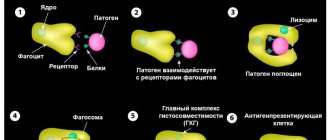
Monocytes adhere to the endothelium of blood vessels at sites of tissue damage, which leads to their activation and migration into the subendothelial space. Upon tissue entry, the monocyte differentiates into a macrophage. (source) Monocytes protect against viral, bacterial, fungal and protozoal infections. They kill microorganisms, absorb foreign particles, remove dead cells and enhance the immune response. (, 1, , )
However, they may also be involved in the development of inflammatory diseases such as arthritis and atherosclerosis. In this article, we'll take a closer look at how monocytes work and how they may be involved in disease. (, , )
Monocyte formation
All blood cells come from common parent cells called hematopoietic stem cells . In adults, blood cells are produced primarily in the bone marrow; this process is called hematopoiesis. The process of producing monocytes in particular is called myelopoiesis . (, )
Mechanisms of myelopoiesis (monocyte formation) during homeostasis and during different phases of infection and inflammation, illustrating important feedback loops between affected tissues and bone marrow.
Myelopoiesis is subject to a complex regulatory system, including such factors as:
- Transcription factor SPI1 (, , , )
- Cytokines: SCF (stem cell factor), GM-CSF (granulocyte-macrophage-colony-stimulating factor), M-CSF (macrophage colony-stimulating factor, CSF1), cytokines IL-3 and IL-6, and interferon IFN-gamma. (, , )
Monocytes live on average 3 days before undergoing apoptosis (programmed cell death). They live longer during periods of high inflammation; as soon as the inflammation disappears, monocyte death occurs. Such programmed death is necessary to prevent aggressive actions of immune cells from continuing inflammation and creating an autoimmune reaction. (, )
Clinical significance of a blood test (excerpt from a lecture by Prof. E.B. Vladimirskaya)
Hematology:
12.03.2009
Doctor of Medical Sciences, Prof. E.B. Vladimirskaya Research Institute of Pediatric Hematology, Ministry of Health of Russia
Blood, being the internal environment of the body, carries within itself the stigmata of the vital functions of various organs and systems, the study of which is of undoubted clinical significance and is necessary for the diagnosis, prognosis of the course and monitoring of therapy of almost all internal human diseases.
The most accessible is the study of the morphological composition of blood; its results are included in the diagnostic algorithm for most pathological processes.
Since the first studies of blood under a microscope without the use of staining (the middle of the last century), blood cells have been divided into red - erythrocytes (based on the color of hemoglobin) and white - leukocytes. Leukocytes, in turn, are divided into cells containing specific granularity in the cytoplasm, and according to the ratio of this granularity to color, they are divided into (neutrophils, eosinophils and basophils) and those without - lymphocytes and monocytes. Based on the shape of the nucleus, the former are often called polymorphonuclear cells, and the latter are called mononuclear cells.
In recent years, clinical blood tests have increasingly been performed on automatic counters, which significantly increases the accuracy of calculations, but, however, does not negate the value of data obtained “manually” using light-optical microscopy. A comparison of the results of these two methods, together with the reference values of the indicators used, is presented in Table 1.
Table 1
| Automatic counting | Units _ | Normal limits | Manual counting |
| hgb -hemoglobin | g/liter | M: 132 - 173 F: 117 - 155 | Hb |
| rbc- red blood cells | 1012 /liter | M: 4.3 – 5.7 F: 3.8 – 5.1 | er. |
| hct-hematocrit | % | M: 39 – 49 F: 35 – 45 | ht |
| mcv - average volume red blood cell | 1 µm3 = 1 femtoliter | 80,0 – 95,0 | Spherical index (3,2-3,4) |
| mch - average hemoglobin content in an erythrocyte | picograms 1 g = 1012 picograms | 27,0 – 31,0 | Color index (0,85 – 1,0) Color.= Nb (g/l) x 3_____ Er (first three digits) |
| mchc – average concentration of HB in 1 erythrocyte | g/dl | 32,0 — 36,0 | |
| rdwwidth of distribution of red blood cells by volume | width histograms | 11,5 – 14,5 | No analogue |
| plt – platelets | *109 /l | 150 — 400 | Platelets |
| wbc- leukocytes | *109 /l | 4,5 –11,0 | Leukocytes |
| neu - neutrophils | *109 /l % | 1,8 – 5,5 47,0 – 72,0 | Neutrophils |
| lym – lymphocytes | *109 /l % | 1,2 – 3,0 19,0 – 37,0 | Lymphocytes |
| mon – monocytes | *109 /l % | 0,1 – 0,9 3,0 – 11,0 | Monocytes |
| eos – eosinophils | *109 /l % | 0,02 – 0,3 0,5 – 5,0 | Eosinophils |
| bas – basophils | *109 /l % | 0,0 – 0,07 0,0 –1,0 | Basophils |
When commenting on the data presented in the table, it is necessary to note:
- the vast majority of automatic counters do not detect young forms of leukocytes, normoblasts and reticulocytes - this data can only be obtained “manually”;
- normative values are never expressed in one figure; there is a limit of permissible fluctuations (it is presented in the table for all indicators), which fits 99.9% of the norm.
Let's analyze the clinical significance of individual blood test indicators.
Red blood counts.
Anemia is a decrease in hemoglobin below 120 g/l: mild - 110-120 g/l; average degree – 90-110 g/l; severe degree – below 90 g/l.
Based on the ratio of red blood indicators, 3 types of anemia are distinguished, which is the starting point for further diagnosis.
Microcytic-hypochromic anemia:
MCV < 80 Color.p. <0.85
MCH < 27; MCHC < 32
- Iron-deficiency anemia
- Anemia due to chronic inflammation
- Congenital spherocytic hemolytic anemia.
- Thalassemia.
Normocytic-normochromic anemias:
MCV 80 – 95
MCH = 27-31; MCHC = 32-36 Color.p. = 0.85-1.0
Acute blood loss.
- Anemia in chronic renal failure
- Anemia due to endocrine pathology.
- Anemia in cancer.
- Hemolytic anemias, immune and non-immune.
- Aplastic anemia.
- Myelodysplastic syndrome.
Hyperchromic macrocytic anemia.
MCV > 95
MCH>31; MCHC = 32-36 Color points > 1.0
- Megaloblastic B-12 is a deficiency (pernicious) anemia.
- Megaloblastic folate deficiency anemia.
- Autoimmune hemolytic anemia.
Thus, when detecting a decrease in hemoglobin, one should first determine the nature of anemia: normo-, hypo- or hyperchromic.
Let us dwell in more detail on iron deficiency anemia and anemia due to chronic inflammation, the diagnosis and treatment of which are the prerogative of general practitioners and do not require special hematological studies.
Iron deficiency anemia (IDA) is detected in 50% of women and 40% of men, representing one of the most common human diseases. The most common cause of IDA is a latent form of bleeding: in men – from the gastrointestinal tract and bronchopulmonary system, in women – menopause and metrorrhagia. Pregnancy is also one of the factors in the development of iron deficiency in women. Insufficient dietary intake of meat products is another significant and at the same time socially determined cause of the development of iron deficiency. In children born with a reserve of iron received from the mother in the last month of intrauterine development, further intake of iron occurs only from complementary foods. Thus, the reasons for the development of iron deficiency in young children may be prematurity, multiple pregnancies, late introduction of complementary foods, infections (increased iron consumption by microbial flora and malabsorption).
The clinical picture of IDA consists of the following clinical syndromes:
1. Anemia: weakness, drowsiness, fatigue, shortness of breath, palpitations, functional systolic murmur, floaters floating before the eyes.
2. Sideropenic syndrome:
· dryness, fragility, hair loss, early gray hair;
· flattening and brittleness of nails;
· dry skin, hyperkeratosis;
· painful, non-healing cracks in the corners of the mouth, tongue, fingers, toes, heels;
· perversion of taste and olfactory addictions;
· frequent infections;
· impaired swallowing, muscle weakness, sphincter weakness (urinary incontinence when laughing, coughing)
The diagnostic algorithm for IDA, in addition to a detailed clinical study to identify hidden bleeding, includes the mandatory determination of basic indicators of iron metabolism. The diagnosis of IDA is confirmed by the following values of these indicators:
- serum iron below 12.5 µmol/l
- total iron binding capacity (TIBC) above 64.4 µmol/l
- serum ferritin below 12 µg/l.
Correction of IDA is carried out by long-term oral treatment with iron preparations (6-8 weeks after hemoglobin restoration).
It is necessary to carry out a differential diagnosis between IDA and anemia in chronic inflammation.
Anemia during chronic inflammation in terms of the morphological composition of the blood is no different from IDA and is also accompanied by a decrease in serum iron content. However, its development is not based on exogenous iron deficiency, but on the impossibility of its utilization. Iron treatment for such anemia is contraindicated. Differential diagnosis is based on the study of iron metabolism indicators and is presented in Table 2.
Table 2. Differential diagnosis between IDA and anemia in chronic inflammation.
| Indicators | ZhDA | Anemia due to chronic inflammation |
| Serum iron | Reduced | Reduced |
| OZhSS | Increased | Normal or reduced |
| Serum ferritin | Reduced | Normal or increased |
To summarize the brief analysis of the clinical significance of the morphological parameters of red blood, we should dwell separately on reticulocytes and normoblasts.
A morphological sign indicating the hemolytic nature of the decrease in hemoglobin is an increase in the number of reticulocytes. With normal hemoglobin, the number of reticulocytes does not exceed 0.5-1.5%. The expected reticulocyte response to hemolytic anemia with intact hematopoiesis is presented in Table 3.
Table 3. Expected reticulocyte response to hemolytic anemia.
| Hematocrit, % | 45 | 40 | 35 | 30 | 25 | 20 | 15 |
| Hemoglobin, g/l | 130 | 120 | 115 | 100 | 83 | 66 | 50 |
| Reticulocytes, % | 0,5 | 1,5 | 5 | 10 | 15 | 20 | 30 |
Dynamic monitoring of reticulocyte levels is also necessary to assess the expected effectiveness of treatment for IDA and pernicious anemia. An increase in reticulocytes is naturally observed on days 5-8 of treatment with iron for IDA and is especially pronounced (up to 60%) on days 5-8 of treatment with vitamin B-12 for pernicious anemia. Such a hematopoietic response to the therapy of these diseases can also be considered as confirmation of the corresponding diagnosis of exjuvantibus.
Normoblastosis in peripheral blood is rare and always indicates a serious pathology. Its appearance is naturally observed in severe forms of hemolytic anemia and in patients who have undergone splenectomy. The detection of normoblasts in the blood of patients who do not suffer from this pathology should be a reason to search for oncological pathology.
about erythrocytosis when the following blood parameters are present: red blood cells above 5.7x10*12/l in men and 5.2x10*12/l in women, hemoglobin above 177 g/l and 172 g/l, respectively, hematocrit above 52% and 48 % respectively.
Primary erythrocytosis is considered to be rare genetically determined familial erythrocytosis and erythremia.
Secondary erythrocytosis, caused by increased formation of erythropoietin in response to arterial hypoxia or in certain tumors, is much more common.
Secondary erythrocytoses can be divided into the following groups:
1. Arterial hypoxia
- Altitude sickness
- Chronic pulmonary failure
- "Blue" heart defects
2. Tumors that produce erythropoietin
- Kidney tumors, hypernephroma
- Adrenal tumor
- Cerebellar hemangioma
- Ovarian cancer
3. Local renal ischemia
- Cyst
- Hydronephrosis
- Renal artery stenosis
4. Harmful production
- Cobalt poisoning
Treatment of secondary erythrocytosis requires elimination of their cause, but can also be symptomatic due to the threat of thrombosis. Symptomatic treatment of erythrocytosis is bloodletting.
White blood counts.
An increase or decrease in the number of different types of peripheral blood leukocytes can be judged only by changes in the absolute number of these formed elements.
Neutrophilia is an increase in the number of neutrophils more than 6x10*9/l.
Less commonly, neutrophilia is a manifestation of chronic myeloid leukemia, accompanied by clinical and hematological features specific to it (enlarged spleen, lymph nodes, blood rejuvenation, anemia, hyperthrombocytosis, bone marrow hyperplasia, the presence of a Ph chromosome and the chimeric c-abl-bcr gene).
Much more often, neutrophilia is a blood reaction to inflammation, the result of exposure to bacterial endotoxin and the release of inflammatory cytokines and chemokines by tissues. Neutrophilic leukocytosis can accompany any inflammation, bacterial, fungal and parasitic infections, necrotic tissue changes, hypoxemia, intoxication and tumors of various locations. With prolonged exposure to factors that induce neutrophilia, the bone marrow granulocyte reserve is depleted and young cells of the neutrophil series (band cells, metamyelocytes and myelocytes) begin to enter the blood. This blood condition is called leukemoid reaction of the neutrophil series. Sometimes it becomes necessary to make a differential diagnosis between such a reaction and the initial form of chronic myeloid leukemia. The absence of anemia, hyperthrombocytosis and a high level of alkaline phosphatase in neutrophils is characteristic of the leukemoid reaction.
Neutropenia is a decrease in the number of neutrophils less than 1.8 x 10*9/l.
Agranulocytosis - a decrease in the number of neutrophils less than 0.5 x 10*9/l.
Neutropenia can be primary (congenital and acquired), associated with blood diseases (acute leukemia, aplasia of hematopoiesis, cyclic neutropenia), and secondary, accompanying diseases, during which destruction and increased consumption of neutrophils occurs.
Secondary, reactive neutropenia includes immune and neutropenia in severe infections. Neutropenia in sepsis is usually accompanied by a rejuvenation of the leukocyte count and is a poor prognostic symptom, indicating depletion of hematopoiesis.
It is necessary to dwell on constitutional, so-called harmless neutropenias. About 4% of people have normal blood composition with a low content of neutrophils. This feature is associated with the genetically determined rapid movement of neutrophils into tissues, where they carry out their inherent protective functions. People with this blood composition are usually less susceptible to intercurrent infections and recover from them faster. However, often such patients, unfortunately, are the subject of close attention of doctors, are subjected to many unnecessary invasive studies, and they develop iatrogenic pathology. Thus, neutropenia, not accompanied by other blood changes and any clinical symptoms, does not require immediate intervention. Such patients require dynamic monitoring.
Separately, I would like to touch upon redistributive neutrophilia and neutropenia. The circulation of neutrophils has its own characteristics: half of the cells circulate with the blood (these cells are to be counted), while the other half is in the “marginal position” at the walls of the vessels. Irritation of the sympathetic system and vasospasm increase the number of circulating cells, while irritation of the parasympathetic system, on the contrary, reduces their number. Hence, stressful conditions contribute to transient neutrophilia (for example, neutrophilia in small children when screaming), and vagotonia - neutropenia.
Eosinophilia – an increase in the number of eosinophils above 0.4 x 10*9/l.
An increased release of eosinophils into the blood occurs under the influence of IL-4 and IL-5, which are formed in increased quantities during immunological tissue damage. Recently, the killer effect of eosinophils has been proven in some helminthiases and parasitic infections. Eosinophilia is a characteristic feature of collagenosis, allergies, many helminthic and parasitic infestations, immunodeficiency, especially hyper-IG-E syndrome, and some tumors.
Monocytosis – the number of monocytes is higher than 0.8 x 10*9/l.
Conditions often, but not always, associated with monocytosis include:
- Infections (especially tuberculosis, endocarditis, syphilis).
- Fever of unknown origin
- Various forms of neoplasia and myeloproliferative diseases.
- Chronic inflammation (especially cholecystitis and rheumatoid polyarthritis)
- Condition after splenectomy.
Lymphocytosis – an increase in the number of lymphocytes more than 4.0 x 10*9/l
Among malignant lymphoproliferative diseases with high lymphocytosis, the most common is chronic lymphocytic leukemia, a disease of people over 45 years of age. The distinctive feature of this lymphocytosis is its monoclonal nature and B-cell origin.
Secondary, reactive lymphocytosis , which is polyclonal in nature, accompanies many viral infections and some inflammatory and immune complex diseases. These include:
1. Lymphotropic viral diseases:
— infectious mononucleosis (atypical mononuclear cells, characteristic clinical picture);
- infectious lymphocytosis (asymptomatic epidemic form in young children - up to 20-30 thousand)
2. Cytomegalovirus infection (atypical mononuclear cells, characteristic clinical picture).
3. Childhood infections: whooping cough, chickenpox, scarlet fever prodrome.
4. Other viral infections: rubella, hepatitis, some respiratory adenoviral infections in the convalescent stage.
5. Inflammatory and immune complex diseases: thyrotoxicosis, ulcerative colitis, Crohn's disease, vasculitis.
Lymphocytopenia - a decrease in the number of lymphocytes below 1.2 x 10*9/l.
It is observed relatively rarely, most often with corticosteroid therapy. May also accompany AIDS, lymphogranulomatosis and various chronic infections (for example, tuberculosis, disseminated lupus erythematosus, sarcoidosis).
Hyperthrombocytosis is considered to be an increase in the number of platelets more than 400.0 x 10*9/l.
Primary hyperthrombocytosis accompanies myeloproliferative diseases and is a consequence of tumor transformation of the megakaryocytic lineage of the bone marrow.
Secondary reactive hyperthrombocytosis is observed:
- After surgical interventions (about 2 weeks).
- After splenectomy (up to 1 year).
- For malignant tumors
- For acute posthemorrhagic and hemolytic anemia.
- For certain inflammations (tuberculosis, acute rheumatism, ulcerative colitis, osteomyelitis).
Thrombocytopenia - a decrease in the number of platelets below 100.0 x 10*9/l most often occurs with tumor diseases of the blood, aplastic anemia and immune thrombocytopenic purpura. Thrombocytopenia is an obligatory component of the hypersplenism syndrome with splenomegaly. It should be borne in mind that a serious threat of bleeding occurs when the platelet count drops below 20.0 x 10*9/l.
Reactive thrombocytopenia is rare and can accompany any immune pathology and disseminated intravascular coagulation.
ESR - erythrocyte sedimentation rate is a nonspecific reaction. Normally, it is 2-15 mm per hour in men under 50 years old, and 2-20 mm per hour in women under 50 years old. After 50 years, men have up to 20 mm per hour, and women – up to 30 mm per hour.
The rate of erythrocyte aggregation depends on their number (it accelerates when their number decreases) and the amount of coarse proteins (inflammatory proteins, fibrinogen, antibodies, gamma globulin, etc.) adsorbed on erythrocytes and accelerating their sedimentation. Based on this, it is clear that there is a wide range of pathologies in which ESR acceleration can be detected.
Thus, analysis of the morphological composition of blood is of great clinical importance, and sometimes is a leading sign in the diagnosis and choice of therapy for many human diseases.
However, it should be remembered that the most important link in such an analysis is the integral assessment of all blood parameters and the mandatory correlation of changes in the blood with the history and clinical manifestations of the disease.
Literature:
1. Guide to Hematology, ed. A.I. Vorobyov, Moscow, 1985
2. Hematology, ed. by WSBeck, London, 1991
3. Manual of Clinical Hematology, ed.by J. Mazza, NY, 1995
Tags: Laboratory, Blood
« Back to article list Share on
Normal monocyte count
Normal ranges for monocytes can be expressed in several different standard units of measurement. Ask your doctor to help you interpret lab results.
Normal Monocyte Ranges:
- 0.2 – 0.8 x10^9/l (laboratory reference values 0.08 – 1.5x10^9/l)
- 1 – 10% (laboratory reference values 4 – 12.5%)
Monocyte counts in normal ranges help reduce the risk of developing and poor outcomes:
- viral, bacterial and fungal infections ()
- heart disease ()
- obesity()
- diabetes ()
- increase in mortality ()
Elevated monocyte count
Monocytosis is a condition in which the number of monocytes circulating in the blood increases to more than 0.8x10^9/L in adults.
Diseases with elevated monocyte levels
- Circulatory disorders (myelodysplastic disorders, acute monocytic, chronic myelomonocytic leukemia, Hodgkin and non-Hodgkin lymphoma) (, , )
- Infections (tuberculosis, viral infections, bacterial endocarditis, brucellosis, malaria, syphilis) (, , , , )
- Autoimmune diseases (systemic lupus erythematosus, rheumatoid arthritis, inflammatory bowel disease) (, , )
- Sarcoidosis ()
- Malignant tumors (ovary, breast, rectum) ()
- Heart attack - infarction ()
- Appendicitis ()
- HIV infection
- Depression()
- Obesity ()
- Severe pneumonia ()
- Alcoholic liver disease ()
Monocytosis most often occurs during and after chronic inflammation or infection. () However, several other problems may be associated with monocytosis: heart disease, depression, diabetes and obesity. (, , )
The most common conditions and diseases associated with high monocyte levels are:
- Chronic (long-term) inflammation of any etiology ()
- Infections such as tuberculosis, malaria and syphilis (, , )
Monocytes are involved in human disease both directly through their functional effects and indirectly through their differentiation into macrophages . Diet influences the number of nonclassical monocytes, monocyte migration, and cytokine production, the effects of which are counteracted by fasting. Additionally, the epigenetic landscape is altered by metabolites in a process called innate immune memory. In atherosclerosis, monocytes differentiate into foam cells, which secrete proinflammatory cytokines and chemokines, accumulate lipids, and possibly participate in calcification. Monocytes exhibit high heterogeneity and their functions can be impaired, as in COPD , while the location of monocytes appears to be critical in lung cancer , with monocytes close to tumors being immunocompromised. Finally, monocytes infiltrate the brain in neurodegenerative diseases such as Alzheimer's disease .
(source) Few symptoms of inflammatory diseases are associated with monocytosis itself. According to many scientists, such symptoms arise from diseases associated with the development of monocytosis. () These symptoms consist of: fever (), pain (), tissue swelling. ()
Interpretation of the INR analysis
The interpretation of the INR analysis is carried out exclusively by the patient’s attending physician, because he knows the characteristics of his body, medical history and medications that the person takes, which can affect the INR value.
Table 2. INR values and their possible interpretation
| The resulting indicator | Decoding |
| 0,85—1,15 | INR norm for a patient who does not have diseases of the blood coagulation system: coagulation processes are normal. |
| 2—3 | Elevated INR: the patient is probably taking anticoagulant therapy (Warfarin) due to the presence of a certain pathology - for example, atrial fibrillation; the person is undergoing treatment for pulmonary embolism; The patient is prescribed treatment due to the occurrence of thrombosis or to prevent thrombosis of large veins. |
| 2,5—3,5 | Such data are observed after installation of a mechanical mitral valve prosthesis in a patient. |
| 3,0—4,5 | This INR indicator indicates the development of diseases of the patient’s vascular bed, as well as myocardial infarction. |
It is recommended to draw conclusions about the state of the blood coagulation system based on INR analysis over time, after a series of studies. It is in this way that the doctor obtains the most complete picture of the patient’s health.
Low monocyte count (monocytopenia)
With monocytopenia, the number of monocytes circulating in the blood decreases to less than 0.2 × 10^9/L in adults. Monocytopenia itself does not appear to cause symptoms, and patients usually experience only symptoms associated with the associated condition. Similar symptoms may include fatigue and fever. (, )
Diseases and conditions with low monocyte levels
- Aplastic anemia ()
- Leukemia (hairy cell leukemia, chronic lymphocytic leukemia) ()
- Carrying out chemotherapy ()
- Radiation therapy ()
- monoMAC syndrome (monocytopenia and mycobacterial infection) ()
- Severe burn injuries ()
- Rheumatoid arthritis ()
- Systemic lupus erythematosus ()
- HIV infection ()
- Vitamin B12 deficiency ()
- Corticosteroid therapy (transient monocytopenia) ()
- Administration of drugs: interferon-alpha and TNF-alpha ()
How to return the leukocyte norm?
The fact of an increase in the volume of monocytes is a signal of viral infections or inflammatory processes in the body. If monocytosis syndrome is confirmed by repeated tests, the help of a therapist or specialists is necessary to eliminate the causes. Painful conditions amenable to drug correction:
- Treatment of a viral infection is the responsibility of the therapist. The doctor prescribes medications to relieve the symptoms of the underlying disease (flu, ARVI), vitamin therapy, and selects a specific type of antibiotics.
- In case of bacterial infection (chickenpox, measles, dysentery, brucellosis), hospitalization with the prescription of complex therapy is required. Simple helminths are treated by taking antiparasitic tablets.
- To relieve signs of monocytosis due to chronic pathologies (granulomatosis, collagenosis), the inflammatory process is suppressed with the help of corticosteroids.
If a malignant neoplasm is detected, the stage of the disease is determined and the location of the tumor is localized. The decision on the choice of treatment method is made by the oncologist. Surgery is used for oncohematological diseases and some types of congenital neutropenia. If the cause of absolute monocytosis is stressful situations, it is recommended to consult a psychologist, and in severe situations, a psychotherapist.
Link between elevated monocyte levels and health status
Atherosclerosis
Monocytes and macrophages are involved in the development and worsening of atherosclerosis, which can lead to various heart diseases and stroke. ()
The accumulation of monocytes and macrophages loaded with cholesterol leads to plaque rupture and thrombus formation. (source)
Monocytes accumulate in blood vessels and promote the development and rupture of atherosclerotic plaques, which block the blood vessels. (, , )
As atherosclerosis progresses, the number of monocytes in the blood tends to increase. ()
Chronic inflammation in diabetes
A clinical study found no association between diabetes mellitus and the number of circulating white blood cells, but monocytes were significantly more numerous in people with complications of diabetes mellitus. (42)
Some scientists believe that monocytes may be responsible for the harmful inflammatory complications of type 2 diabetes. Studies have shown that monocytes secrete the cytokines TNF-alpha, IL-6 and IL-1 in both type 1 and type 2 diabetic . These pro-inflammatory molecules (cytokines) are thought to contribute to the development of blood vessel diseases (such as atherosclerosis). (, , , , , )
Mortality rates and monocyte counts
In a large study of more than 4,000 adult patients in a Swiss hospital, people with monocytosis had more and more severe than those who did not have elevated monocyte counts. In addition, patients with monocytosis had lower survival rates during their hospital stay. ()
In a separate study of older Korean men and women, monocytosis was associated with an increased risk of cardiovascular and cancer death. ()
But this relationship has not been studied well enough to say for sure that monocytosis can predict mortality rates. However, modern research has largely supported this association. (, , )
Heart recovery after a heart attack
After a heart attack (infarction), monocytes repair damage to the heart by destroying damaged and dead heart cells. However, animal studies suggest that increased monocyte counts are associated with impaired healing after infarction. (, )
Features of monocytosis
An increase in the number of monocytes that have become macrophages in a general blood test indicates the activation of a protective mechanism as a response to an attack by pathogens. In addition to phagocytosis (capture and digestion of solid particles), large leukocytes are involved in the creation of various types of immunity along with lymphocytes. After blood sampling, the volume of corpuscles is calculated based on a special leukocyte formula. The norm of monocytes in the analysis sheet is represented by two indicators:
- Absolute number - the result is calculated as the average number of cells per 1 μl (microliter). The content of leukocytes of this type per liter of blood is designated by the term “abs”.
- The relative value is represented by the percentage of the number of cells in the total number of leukocytes. The result is indicated in millions per liter.
The normal analysis result for adult patients is considered to be 2-11%. By analogy with the indicators, monocytosis is divided into absolute and relative varieties. When the absolute level of young cells exceeds 1.0 * 109 cells, we are talking about absolute syndrome. By increasing only the percentage of corpuscles, the syndrome becomes relative. From the point of view of the pathogenic mechanism, reactive type monocytosis (infectious inflammation) is distinguished, as well as a neoplastic type (malignant).
Hormones and increased monocytes
Leptin
The hormone leptin increases the number of monocytes and their production of cytokines, since leptin is a pro-inflammatory cytokine. () Leptin levels correlate with body weight: in obesity, leptin is often elevated.
Decrease in estrogen during menopause
An increase in the number of monocytes in the blood of women has been found during menopause, when estrogen levels decrease. In addition, the number of monocytes decreases after estrogen replacement therapy. ()
A growth hormone
Growth hormone increases the number of white blood cells, including monocytes. ()
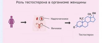
Testosterone
Testosterone injections increased the number of monocytes, granulocytes and large lymphocytes in mice. ()
Ways to lower monocyte levels
Monocytosis is a condition that requires diagnosis and treatment by a healthcare professional. Talk to your doctor before trying any strategies to reduce your monocyte count.
Regular exercise
Regular physical activity has an anti-inflammatory effect . In one study, monocytes were significantly reduced by 6 weeks of cycling training in overweight women who did not exercise regularly.
As
we age , there is an increase in general inflammation and a decrease in immune capabilities. In particular, the number of monocytes increases . But physical activity and diet can reduce the level of monocytes, and transfer macrophages from the M1 type to the less inflammatory M2 type.
(source) Monocyte count (decrease in monocyte count) was also significantly associated with decreased triglyceride levels, increased insulin sensitivity, and decreased body mass index (weight loss). All these changes in the body occur under the influence of physical activity. ()
Weight loss
In obese people, weight loss was accompanied by a significant decrease in the number of monocytes and neutrophils . A decrease in the number of circulating monocytes correlated with better insulin sensitivity of body tissues, which prevented the development of metabolic syndrome. ()
Omega-3 fatty acids
Regular consumption of omega-3 fatty acids, found in fatty fish such as mackerel and salmon, or in fish oil supplements, may protect against atherosclerosis and heart disease. ()
People taking fish oil supplements were less likely to show inflammation in the walls of blood vessels caused by monocyte activity. This effect was not as pronounced in people already taking special medications to treat peripheral blood vessel disease. ()
Moderate alcohol consumption
Alcohol affects monocyte function. In one study, the monocytes of people who drank moderate amounts of alcohol were less active, even after just one drink. Monocytes directly exposed to alcohol also had a reduced inflammatory response to proinflammatory proteins. (, )
Moderate alcohol consumption, about 1 or 2 standard drinks per day, is associated with significantly reduced monocyte production of the inflammatory cytokines TNF-alpha and IL-1beta. Alcohol is also associated with increased production of the cytokine IL-10, which is an anti-inflammatory protein. (, )
However, we do not recommend increasing alcohol consumption in order to relieve inflammation. Talk to your doctor about more appropriate anti-inflammatory actions.
Mediterranean diet
Some studies demonstrate that the Mediterranean diet can reduce inflammation caused by monocytes. (, )
PYRAMID OF THE MEDITERRANEAN DIET
The Mediterranean diet consists of foods such as seeds, nuts, vegetables, fruits, whole grains and monounsaturated fats from olive oil.
Starvation
Intermittent or prolonged fasting can reduce the proinflammatory activity of monocytes. For example, the short-chain fatty acid β-hydroxybutyrate, which when released from the liver during prolonged fasting , has been shown to suppress the production of the cytokines IL-1β and IL-18 by human monocytes, which was induced by the NLRP3 inflammasome. ()
Calorie restriction has beneficial effects on many chronic metabolic disorders such as type 2 diabetes, nonalcoholic fatty liver disease, and cardiovascular disease, and intermittent fasting is sufficient to reduce the number of all monocyte populations in healthy individuals. ()
Taking cortisol
When a single dose of cortisol is administered, the number of monocytes decreases by 90% 4-6 hours after receiving the drug. This decline in numbers lasted for about 24 hours. Subsequently, monocyte levels return to normal 24 to 72 hours after cortisol treatment. ()
Taking estrogen
According to one study, estrogen (and possibly also progesterone) reduces the number of monocytes by preventing their production. This mechanism could explain why cell-mediated immunity appears to be suppressed during pregnancy . ()
Medicine: infliximab
Infliximab (monoclonal antibody TNF-alpha) is an immunosuppressive drug prescribed to treat inflammatory diseases such as Crohn's disease, ulcerative colitis, and rheumatoid arthritis. (, , )
Infliximab inhibits monocytes, which may help reduce inflammation in patients with chronic inflammatory diseases. ()
Link between low monocyte levels and health status
Cardiovascular diseases
Of all white blood cells, monocyte counts have the strongest association with the incidence of cardiovascular disease in people without any symptoms. Lower monocyte levels are directly associated with lower cardiovascular risk of disease and death. ()
Link of monocytes (increased levels) in cardiovascular diseases (source)
Susceptibility to infections
Monocytes are involved in the immune response to infection , so it is not surprising that low monocyte counts are associated with increased infection rates. Monocytopenia is directly associated with monoMAC syndrome: increased susceptibility to mycobacterial infections, fungal infections, and human papillomavirus (HPV). (, )
Hematopoietic disorders
Monocytopenia is associated with blood disorders such as myelodysplasia, acute myelogenous leukemia, chronic myelomonocytic leukemia, and lymphomas. If you are concerned about your low monocyte count and your possible risk of these blood disorders, talk to your doctor about additional tests available. ()
Cervical cancer
Patients with primary immunodeficiency have insufficient numbers of monocytes and are susceptible to severe, persistent human papillomavirus (HPV) infections, which can cause cervical cancer. If you have problems with your monocytes, talk to your doctor about strategies to prevent cervical cancer, especially if you have not been vaccinated against HPV. (, )
INR norm
The standardized INR norm is 0.85-1.15 units. If a person takes anticoagulants, then his INR is increased , which is natural: the patient’s blood contains fewer coagulation factors. In this case, the INR norm for the patient is 2-3 units.
The blood INR indicator is influenced by many factors, ranging from the patient’s gender and age to the characteristics of his medical history. Depending on the age of the patients, the following limits of normal blood INR are distinguished:
Table 1. INR indicator depending on the patient’s age
| Patient age | Normal INR (in units) |
| Newborn period, as well as the first year of life | 0,9— 1,25 |
| 1 year—6 years | 0,95— 1,1 |
| 12—16 years | up to 1.35 |
| over 16 years | 0,85— 1,3 |
As you can see, blood INR by age is an indicator that varies significantly depending on the category in which the patient belongs.