Hemolytic anemia
Hereditary membranopathies, enzymopenias and hemoglobinopathies
The most common form of this group of anemias is microspherocytosis, or Minkowski-Choffard disease.
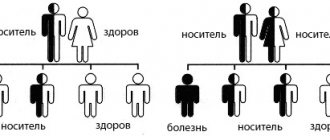
Inherited in an autosomal dominant manner; usually traced to several members of the family. The defectiveness of erythrocytes is caused by a deficiency of actomyosin-like protein and lipids in the membrane, which leads to a change in the shape and diameter of erythrocytes, their massive and premature hemolysis in the spleen. Manifestation of microspherocytic HA is possible at any age (in infancy, adolescence, old age), but usually manifestations occur in older children and adolescents. The severity of the disease varies from subclinical to severe forms characterized by frequently recurring hemolytic crises. At the moment of crisis, body temperature, dizziness, and weakness increase; Abdominal pain and vomiting occur. The main symptom of microspherocytic hemolytic anemia is jaundice of varying degrees of intensity. Due to the high content of stercobilin, feces become intensely dark brown. In patients with Minkowski-Choffard disease, there is a tendency to form stones in the gall bladder, therefore, signs of exacerbation of calculous cholecystitis often develop, attacks of biliary colic occur, and when the common bile duct is blocked by a stone, obstructive jaundice occurs. With microspherocytosis, in all cases the spleen is enlarged, and in half of the patients the liver is also enlarged. In addition to hereditary microspherocytic anemia, other congenital dysplasias often occur in children: tower skull, strabismus, saddle nose deformation, malocclusion, gothic palate, polydactyly or bradydactyly, etc. Middle-aged and elderly patients suffer from trophic ulcers of the leg, which arise as a result of hemolysis of red blood cells in the capillaries of the extremities and are difficult to treat.
Enzymopenic anemia is associated with a deficiency of certain red blood cell enzymes (usually G-6-PD, glutathione-dependent enzymes, pyruvate kinase, etc.). Hemolytic anemia may first manifest itself after suffering an intercurrent illness or taking medications (salicylates, sulfonamides, nitrofurans). Usually the disease has a smooth course; “pale jaundice”, moderate hepatosplenomegaly, and heart murmurs are typical. In severe cases, a pronounced picture of a hemolytic crisis develops (weakness, vomiting, shortness of breath, palpitations, collapsing state). Due to intravascular hemolysis of erythrocytes and the release of hemosiderin in the urine, the latter acquires a dark (sometimes black) color. Independent reviews are devoted to the features of the clinical course of hemoglobinopathies - thalassemia and sickle cell anemia.
Acquired hemolytic anemia
Among the various acquired variants, autoimmune anemia is the most common. For them, a common trigger is the formation of antibodies to antigens of their own red blood cells. Hemolysis of erythrocytes can be both intravascular and intracellular in nature. Hemolytic crisis in autoimmune anemia develops acutely and suddenly. It occurs with fever, severe weakness, dizziness, palpitations, shortness of breath, pain in the epigastrium and lower back. Sometimes acute manifestations are preceded by precursors in the form of low-grade fever and arthralgia. During a crisis, jaundice rapidly increases, not accompanied by itching, and the liver and spleen enlarge. In some forms of autoimmune anemia, patients do not tolerate cold well; in low temperatures they may develop Raynaud's syndrome, urticaria, and hemoglobinuria. Due to circulatory failure in small vessels, complications such as gangrene of the toes and hands are possible.
Toxic anemia occurs with progressive weakness, pain in the right hypochondrium and lumbar region, vomiting, hemoglobinuria, and high body temperature. From 2-3 days jaundice and bilirubinemia develop; on days 3-5, liver and kidney failure occurs, the signs of which are hepatomegaly, fermentemia, azotemia, and anuria. Certain types of acquired hemolytic anemia are discussed in the relevant articles: “Hemoglobinuria” and “Thrombocytopenic purpura”, “Hemolytic disease of the fetus”.
A little about red blood cells
Erythrocytes, or red blood cells, are blood cells whose main function is to transport oxygen to organs and tissues.
- Red blood cells are formed in the red bone marrow, from where their mature forms enter the bloodstream and circulate throughout the body. The lifespan of red blood cells is 100-120 days. Every day, about 1% of them die and are replaced by the same number of new cells.
- If the lifespan of red blood cells is shortened, more of them are destroyed in the peripheral blood or spleen than have time to mature in the bone marrow - the balance is disrupted. The body responds to a decrease in the content of red blood cells in the blood by increasing their synthesis in the bone marrow, the activity of the latter increases significantly - 6-8 times.
As a result, an increased number of young red blood cell precursor cells - reticulocytes - is determined in the blood. The destruction of red blood cells with the release of hemoglobin into the blood plasma is called hemolysis.
4.Treatment
As shown above, in some cases, autoimmune hemolytic anemia is practically asymptomatic or minimally symptomatic, resolves spontaneously and therefore does not require treatment. In more severe cases, the treatment of choice is corticosteroid hormones, sometimes in combination with immunosuppressants. Because the hemoglobin released when red blood cells are destroyed is toxic, detoxification measures may be required. A purely symptomatic treatment is blood transfusion. The last resort in some cases is splenectomy (removal of the spleen); if this, in combination with hormonal therapy, turns out to be ineffective, cytostatics are prescribed.
In general, it is usually possible to select a fairly effective therapeutic regimen, but autoimmune hemolytic anemia is a serious and dangerous condition, so the prognosis often remains unfavorable.
Symptoms
{banner_banstat4}
If a patient is diagnosed with hemolytic anemia, its symptoms are divided into two types: anemic and hemolytic. It is worth noting that in the case when the destruction of red blood cells is a complication of another disease, then this type of anemia is supplemented by the symptoms of the underlying disease.
The anemic type is characterized by the following:
- pale, bluish skin;
- frequent dizziness;
- increased fatigue;
- severe shortness of breath during normal physical activity;
- rapid heartbeat;
More typical for the hemolytic type:
- yellow color of the skin and mucous membranes;
- the color of the urine is red, brown or cherry, sometimes even the color of the urine is black;
- enlargement of the liver or spleen, often patients experience severe protrusion of the liver;
- pain under the left rib;
- high temperature, at this moment the process of destruction of red blood cells is at its maximum.
Symptoms
Hemolytic anemia manifests itself more prominently during crises. With sluggish processes, the symptoms are blurred. The specific type of pathology can only be determined with the help of additional diagnostics.
The main symptoms include:
| Anemia syndrome | manifests itself in pallor of the skin and mucous membranes. Accompanied by symptoms of lack of oxygen in the form of shortness of breath, dizziness, weakness, and increased heart rate. |
| Jaundice syndrome | An increase in the concentration of bilirubin as a breakdown product of red blood cells is expressed by yellowness of the skin and a change in the color of urine. |
| Hepatosplenomegaly | the enlargement of the spleen occurs due to intense hemolysis and can reach significant sizes. The liver is less susceptible to changes. But in some cases there is an increase in it, accompanied by heaviness in the hypochondrium. |
Hemolytic anemia may manifest itself with additional symptoms such as:
- abdominal pain;
- loose stools;
- bone soreness;
- elevated temperature;
- pain in the chest and kidney area.
Even if there are only 2 or 3 of the listed symptoms, this should be a reason for a medical examination. After all, toxic bilirubin with prolonged exposure to tissues and organs can disrupt their functions.
Diagnostics
In the diagnosis of hemolytic anemia, a general clinical blood test is used (anemia and changes in the size and shape of red blood cells are detected), a biochemical blood test (including serum bilirubin, ALT, LDH), serum haptoglobin, hemosiderin and urine hemoglobin. To confirm the diagnosis, bone marrow puncture can be used (active processes of erythropoiesis are determined in the biopsy specimen).
The most characteristic of active intravascular hemolysis is spherocytosis of erythrocytes (with transfusion reactions, hereditary spherocytosis, with hemolytic anemia with warm antibodies). Schistocytosis (with intravascular prosthetics, microangiopathies), sickle erythrocytes (with sickle cell anemia), target erythrocytes (with liver pathologies, hemoglobinopathies), nucleated erythrocytes and basophilia (with beta thallasemia major) may also be observed.
Heinz bodies are found in unstable hemoglobin, activation of peroxidation, acanthocytes in anemia with spur-shaped red blood cells, agglutinated cells in cold agglutinin disease.
2. Reasons
The name itself indicates that the basis of autoimmune hemolytic anemia is a failure of the so-called. immune tolerance: in this case, protective factors attack their own red blood cells, which for some reason begin to be recognized as foreign objects and, as such, are “sentenced” to destruction.
Autoimmune diseases, generally speaking, are difficult to understand and etiopathogenetic analysis: there is always the possibility that the immune system actually becomes aggressive not towards its own cells, but towards something very similar to them, masquerading - and it can be very difficult to identify this a true insidious “enemy”, along with which healthy cells are destroyed. Occasionally, autoimmune hemolytic anemia is recorded, in which antibodies appear in the body that are aggressive towards bone marrow erythroblast cells (one of the precursors of mature red blood cells). However, the vast majority of such anemias are somehow related to the temperature factor - cold or, conversely, heat. Immune failure consists in the fact that one or more types of “thermal”, warm or cold antibodies (agglutinins, hemolysins) open a systematic “hunt” for red blood cells.
Such an immune failure towards auto-aggression may be due to various reasons. It is known that hemolytic anemia of this type sometimes develops as secondary, symptomatic, i.e. as part of other systemic diseases with an autoimmune component (lupus erythematosus, rheumatoid arthritis, etc.), and sometimes occurs against the background and, possibly, as a result of taking medications (usually antibiotics) or other chemical compounds (especially acetic acid, lead, etc.). etc.), after the bite of poisonous snakes, with hypovitaminosis E, intracellular parasitosis, implantation of one or more artificial valves, untreated vascular pathology.
At least half of cases of autoimmune hemolytic anemia remain completely unclear in terms of causes and triggers; such variants are classified as idiopathic anemia.
Visit our Therapy page


