- home
- general surgery
- Esophageal diverticula
To identify a diverticulum of the esophagus, its localization, as well as choose the correct surgical treatment tactics, you must send me a complete description of gastroscopy, X-ray of the esophagus and stomach with barium, preferably an ultrasound of the abdominal organs, by indicating your age and main complaints. Then I will be able to give a more accurate answer to your situation.
Esophageal diverticulum
- this is a protrusion of the submucosal layer and mucous membrane of the esophageal wall through a defect in its muscular lining.
The first description of esophageal diverticula was made by the English anatomist Ludlow (1764). Our Russian scientist Rokitansky (1840) divided all esophageal diverticula into pulsion and traction and described epibronchial diverticula. The German scientist Zenker (1877) characterized esophagopharyngeal diverticula.
Diverticula can be congenital or acquired (they are much more common), the latter arise as a result of inflammatory changes in the wall of the organ and surrounding tissues.
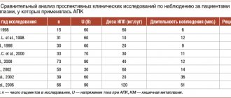
Fig. 1 Location of esophageal diverticula (1- left main bronchus, 2- peribronchial diverticulum, 3- trachea, 4- lower pharyngeal constrictor, 5- Zenker diverticulum, 6- right main bronchus, 7- esophagus, 8- epiphrenic diverticulum, 9- diaphragm, 10- abdominal part of the esophagus).
In their origin, they can be pulsational (based on an increase in pressure in the lumen of the organ with subsequent protrusion of the submucosal layer and mucous membrane) and traction (the inflammatory process around the esophagus pulls its wall towards itself with the formation of a protrusion).
Depending on the location, diverticula of the esophagus are divided into (Fig. 1): - pharyngoesophageal (the so-called Zenker diverticula), - epibronchial (located in the middle section of the esophagus, in the bifurcation area - the most common localization) - epiphrenal (located above the diaphragm).
Diverticula are observed more often after 50 years, mainly in men.
Pharyngeal-esophageal (Zenker's) diverticulum.
The main role in the pathogenesis of the formation of this type of diverticula is achalasia (inability to relax) of the cricopharyngeal muscles, which results in a violation of the opening of the upper esophageal sphincter in response to swallowing. Excessive intraluminal pressure pushes the submucosal layer into the resulting muscle defect. The diverticulum then descends between the posterior wall of the esophagus and the spine. The inner surface of the diverticulum is covered with a mucous membrane; it may have superficial erosions, foci of inflammation and scars.
Features of the esophagus affecting the course of diverticulitis
The upper boundary of the thoracic section of the esophageal tube is the conventional line of the second thoracic vertebra from the posterior mediastinum (the space surrounding the heart). The lower one coincides with the esophageal opening of the diaphragm. The entire segment is 16–18 cm long in an adult. It is separated from the spine by a thin layer of fatty tissue.
It is in close contact with the inner layer of the pleura (mediastinal region). Passing from top to bottom, the esophagus is first located to the left of the trachea, then passes the area of the aorta and azygos vein, at the level of the fourth thoracic vertebra, located next to the left main bronchus and the bifurcation of the trachea.
Here, the left atrium of the heart and the pericardial wall, the aortic arch, and the subclavian artery are adjacent to the esophagus in front. Along its entire course, the esophagus is accompanied by the recurrent nerve and multiple groups of lymph nodes. The close proximity to important organs of the chest leads to their damage due to diverticula of the esophagus.
Clinical presentation and diagnosis of pharyngoesophageal diverticulum.
Small diverticula are manifested by a sore throat, dry cough, foreign body sensation, and increased salivation. When it is filled with food, swallowing problems (dysphagia) may develop. A protrusion may appear in the neck, especially when the head is pulled back. As the size of the diverticulum increases, spontaneous reflux of undigested food appears from the lumen of the diverticulum into the esophagus. As a result of compression of the trachea, difficulty breathing may occur, and when the recurrent nerve is compressed, hoarseness may occur. When food is retained for a long time in the diverticulum, a putrid odor from the mouth appears. All these manifestations lead to eating disorders and weight loss.
Puchkov K.V., Ivanov V.V. and others. Technology of dosed ligating electrothermal effects at the stages of laparoscopic operations: monograph. - M.: ID MEDPRACTIKA, 2005. - 176 p.
Puchkov K.V., Bakov V.S., Ivanov V.V. Simultaneous laparoscopic surgical interventions in surgery and gynecology: Monograph. - M.: ID MEDPRACTIKA, 2005. - 168 p.
Puchkov K.V., Rodichenko D.S. Manual suture in endoscopic surgery: monograph. - M.: MEDPRACTIKA, 2004. - 140 p.
Zenker's diverticula can be complicated by the development of diverticulitis (inflammation of the wall), then phlegmon of the neck, mediastinitis, development of an esophageal fistula, etc. Regurgitation (reflux) and aspiration (entry into the respiratory tract) of the contents of the diverticulum lead to chronic bronchitis, repeated pneumonia, and lung abscesses. Bleeding from the eroded mucous membrane of the diverticulum may occur, as well as the development of polyps and mucosal cancer.
Statistics
The frequency of diverticula of the thoracic esophagus is 20 times higher than similar lesions in the cervical and abdominal parts. The overall prevalence is 40% of all diverticula of the digestive system. During X-ray examination, pathology is found in 2% of individuals.
Most often, esophageal diverticulum appears as a single formation (90% of cases), but in 1/10 of the patients multiple protrusions are found. It has been established that men over fifty years of age who suffer from chronic diseases of the digestive system (gastritis, peptic ulcer, pathology of the biliary tract) are more likely to get sick.
There are 3 possible areas of localization of diverticula (4 - subdiaphragmatic space)
Diagnosis of Zenker's diverticula.
Pharyngeal-esophageal diverticula can sometimes be detected by inspection and palpation of the neck. The protrusion has a soft consistency, decreases with pressure; after drinking water, a splashing noise can be detected by percussion over it.
The main diagnostic method is a contrast X-ray examination, which establishes the presence of a diverticulum, the width of the neck, the duration of barium retention in it, the degree of obstruction of the esophagus, signs of the development of a polyp and cancer in the diverticulum, the formation of esophageal-bronchial and esophageal-mediastinal fistulas. Fiberendoscopic examination makes it possible to establish the presence of a diverticulum, detect ulceration of its mucous membrane, the presence of bleeding, and diagnose a polyp or cancer in the diverticulum. Due to the high risk of perforation of the esophageal diverticulum, esophagoscopy should be performed with great caution.
It is optimal to perform esophagomanometry - a study of esophageal motility. Patients with coronary symptoms undergo a consultation with a cardiologist, an ECG study, Holter monitoring, and echocardiography.
Differential diagnosis of esophageal diverticulum is carried out with GERD, esophagospasm, hiatal hernia, esophageal strictures, achalasia cardia, esophageal cancer, mediastinal cyst, angina pectoris, and ischemic heart disease.
Which doctor should I contact?
If the described symptoms occur, you need to urgently make an appointment with a gastroenterologist. Our gastroenterologists are true professionals. They will listen to your complaints, conduct an examination, and prescribe therapy. At the Kuntsevskaya clinic you will receive truly qualified assistance.
IMPORTANT! Esophageal diverticulum is a dangerous disease that requires careful medical supervision and control. Often the diverticulum does not manifest itself for a long time.
However, if a person experiences symptoms of esophageal diverticulum, it is imperative to consult a gastroenterologist. Make an appointment with a gastroenterologist at the Kuntsevo Medical and Rehabilitation Center, who will conduct a full diagnosis and prescribe a complete detailed treatment plan for the person. This is followed by the rehabilitation phase, during which you will also be accompanied by our gastroenterologist.
Sign up
Treatment of esophageal diverticula
If the diverticula are small in size, there are no complications, and there are absolute contraindications to surgical treatment, conservative therapy is carried out aimed at preventing the retention of food masses in the diverticulum and reducing the possibility of developing diverticulitis.
Conservative therapy includes adherence to nutrition and diet. Food should be warm, pureed, and not irritate the mucous membrane. It is advisable to eat food in fractional portions, up to 6 times a day. After each meal, the patient drinks up to 100-200 ml of mineral water or other heated liquid and performs postural drainage of the diverticulum (drainage by body position). If esophagitis is present, drug therapy is added.
What complications does diverticulitis cause?
Inflammation of the diverticulum over a long period of time can lead to:
Hiatus hernia
- to attacks of suffocation;
- phlegmon of the neck;
- chronic bronchitis;
- aspiration pneumonia, lung abscess;
- bleeding;
- abscess formation of a diverticulum;
- mucosal erosion;
- perforation into surrounding tissues;
- mediastinitis with esophageal-mediastinal fistula;
- esophageal polyps;
- cancerous degeneration.
Bifurcation diverticulitis has the rarest tendency to complications.
Surgical treatment of pharyngeal-esophageal (Zenker) diverticula.
Indications for surgical treatment are as follows: complications (perforation, penetration, bleeding, stenosis of the esophagus, cancer, development of fistulas), large diverticula complicated by at least short-term retention of food masses in them, long-term retention of food in the diverticulum, regardless of its size. Ineffectiveness of conservative therapy.
The essence of surgical treatment is complete removal of the diverticulum.
(cutting off from the esophagus, followed by suturing the organ wall) - diverticulectomy: the diverticulum is isolated from the surrounding tissues to the neck, a myotomy is performed, it is excised and the hole in the wall of the esophagus is sutured.
We isolate pharyngoesophageal diverticula from the cervical approach. A cosmetic skin incision is made in the projection of the anterior edge of the left sternocleidomastoid muscle. We mobilize the left lobe of the thyroid gland, retract it medially, and the neurovascular bundle laterally. Before surgery, a thick probe is inserted into the esophagus, which greatly facilitates the surgical treatment of esophageal diverticula. Then we perform extramucosal esophagomyotomy
several centimeters long, making sure that all the fibers of the m. cricopharyngeus (cricopharyngeal myotomy). We isolate the diverticulum up to the neck and stitch its base with an EndoGia-30 endoscopic stapler (Switzerland). The specimen is sent for pathohistological examination. The duration of stay in the clinic is 2-3 days. You can eat after 1-2 days.
Watch a video of operations performed by Professor K.V. Puchkov. You can visit the website “Video of operations of the best surgeons in the world.”
Treatment results and lifestyle after recovery
The effectiveness of treatment will depend on how well the patient follows the points of the therapeutic program. An esophageal diverticulum is like a time bomb; anything can happen. Therefore, after treatment, strictly follow the recommendations of the gastroenterologist. Visit a specialist from time to time.
Learn a few rules for yourself:
- walk more in the fresh air;
- follow a diet;
- establish a work and rest schedule;
- get rid of bad habits;
- play sports.
We are talking about light exercises. Kuntsevo has its own exercise therapy room. Under the guidance of experienced instructors, do gentle exercises.
Epiphrenic diverticula
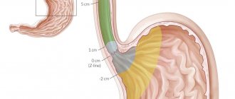
Epiphrenic diverticula, usually pulsational, are located along the posterior or right wall of the esophagus 2-11 cm above the diaphragm. Diverticula are spherical or mushroom-shaped. The leading factors in the formation of epiphrenic diverticula are the following factors: weakness of the muscular wall, increased intra-esophageal pressure and pressure of the food bolus on weak areas of the esophageal wall. Muscular weakness can be congenital or acquired. An increase in intra-esophageal pressure occurs due to uncoordinated peristalsis of the esophagus and its lower sphincter.
Causes
Based on their origin, esophageal diverticula are divided into congenital and acquired. Congenital formation is formed due to a violation of the formation of layers of the wall of the esophageal tube. In a certain area, insufficiently dense muscle tissue appears, which cannot withstand the load and leads to protrusion.
The process of formation of diverticula is also called diverticulosis. Acquired diverticula develop due to inflammatory processes in neighboring organs (lungs, pleura, pericardium) and in the esophagus itself, injuries. Diverticula appear over a long period of time:
- esophagitis;
- mediastinitis;
- gastroesophageal reflux disease;
- fungal infection;
- tuberculosis of regional lymph nodes;
- esophagospasm;
- achalasia;
- cicatricial narrowing of the esophagus.
The mechanism of protrusion formation can be pulsation, traction or mixed.
- Pulsion diverticulum is always associated with dysfunction of the motility of the esophageal wall, spastic contractions of the muscle layer, and a subsequent increase in intraesophageal pressure. This leads to stretching and bulging at the weakest point.
- Traction mechanism - caused by fusion and abnormal fixation of the esophageal wall to the inflamed lymph nodes of the mediastinum. As a result, the muscle layer stretches and then bulges.
With a mixed type, both mechanisms of action take place.
The surgeon diagnoses and treats pathology
Symptoms and clinical manifestations of epiphrenic diverticula
Almost half of epiphrenic diverticula are detected incidentally as a radiological finding. Usually these are diverticula with a diameter of up to 2-3 cm. With epiphrenic diverticula, patients complain of a feeling of heaviness, pain in the lower part of the sternum or in the area of the xiphoid process, and progressive difficulty swallowing. Some patients experience aerophagia, putrid breath, and regurgitation of old decomposed food. In some cases, attacks of bronchial asthma and angina may occur. The severity of the symptoms of the disease depends on the size of the diverticulum and the degree of its filling with food masses.
Indications for surgical intervention for epiphrenic diverticula do not differ from the treatment of Zenker's diverticula. The operation is indicated for complications: perforation, penetration, bleeding, stenosis of the esophagus, cancer, development of fistulas, large diverticula complicated by at least short-term retention of food masses in them, long-term retention of food in the diverticulum, regardless of its size. Lack of effectiveness from conservative therapy.
Very important! A feature of my approach to choosing treatment tactics for patients with esophageal diverticula is the earlier determination of indications for surgical treatment using the laparoscopic method (through several punctures of the abdominal wall), without waiting for complications to develop and without wasting time on useless conservative treatment methods.
Typically, the traditional approach, which most clinics use, to epiphrenic diverticula is to perform a left-sided thoracotomy along the VII - VIII intercostal space.
Currently, in the surgery of esophageal diverticula, methods for their removal using video thoracoscopic or laparoscopic (which is even less traumatic) techniques are becoming increasingly widespread.
My experience in treating patients with esophageal diverticula is more than 14 years
.
During this time, I was able to successfully operate and treat more than 100 patients
.
Very important! Unlike other clinics, I use a minimally invasive laparoscopic approach, which allows, without opening the pleural cavity, to isolate the epiphrenic diverticulum at a distance of up to 8-10 cm from the cardia, remove it and reliably close the esophageal wound. To do this, I stitch the base of the diverticulum with an endoscopic stapler EndoGia-30 (Switzerland), which allows you to apply a 3-row titanium suture.
The basis of a precision technique for bloodless isolation of a diverticulum from surrounding tissues is the use of the LigaSure dosed vessel ligation system (Switzerland).
Very important! I always remove the esophageal diverticulum under the intraoperative control of fibroesophagoscopy; this allows during the operation (by means of illumination) to give the surgeon an excellent reference point in the surrounding tissues and, most importantly, to excise the diverticulum under double visual control (laparoscopy and fibroisophagoscopy). This double control, in turn, guarantees the completeness of its excision and the absence of narrowing of the lumen of the esophagus after suturing the diverticulum stalk with a stapler.
In the case of a combination of epiphrenic diverticula and achalasia cardia, hiatal hernia, chronic reflux esophagitis
I perform simultaneous simultaneous surgical intervention: diverticulectomy and correction of the hiatal hernia or cardiomyotomy in my own modification.
The patient begins to walk the next day after surgery and eat food 1-2 days later. Discharged on the 3rd day, after a control chest x-ray.
Thus, the earlier (before complications develop) surgery is performed for esophageal diverticulum, the better the treatment results.
Pharyngeal-esophageal diverticula are operated on by a thoracic surgeon using access to the neck, through a cosmetic incision using the LigaSure dosed vascular ligation system (Switzerland) to isolate the diverticulum and the endoscopic stapler EndoGia-30 (Switzerland), which allows you to apply a safe 3-row titanium suture to the wall esophagus.
Rice. 2. Removal of esophageal diverticulum using an endoscopic stapler using laparoscopic access.
Epiphrenic diverticula (from 1 to 10 cm from the cardia) I operate laparoscopically
through punctures, also using the LigaSure dosed vessel ligation system (Switzerland) to isolate the diverticulum and the EndoGia-30 endoscopic stapler (Switzerland) (Fig. 2).
In this case, I perform all stages under double visual control - laparoscopic and fibroesophagoscopic from the side of the lumen of the esophagus.
All of the above measures make it possible to perform surgical interventions for esophageal diverticula reliably, with a minimum number of complications, with a short hospitalization period and a quick return to normal life.
Prognosis and prevention
The prognosis of the disease is generally favorable: after surgical removal of the diverticulum, the disease, as a rule, does not recur. Complications - diverticulitis, bleeding, perforation - can be life-threatening, so a patient with esophageal diverticulum should carefully follow dietary and lifestyle recommendations and be regularly examined by a gastroenterologist.
To prevent esophageal diverticula, it is necessary to promptly treat diseases that may contribute to their occurrence.
[1] Maev I.V., Dicheva D.T., Andreev D.N. Diverticula of the gastrointestinal tract: educational method. manual for doctors. Moscow, 2015.
Postoperative follow-up
After the operation, 3-4 incisions 5-10 mm long remain on the skin of the abdomen. From the first day, patients begin to get out of bed, drink, and on the second day take liquid warm food. Discharge from the hospital is carried out on 3-4 days, depending on the severity of the disease. The patient can begin work in 2 - 3 weeks. A strict diet should be followed for one and a half to two months, a softer diet should be followed for 2-3 months. Further, as a rule, the patient leads a normal lifestyle - without medications or diet.
At the request of patients, the clinic can undergo a full examination before surgery to determine the optimal treatment tactics and choose the method of surgical intervention.
Why is it better to seek treatment from us?
Early detection of esophageal diverticulum and a complete assessment of the risk of the disease for a particular patient allows doctors at the Kuntsevo Treatment and Rehabilitation Center to fully assess the person’s condition and choose the most effective treatment tactics in each individual case. In addition, modern diagnostic methods available at the Kuntsevo Treatment and Rehabilitation Center (gastroscopy, MRI of the esophagus, MRI of the esophagus “in sleep”) allow the doctor to monitor the patient’s dynamics, evaluate the effectiveness of therapy and make changes to speed up the person’s recovery process.
IMPORTANT! You should not self-medicate if you discover this symptom. Cold hands can be a manifestation of both the physiological norm of your body and a pathological process that develops in the body.
The Kuntsevo Multidisciplinary Center has doctors of the highest category and doctors of medical sciences. Using modern equipment and high-tech devices, diagnosing esophageal diverticulum has become easier and more effective. You will be pleasantly surprised by the best prices and friendly staff. Don't delay treatment - make an appointment!
The contents of this article have been checked and confirmed for compliance with medical standards by gastroenterologist-nutritionist Tatyana Vladimirovna Lucheninova