- home
- general surgery
- Esophageal diverticula
To identify a diverticulum of the esophagus, its localization, as well as choose the correct surgical treatment tactics, you must send me a complete description of gastroscopy, X-ray of the esophagus and stomach with barium, preferably an ultrasound of the abdominal organs, by indicating your age and main complaints. Then I will be able to give a more accurate answer to your situation.
Esophageal diverticulum
- this is a protrusion of the submucosal layer and mucous membrane of the esophageal wall through a defect in its muscular lining.
The first description of esophageal diverticula was made by the English anatomist Ludlow (1764). Our Russian scientist Rokitansky (1840) divided all esophageal diverticula into pulsion and traction and described epibronchial diverticula. The German scientist Zenker (1877) characterized esophagopharyngeal diverticula.
Diverticula can be congenital or acquired (they are much more common), the latter arise as a result of inflammatory changes in the wall of the organ and surrounding tissues.
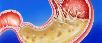
Fig. 1 Location of esophageal diverticula (1- left main bronchus, 2- peribronchial diverticulum, 3- trachea, 4- lower pharyngeal constrictor, 5- Zenker diverticulum, 6- right main bronchus, 7- esophagus, 8- epiphrenic diverticulum, 9- diaphragm, 10- abdominal part of the esophagus).
In their origin, they can be pulsational (based on an increase in pressure in the lumen of the organ with subsequent protrusion of the submucosal layer and mucous membrane) and traction (the inflammatory process around the esophagus pulls its wall towards itself with the formation of a protrusion).
Depending on the location, diverticula of the esophagus are divided into (Fig. 1): - pharyngoesophageal (the so-called Zenker diverticula), - epibronchial (located in the middle section of the esophagus, in the bifurcation area - the most common localization) - epiphrenal (located above the diaphragm).
Diverticula are observed more often after 50 years, mainly in men.
Pharyngeal-esophageal (Zenker's) diverticulum.
The main role in the pathogenesis of the formation of this type of diverticula is achalasia (inability to relax) of the cricopharyngeal muscles, which results in a violation of the opening of the upper esophageal sphincter in response to swallowing. Excessive intraluminal pressure pushes the submucosal layer into the resulting muscle defect. The diverticulum then descends between the posterior wall of the esophagus and the spine. The inner surface of the diverticulum is covered with a mucous membrane; it may have superficial erosions, foci of inflammation and scars.
Classification
Classification of esophageal adenocarcinoma is carried out according to the criteria of the international classification of malignant neoplasms TNM (8th revision):
T
- Tis carcinoma in situ/high grade dysplasia;
- T1 tumor growth into the lamina propria or submucosal layer: T1a lamina propria or muscularis lamina of the mucosa;
- T1b submucosal layer.
- T4a pleura, peritoneum, pericardium, diaphragm;
N
- N0 no metastases in regional lymph nodes;
- N1 damage to 1-2 regional lymph nodes;
- N2 damage to 3-6 regional lymph nodes;
- N3 damage to 7 or more regional lymph nodes.
M
- M1 presence of distant metastases.
The following groups of lymph nodes are regional:
- disgraced,
- internal jugular,
- upper and lower cervical,
- cervical paraesophageal,
- supraclavicular (bilateral),
- pretracheal (bilateral),
- lymph nodes of the lung root (bilateral),
- upper paraesophageal (above v. azygos),
- bifurcation,
- lower paraesophageal (below v. azygos),
- posterior mediastinal,
- diaphragmatic,
- perigastric (right and left cardiac, lymph nodes along the lesser curvature of the stomach, along the greater curvature of the stomach, suprapyloric, infrapyloric, lymph nodes along the left gastric artery).
Damage to the celiac lymph nodes is not a contraindication to chemoradiation therapy or the issue of surgical treatment.
Degree of tumor differentiation
GX – the degree of tumor differentiation cannot be determined;
G1 – well-differentiated tumor;
G2 – moderately differentiated tumor;
G3 – poorly differentiated tumor;
G4 – undifferentiated tumor.
In the classification of esophageal adenocarcinoma, cardioesophageal cancer , i.e. cancer developing in the area of the esophagogastric junction and cardia.
Sievert classification
Adenocarcinoma of the esophagogastric junction, according to Sievert's classification, is divided into 3 types:
Type I - adenocarcinoma of the distal esophagus (often associated with Barrett's esophagus), the center of the tumor is located within 1-5 cm above the cardia (dentate line);
Type II – true adenocarcinoma of the esophagogastric junction (true cancer of the cardia), the center of the tumor is located within 1 cm above and 2 cm below the cardia (dentate line);
Type III - cancer with the localization of the main mass of the tumor in the subcardial part of the stomach within 2-5 cm below the dentate line and possible involvement of the distal parts of the esophagus.
Tumors of the esophagogastric junction of types I and II are subject to treatment according to algorithms corresponding to the RP. Type III tumors are treated according to algorithms corresponding to stomach cancer.
Classification of cancer of the esophagogastric junction according to Siewert
Classification of esophageal adenocarcinoma by stages
Clinical presentation and diagnosis of pharyngoesophageal diverticulum.
Small diverticula are manifested by a sore throat, dry cough, foreign body sensation, and increased salivation. When it is filled with food, swallowing problems (dysphagia) may develop. A protrusion may appear in the neck, especially when the head is pulled back. As the size of the diverticulum increases, spontaneous reflux of undigested food appears from the lumen of the diverticulum into the esophagus. As a result of compression of the trachea, difficulty breathing may occur, and when the recurrent nerve is compressed, hoarseness may occur. When food is retained for a long time in the diverticulum, a putrid odor from the mouth appears. All these manifestations lead to eating disorders and weight loss.
Puchkov K.V., Ivanov V.V. and others. Technology of dosed ligating electrothermal effects at the stages of laparoscopic operations: monograph. - M.: ID MEDPRACTIKA, 2005. - 176 p.
Puchkov K.V., Bakov V.S., Ivanov V.V. Simultaneous laparoscopic surgical interventions in surgery and gynecology: Monograph. - M.: ID MEDPRACTIKA, 2005. - 168 p.
Puchkov K.V., Rodichenko D.S. Manual suture in endoscopic surgery: monograph. - M.: MEDPRACTIKA, 2004. - 140 p.
Zenker's diverticula can be complicated by the development of diverticulitis (inflammation of the wall), then phlegmon of the neck, mediastinitis, development of an esophageal fistula, etc. Regurgitation (reflux) and aspiration (entry into the respiratory tract) of the contents of the diverticulum lead to chronic bronchitis, repeated pneumonia, and lung abscesses. Bleeding from the eroded mucous membrane of the diverticulum may occur, as well as the development of polyps and mucosal cancer.
Diagnosis of Zenker's diverticula.
Pharyngeal-esophageal diverticula can sometimes be detected by inspection and palpation of the neck. The protrusion has a soft consistency, decreases with pressure; after drinking water, a splashing noise can be detected by percussion over it.
The main diagnostic method is a contrast X-ray examination, which establishes the presence of a diverticulum, the width of the neck, the duration of barium retention in it, the degree of obstruction of the esophagus, signs of the development of a polyp and cancer in the diverticulum, the formation of esophageal-bronchial and esophageal-mediastinal fistulas. Fiberendoscopic examination makes it possible to establish the presence of a diverticulum, detect ulceration of its mucous membrane, the presence of bleeding, and diagnose a polyp or cancer in the diverticulum. Due to the high risk of perforation of the esophageal diverticulum, esophagoscopy should be performed with great caution.
It is optimal to perform esophagomanometry - a study of esophageal motility. Patients with coronary symptoms undergo a consultation with a cardiologist, an ECG study, Holter monitoring, and echocardiography.
Differential diagnosis of esophageal diverticulum is carried out with GERD, esophagospasm, hiatal hernia, esophageal strictures, achalasia cardia, esophageal cancer, mediastinal cyst, angina pectoris, and ischemic heart disease.
Prevention
The main point of preventing Barrett's esophagus is timely diagnosis and treatment of reflux. For early detection of BE, it is recommended to perform esophagoscopy with clarifying diagnostics and targeted biopsy in patients with GERD for more than 5 years. Particular attention should be paid to men over 50 years of age, patients with obesity, GERD for more than 10 years and heartburn for more than 5 years.
General preventive measures, although trivial, also have a positive effect:
- Weight reduction to normal.
- To give up smoking.
- A balanced diet with sufficient amounts of fresh vegetables, fruits, vitamins C and E.
- Refusal of alcohol abuse, especially strong alcoholic drinks.
Treatment of esophageal diverticula
If the diverticula are small in size, there are no complications, and there are absolute contraindications to surgical treatment, conservative therapy is carried out aimed at preventing the retention of food masses in the diverticulum and reducing the possibility of developing diverticulitis.
Conservative therapy includes adherence to nutrition and diet. Food should be warm, pureed, and not irritate the mucous membrane. It is advisable to eat food in fractional portions, up to 6 times a day. After each meal, the patient drinks up to 100-200 ml of mineral water or other heated liquid and performs postural drainage of the diverticulum (drainage by body position). If esophagitis is present, drug therapy is added.
Self-administered chemoradiotherapy
An alternative to surgical treatment of resectable locally advanced forms of esophageal cancer is chemoradiotherapy, which achieves a comparable 5-year overall survival rate of 20-27%. In a head-to-head study comparing cisplatin-based chemoradiotherapy with 5-fluorouracil infusion versus surgery alone, there was no significant difference in long-term outcomes, and toxicity and mortality were significantly lower with conservative treatment.
It is highly desirable to carry out conformal 3D CRT external beam radiation therapy on linear accelerators with an energy of 6-18 Mev, as well as on proton complexes operating with an energy of 70-250 Mev. A single focal dose is 1.8-2 Gy, the total dose is up to 50-55 Gy. Increasing SOD does not lead to improved results, only increasing mortality.
During radiation therapy, chemotherapy is administered , often based on cisplatin and infusions of 5-fluorouracil. In the presence of severe dysphagia, endoscopic electrorecanalization of the esophagus or puncture microgastrostomy is performed before irradiation. Chemoradiation therapy is often complicated by the development of radiation esophagitis and an increase in the severity of dysphagia, which aggravates the patient’s nutritional deficiency and worsens the tolerability of treatment. In such situations, a partial or complete transition to parenteral nutrition and placement of a temporary puncture microgastrostomy is possible.
The choice between independent chemoradiotherapy or surgical treatment (with or without preoperative chemoradiotherapy) depends on the location of the primary tumor, the patient's functional status, and the experience of the surgeon. Thus, in intact patients with a tumor localized in the middle or lower third of the esophagus, it is preferable to include surgery in the treatment plan.
If the tumor remains viable after chemoradiotherapy or local relapse, a so-called “salvage esophagectomy” can be performed.
Surgical treatment of pharyngeal-esophageal (Zenker) diverticula.
Indications for surgical treatment are as follows: complications (perforation, penetration, bleeding, stenosis of the esophagus, cancer, development of fistulas), large diverticula complicated by at least short-term retention of food masses in them, long-term retention of food in the diverticulum, regardless of its size. Ineffectiveness of conservative therapy.
The essence of surgical treatment is complete removal of the diverticulum.
(cutting off from the esophagus, followed by suturing the organ wall) - diverticulectomy: the diverticulum is isolated from the surrounding tissues to the neck, a myotomy is performed, it is excised and the hole in the wall of the esophagus is sutured.
We isolate pharyngoesophageal diverticula from the cervical approach. A cosmetic skin incision is made in the projection of the anterior edge of the left sternocleidomastoid muscle. We mobilize the left lobe of the thyroid gland, retract it medially, and the neurovascular bundle laterally. Before surgery, a thick probe is inserted into the esophagus, which greatly facilitates the surgical treatment of esophageal diverticula. Then we perform extramucosal esophagomyotomy
several centimeters long, making sure that all the fibers of the m. cricopharyngeus (cricopharyngeal myotomy). We isolate the diverticulum up to the neck and stitch its base with an EndoGia-30 endoscopic stapler (Switzerland). The specimen is sent for pathohistological examination. The duration of stay in the clinic is 2-3 days. You can eat after 1-2 days.
Watch a video of operations performed by Professor K.V. Puchkov. You can visit the website “Video of operations of the best surgeons in the world.”
Epiphrenic diverticula
Epiphrenic diverticula, usually pulsational, are located along the posterior or right wall of the esophagus 2-11 cm above the diaphragm. Diverticula are spherical or mushroom-shaped. The leading factors in the formation of epiphrenic diverticula are the following factors: weakness of the muscular wall, increased intra-esophageal pressure and pressure of the food bolus on weak areas of the esophageal wall. Muscular weakness can be congenital or acquired. An increase in intra-esophageal pressure occurs due to uncoordinated peristalsis of the esophagus and its lower sphincter.
Symptoms and clinical manifestations of epiphrenic diverticula
Almost half of epiphrenic diverticula are detected incidentally as a radiological finding. Usually these are diverticula with a diameter of up to 2-3 cm. With epiphrenic diverticula, patients complain of a feeling of heaviness, pain in the lower part of the sternum or in the area of the xiphoid process, and progressive difficulty swallowing. Some patients experience aerophagia, putrid breath, and regurgitation of old decomposed food. In some cases, attacks of bronchial asthma and angina may occur. The severity of the symptoms of the disease depends on the size of the diverticulum and the degree of its filling with food masses.
Indications for surgical intervention for epiphrenic diverticula do not differ from the treatment of Zenker's diverticula. The operation is indicated for complications: perforation, penetration, bleeding, stenosis of the esophagus, cancer, development of fistulas, large diverticula complicated by at least short-term retention of food masses in them, long-term retention of food in the diverticulum, regardless of its size. Lack of effectiveness from conservative therapy.
Very important! A feature of my approach to choosing treatment tactics for patients with esophageal diverticula is the earlier determination of indications for surgical treatment using the laparoscopic method (through several punctures of the abdominal wall), without waiting for complications to develop and without wasting time on useless conservative treatment methods.
Typically, the traditional approach, which most clinics use, to epiphrenic diverticula is to perform a left-sided thoracotomy along the VII - VIII intercostal space.
Currently, in the surgery of esophageal diverticula, methods for their removal using video thoracoscopic or laparoscopic (which is even less traumatic) techniques are becoming increasingly widespread.
My experience in treating patients with esophageal diverticula is more than 14 years
.
During this time, I was able to successfully operate and treat more than 100 patients
.
Very important! Unlike other clinics, I use a minimally invasive laparoscopic approach, which allows, without opening the pleural cavity, to isolate the epiphrenic diverticulum at a distance of up to 8-10 cm from the cardia, remove it and reliably close the esophageal wound. To do this, I stitch the base of the diverticulum with an endoscopic stapler EndoGia-30 (Switzerland), which allows you to apply a 3-row titanium suture.
The basis of a precision technique for bloodless isolation of a diverticulum from surrounding tissues is the use of the LigaSure dosed vessel ligation system (Switzerland).
Very important! I always remove the esophageal diverticulum under the intraoperative control of fibroesophagoscopy; this allows during the operation (by means of illumination) to give the surgeon an excellent reference point in the surrounding tissues and, most importantly, to excise the diverticulum under double visual control (laparoscopy and fibroisophagoscopy). This double control, in turn, guarantees the completeness of its excision and the absence of narrowing of the lumen of the esophagus after suturing the diverticulum stalk with a stapler.
In the case of a combination of epiphrenic diverticula and achalasia cardia, hiatal hernia, chronic reflux esophagitis
I perform simultaneous simultaneous surgical intervention: diverticulectomy and correction of the hiatal hernia or cardiomyotomy in my own modification.
The patient begins to walk the next day after surgery and eat food 1-2 days later. Discharged on the 3rd day, after a control chest x-ray.
Thus, the earlier (before complications develop) surgery is performed for esophageal diverticulum, the better the treatment results.
Pharyngeal-esophageal diverticula are operated on by a thoracic surgeon using access to the neck, through a cosmetic incision using the LigaSure dosed vascular ligation system (Switzerland) to isolate the diverticulum and the endoscopic stapler EndoGia-30 (Switzerland), which allows you to apply a safe 3-row titanium suture to the wall esophagus.
Rice. 2. Removal of esophageal diverticulum using an endoscopic stapler using laparoscopic access.
Epiphrenic diverticula (from 1 to 10 cm from the cardia) I operate laparoscopically
through punctures, also using the LigaSure dosed vessel ligation system (Switzerland) to isolate the diverticulum and the EndoGia-30 endoscopic stapler (Switzerland) (Fig. 2).
In this case, I perform all stages under double visual control - laparoscopic and fibroesophagoscopic from the side of the lumen of the esophagus.
All of the above measures make it possible to perform surgical interventions for esophageal diverticula reliably, with a minimum number of complications, with a short hospitalization period and a quick return to normal life.
Postoperative follow-up
After the operation, 3-4 incisions 5-10 mm long remain on the skin of the abdomen. From the first day, patients begin to get out of bed, drink, and on the second day take liquid warm food. Discharge from the hospital is carried out on 3-4 days, depending on the severity of the disease. The patient can begin work in 2 - 3 weeks. A strict diet should be followed for one and a half to two months, a softer diet should be followed for 2-3 months. Further, as a rule, the patient leads a normal lifestyle - without medications or diet.
At the request of patients, the clinic can undergo a full examination before surgery to determine the optimal treatment tactics and choose the method of surgical intervention.
Forecast
The prognosis for esophageal adenocarcinoma is determined by the stage of the disease. Unfortunately, the structural features of the esophagus, the high risk of metastasis, and the lack of a specific clinical picture in the early stages of the disease lead to the fact that 2/3 of patients have stage 3 or 4 at the time of diagnosis. This is either a locally advanced inoperable process or distant metastases in the lungs, liver, and bones. In this case, the 12-month survival rate is only 38%.
With localized stages, 5-year survival rate can reach 47%, with damage to regional lymph nodes - 25%, in the presence of distant sites of stasis does not exceed 5%.