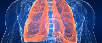
Computed tomography (CT) of the lungs is considered the “gold standard” for diagnosing pneumonia, in particular pneumonia associated with COVID-19. Tomograms—multiple scans of the respiratory organ in three planes—visualize non-functional areas of compaction or infiltration of the lung tissue.
When they talk about lung damage during pneumonia, they mean that the alveoli - small bubble-shaped cavities of the lungs that are responsible for storing air and gas exchange, fill with liquid, mucus, fibrous tissue and “fail.”
In the early stages, pneumonia can be virtually asymptomatic or cause minor discomfort: cough, difficulty breathing, fever. However, it quickly turns into a more severe form and the person begins to feel a lack of air, a spasm in the chest caused by pulmonary edema, or acute respiratory distress syndrome - a widespread inflammatory process that causes complications in the heart and in some cases leads to death.
In this regard, it is very important to recognize pneumonia in time and begin treatment. Lung CT is the only diagnostic method that allows you to identify foci of infiltration and assess the degree of their severity, even if less than 5% of the lungs are affected.
After a computed tomography scan of the lungs, especially if there is suspicion of viral pneumonia, patients are primarily interested in the results and interpretation of the examinations. In this article we will talk about what CT1, CT2, CT3, CT4 mean in conclusion, and what you should pay attention to if pneumonia is still detected.
What organs are visible on an X-ray of the lungs?
The X-ray image focuses mainly on the respiratory organs: larynx, trachea, bronchi, lungs, pleura, alveoli, chest, diaphragm. The image is performed in such a way that the organs of the circulatory, lymphatic and digestive systems are additionally visible on the X-ray of the lungs. For example, the contours of the heart, the shadow of the esophagus, aorta, and intrathoracic lymph nodes are visible. Organs are partially visualized because shadows overlap and overlap each other in a two-dimensional X-ray image. This is a feature of the diagnostic method, in contrast to CT and MRI. The term “mediastinum” often appears in the conclusions of specialists. This name hides a whole complex of organs running down the center of the human body between the upper chest and the diaphragm. In the radiograph of the lungs, part of the mediastinum falls into the field of view of the radiologist:
- trachea
- bronchi
- aorta
- pulmonary arteries and veins
What else can be seen on an X-ray of the lungs, besides the organs of the respiratory system? Bones of the chest: sternum, collarbone, ribs, shoulder blades, thoracic vertebrae.
Rice. 2: Anatomical picture of the internal organs of the chest Thus, from the X-ray image one can more or less clearly see the pathologies of the bones of the upper part of the human body, lungs and bronchi. Along the way, a chest x-ray can reveal cardiovascular abnormalities. However, in some cases, it is difficult to draw full conclusions about the functioning of the heart and blood vessels based on the X-ray picture due to the low ray penetration in this area. An x-ray gives the doctor the opportunity to assess, perhaps, the location and size, since more than half of the heart projectionally covers the mediastinum. The image shows only the left part of the main organ, two aortic arches, the superior and inferior vena cava, and the pulmonary trunk. This is the so-called vascular bundle. The upper part of the mediastinum, or more precisely, the width in this area, allows us to draw conclusions about some deviations in the functioning of the cardiovascular system.
For example, an increased width of the mediastinum is used to diagnose an aortic aneurysm.
Often, if there are X-ray signs of pathology, an in-person appointment with a cardiologist and additional research using other more informative methods are required. It is possible to assess the condition of the esophagus only by performing an X-ray with contrast, which is prescribed strictly according to indications. Also, during the interpretation of the x-ray, doctors evaluate soft tissues - neck, trachea.
When scanning is prohibited
The main danger of the technology is the presence of an X-ray field during the examination. It is minimized to low-dose levels, which is considered safe for relatively healthy people. However, this type of diagnosis is not allowed:
- young children;
- women at any stage of pregnancy;
- cancer patients who have recently undergone radiation therapy;
- patients living or working in places with increased background radiation, often requiring radiography.
In some cases, limitations may be associated with the use of contrast. To enhance the clarity of the images, an iodine-based staining agent is injected intravenously. For more information about contrast enhancement, please follow the link: Lung CT with Contrast.
How to read a lung x-ray
The image reading algorithm is associated with clear knowledge in the field of x-ray anatomy and extensive experience, on the basis of which doctors have to make complex conclusions. We strongly recommend that our patients and the reader of the article abandon the idea of independently describing X-ray images, since in addition to the picture there are age-related deviations, defects in the apparatus, physiological features of the structure of internal organs, the consequences of diseases and other aspects that must be taken into account. Among other things, the specialist takes into account the peculiarities of the patient’s positioning. For example, the intensity of the pulmonary pattern in different sections varies depending on the position - horizontal/vertical. To obtain the right to describe images, a radiologist, in addition to general medical education, undergoes specialization training. A physician who is not trained in radiology cannot correctly interpret a diagnostic image.
X-ray interpretation by medical university students is carried out using standard algorithms. The schemes do not take into account many rare nosological forms, but for most situations, reading images using a standard is quite suitable. X-ray researcher William Conrad Roentgen himself did not know human x-ray anatomy, so he did not conduct experiments with anatomical images. Metal objects were used to evaluate the properties of the invention. The first equipment, created in 1896, did not have modern digital quality, so it was impossible to correctly decipher an X-ray image. Some aspects of normal x-ray anatomy of internal organs were provided to medicine by Hoffmans and Maastricht. Studies were conducted to determine the radiation dose that a person receives during X-rays. In order for humanity to receive a high-quality type of diagnosis, the first X-ray researchers conducted experiments in absolute darkness, sacrificing health due to ionizing radiation. Doses from historic X-ray equipment were 100 times higher than those generated by modern digital machines. The image was not of high quality, but small details could be seen in it. High-quality interpretation of lung X-rays has become possible due to the introduction of scattering gratings, reduction of exposure time, and the use of software. Readers will be interested to know that X-rays were discovered in 1895.
Let's look at some aspects of interpreting lung images using radiographs as an example. Rice. 5: X-ray of the chest organs in a direct projection: large black areas on both sides of the image - pulmonary fields Correct interpretation of a chest x-ray IN THE PRESENCE OF DISEASE begins with identifying the leading pathological syndrome. Then a thorough intrasyndromic diagnosis is carried out. In the presence of several X-ray syndromes, the diseases should be differentiated according to a certain criterion. Most often, when trying to decipher an x-ray, doctors encounter three pathomorphological forms:
- Inflammation;
- Cyst;
- Tumor.
When identifying the leading X-ray syndrome, we propose to classify the pathology as one of the groups described above. Then the structure of clearing or darkening is studied. An alternative algorithm for interpreting lung x-rays is based on “backward elimination.” The rationality of using the scheme is relevant in the presence of many diseases characterized by similar radiological manifestations. For example, with miliary dissemination, the following nosological forms must be excluded:
- Tuberculosis;
- Silicosis;
- Carcinomatosis.
If the diseases are not confirmed by the specificity of X-ray syndromes, the following conditions should be assumed:
- Interstitial viral pneumonia;
- Hemosiderosis (deposition of iron inclusions in lung tissue).
Other diseases must be excluded when the x-ray picture of the above-described nosology is not confirmed. Correct interpretation of the radiograph is impossible if clinical data are excluded from the analysis. The radiologist's conclusion must be clinical and radiological! That is why the patient is referred for an X-ray by a general practitioner, who sets out in detail the purpose of the study, so that the laboratory assistant can choose the correct placement option, and the radiologist understands how to interpret the X-ray of the lungs in accordance with the attending physician’s expectations from the diagnosis. Rice. 6: Digital X-ray of the lungs in a direct projection: the leading syndrome is expansion of the cardiac shadow to the left
Operating principle of CT
The technology is based on the use of low-dose X-ray radiation, which can be absorbed by body tissues with varying degrees of intensity. The degree of reflection of the ionizing field from hard and soft fibers is recorded by equipment sensors, displaying them in photographs in black and white. Despite the monochrome images, the equipment is capable of distinguishing more than 500 shades of gray inherent in different tissues and organs. Any deviations from the norm in their structure, density, or size are immediately recorded by equipment.
Scans are made layer by layer with a minimum offset of 1 mm. Thus, up to 1000 sections can be obtained in one procedure without white spots or blind spots. From these scans, a multidimensional model of the organ is compiled, each section of which can be viewed from different angles and with maximum approximation. Since computed tomography involves X-ray radiation (radiation exposure is present), it can only be performed in consultation with the attending physician and no more than 5-8 times a year for an adult patient. Changing the frequency of repeated procedures is available only as prescribed by a specialist.
Medical X-ray Reading Algorithm
Qualified radiologists interpret chest x-rays according to the following scheme:
- Studying the quality of the image;
- Identification of the suspicious area;
- Determination of “normality” or “pathology” of an image;
- Assessment of the localization of the primary lesion (pleura, lungs, bone tissue);
- Description of the structure of the X-ray syndrome.
To describe the pathological picture, radiology doctors identify 10 radiological syndromes:
- Total darkening of the pulmonary field;
- Limited shade;
- Dissemination;
- Round shadow;
- Cavities;
- Enlightenment (subtotal, total);
- Focal dissemination or limited focus;
- Pathology of pulmonary pattern;
- Root changes;
- Disturbances in the patency of the bronchial tree.
The most common are diseases of the cardiovascular system. They provoke disability and premature mortality. Early detection of heart pathology makes it possible to save lives. A qualified interpretation of a lung x-ray should include an assessment of the condition of the pulmonary fields, cardiovascular system, and soft tissues. Rice. 7: X-ray of the OGK (direct projection): high standing of the right dome of the diaphragm. The optimal combination of radiography with other clinical instrumental methods allows for the timely detection of hemodynamic, pulmonary and other organ dysfunctions. The knowledge of a radiologist is valuable not only because this specialist knows how to correctly read X-ray images of the lungs and correctly formulate descriptions with a conclusion. A competent expert will definitely offer a recommendation for a more thorough diagnosis or dynamic monitoring of the patient over a certain period of time.
What may be in the results
The scans produced by the equipment are immediately sent to the doctor’s computer, who studies them in detail after the procedure. The interpretation takes from half an hour to a day, so it is not necessary to wait for the results in the clinic. It is enough to leave your contacts at the reception so that you will be notified of readiness or a digital version of the conclusion and photographs will be sent to your email address.
In the photographs you can see clear boundaries of the internal organs, blood vessels and bones. The denser the fabric, the lighter it appears in the images. The pulmonary parenchyma is normally homogeneous, gray, but with the development of inflammation, fibrosis, and filling of the alveoli with fluid, the pattern changes. Foci of compaction are displayed in the form of a lightened network, “frosted glass”. Tumors and other neoplasms are also visible with precise sizes and configurations; they are indicated by a developed vascular network around the pathological object.
What diseases can X-rays of the lungs detect?
Next, we will give an idea of what the lung x-ray method reveals. An X-ray image of the chest can do a lot, including visualizing diseases of the circulatory, lymphatic, and immune systems. Of course, a beginner in radiology and a person without special knowledge cannot see the cause-and-effect relationships of certain diseases of systems adjacent to the respiratory system. However, an experienced specialist will draw the right conclusions and determine the pathology for sure. It is possible to see cardiovascular disease using a chest x-ray, and this method is often the first step towards identifying a problem with “heart” health. From the image you can determine the following pathologies of the heart, blood vessels and aorta:
- Hypertonic disease
- Excess cholesterol in the blood
- Heart failure
- Cardiomegaly
- Aneurysm
- Accumulation of fluid in the pericardial sac
Of course, the target of diagnosis is most often the respiratory system. So, what can an X-ray determine in the lung area:
- Bronchitis
- Pneumonia
- Tuberculosis
- Atelectasis
- Cancer
- Pneumothorax
- Pleurisy
- Pleural empyema
- Hydrothorax
- Asthma
- Pneumosclerosis
- Sarcoidosis
- Emphysema
- Abscess
- Bronchopneumonia
- COPD
- Pulmonary embolism
- And other important diseases of the lungs, pleura, bronchi
A chest X-ray image visualizes fractures of bones and the thoracic spine, dislocations, osteomyelitis, osteochondrosis, ankylosing spondylitis, osteosarcoma, osteoporosis and other bone pathologies. What else does a lung x-ray see... An x-ray of the chest X-ray visualizes signs of autoimmune diseases, such as substernal goiter, hyperthyroidism. Abnormalities in the circulatory system such as anemia. Detects parasites in the lungs, a disease called echinococcosis.
Do I need to prepare for the procedure?
There is no need to prepare specifically or for a long time for a tomography session, but when going for a scan, it is important to consider the following points:
- Interaction with the device occurs in a lying position, so it is better to wear loose, comfortable clothes that do not restrict movement when lying on a high couch.
- All foreign objects, metal jewelry, and small change must be removed from the body and pockets, as distortions and artifacts may appear in the photographs.
- Tell the specialist about chronic pathologies, possible pregnancy, the presence of built-in implants, electronic stimulators.
- To register or conclude an agreement for the service, bring your passport, as well as the results of other studies and a referral from the treating doctor.
How much does a CT scan of the lungs cost in St. Petersburg and what does the price depend on?
In St. Petersburg, the cost of lung tomography starts from 2800 rubles and depends on the following factors:
- type, quality and year of manufacture of the tomograph;
- price level in the medical center (economy, average or premium);
- time of day at which the study is carried out (often it can be done cheaper at night, for more details - CT scan of the lungs at night);
- the need to obtain film with a photograph (usually pictures are recorded on disk, and film costs extra);
- the need to administer contrast, read more here - CT scan of the lungs with contrast;
What to do if you need to undergo a test urgently
To urgently undergo the study, call: +7 (812) 385-77-56. Our operators see a summary table of available places in most diagnostic clinics in St. Petersburg, so they can make an appointment in the near future.
Sources used:
- Gedymin L.E., Filippov V.P., Evgushchenko G.V., et al. The role of biopsy in the diagnosis of pulmonary diseases at the prehospital level of observation // Clinical Medicine. - 2009. - No. 4. — P.41-44.
- Ivanova E.V., Bilichenko T.N., Chuchalin A.G. Morbidity and mortality of the working age population in Russia due to respiratory diseases in 2010 -2012. // Pulmonology. - 2015. - T. 25. - No. 3. - P.291-297.
- Karashchuk N.P., Kiseleva M.V. Lung cancer and tuberculosis // Scientific Medical Bulletin of Ugra. -2014. — No. 1-2(5-6). — P.71-73.
- Kartashov M.V., Kartashova O.M., Kotlyarov P.M. First experience of using diffusion-weighted magnetic resonance imaging for small cell lung cancer // Medical visualization. – 2011. – No. 4. – P.28-33.
- Kotlyarov P.M. Post-processing of multislice computed tomography data in the refined diagnosis of pathological changes in diffuse lung diseases // Pulmonology. - 2021. - No. 4. — P.472-477.
- Kotlyarov P.M., Sergeev N.I. Radiation research methods in the differential diagnosis of parasitic and tumor lesions of the lungs // Siberian Journal of Oncology. – 2021. – No. 15(4). – P.33-39.
- Ternovoy S.K., Nasnikova I.Yu., Morozov S.P. Modern computed tomography in clinical medicine // Kremlin Medicine. Clinical Bulletin. - 2008. - No. 2. — P.9-13.
- Tyurin I.E. Differential diagnosis of single lesions in the lungs // Polyclinic. - 2014. - No. 3-1. — P.28-32.
- Yudin A.L., Afanasyeva N.I., Abovich Yu.A., et al. High-resolution computed tomography in the diagnosis of interstitial pneumonia // Medical visualization. -2002. - No. 4. — P.40-48.
Text approved by therapist Laeva Alina Vadimovna
CT lungs Admiralty CT lungs Bucharest CT lungs Vasileostrovskaya CT lungs Volkovskaya CT lungs Gorky CT lungs Gostiny Dvor CT lungs Dostoevskaya CT lungs Elizarovskaya CT lungs Zvenigorodskaya CT lungs Zvezdny CT lungs Komendantsky Prospekt CT lungs Krestovsky Island CT lungs Kupchino CT lungs Ladozhskaya CT lungs Ligovsky Prospect CT lungs Lomonosov CT lungs Mayakovskaya CT lungs International CT lungs Moscow CT lungs Moscow Gate CT lungs Nevsky Prospekt CT lungs Novocherkasskaya CT lungs Bypass channel CT lungs Obukhovo CT lungs Pobeda Park CT lungs Alexander Nevsky Square CT lungs Primorskaya CT lungs Proletarskaya CT lungs Bolshevikov Avenue CT lungs Rybatskoe CT lungs Sadovaya CT lungs Sennaya Square CT lungs Spasskaya CT lungs Sports CT lungs Staraya Derevnya CT lungs Technological Institute CT lungs Dybenko Street CT lungs Frunzenskaya CT lungs Chkalovskaya CT lungs Elektrosila show all metro show more