X-ray of the upper gastrointestinal tract and colon is one of the mandatory stages in diagnosing the digestive canal. X-ray of the stomach and intestines It is carried out with contrast, which allows you to see the patency of the esophagus, the condition of the mucous membrane - its relief, peristalsis, tone. Key objectives of the survey:
- search for any motility disorders of the digestive canal;
- diagnosis of hiatal hernia;
- detection or confirmation of varicose veins of the upper digestive canal;
- search for benign and malignant formations (polyps, ulcers, tumors);
- In the diagnosis of gastritis, radiography of the stomach is also used, but plays a secondary role.
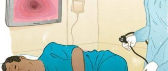
Features of colonoscopy
The procedure is carried out using an endoscopic device - a colonoscope. The main feature is the need for careful preparation, including a special diet and taking medications that help cleanse the intestinal mucosa. Preparation lasts at least 2 days.
The study reveals:
- Oncological diseases.
- Sources of intestinal bleeding.
- Assess intestinal motility.
- Diagnose inflammatory processes in the intestine.
Indications for the procedure
A referral for a barium X-ray of the intestines is issued by an oncologist or gastroenterologist; in some cases, a general practitioner can refer you for the procedure. An examination is prescribed in cases where the doctor suspects a disease that poses a threat. Therefore, a referral for an X-ray of the intestines with barium should be taken with particular seriousness.
There is a whole range of signs of dangerous diseases of the digestive tract, in which a specialist prescribes an examination of the intestine with a barium-based contrast agent using radiography.
Serious weight loss for no significant reason
Severe weight loss without lifestyle or diet changes clearly indicates pathology. The patient often experiences lethargy and apathy. The source of the symptom may be a disruption of the intestines. To find out the cause, your doctor may order a barium X-ray of your small or large intestine.
The occurrence of loose stools for no reason
Loose stools for no apparent reason indicate a disruption in the digestive process. With long-term problems, it can cause intestinal damage. Therefore, the symptom requires prompt diagnosis and proper treatment.
Change in stool color
Loose, black stools are also called “tarry” stools. The discharge resembles a black paste with a pungent, unpleasant odor. The symptom indicates bleeding in the initial part of the duodenum or in the small intestine. If the bleeding is heavy, the patient may show signs of blood loss.
These conditions are extremely dangerous. Therefore, at the first appearance of tarry stools, it is better to immediately contact a specialist and undergo an intestinal examination with barium as prescribed; X-rays will show the source of bleeding and help the doctor make a diagnosis.
The appearance of severe pain in the abdominal area
Pain in the abdominal area can be caused by intestinal deformation or dysfunction. Often pain in the abdominal area indicates an ulcer. To determine the source of pain, various procedures are prescribed to examine the intestines, including the use of barium during x-rays.
Serious pathologies (emerging tumor processes)
Various neoplasms can interfere with normal intestinal motility, leading to pain, impaired defecation, loss of appetite, etc. To localize the neoplasm and establish its nature, the patient is prescribed an intestinal X-ray; the use of barium in an X-ray helps to obtain a contrast and informative image.
Contraindications
Regardless of what examination is planned to be performed - an X-ray examination of the intestines or a colonoscopy - both methods have similar contraindications, and they are not associated with barium, since this contrast agent is well tolerated by almost all patients. The most serious contraindications include:
- Pregnancy.
- Severe diseases of the cardiovascular system, blood clotting disorders.
- Deep intestinal ulcers accompanied by heavy bleeding.
Important
Keep in mind that during irrigoscopy there is a certain radiation exposure to the body, so the procedure cannot be prescribed too often.
Intestinal irrigography: what it is, how it is carried out, preparation - MEDSI
Table of contents
- What is irrigoscopy?
- What does the study show?
- Stages of irrigography
- Indications for irrigography
- Contraindications
- Preparing for the examination
- Advantages of carrying out the procedure at MEDSI
The intestines are an important part of the human body, which is responsible for both the digestion process and the removal of feces from the body.
It consists of several parts:
- Small intestine:
- Duodenum - food eaten from the stomach enters here
- Small intestine - here food is digested, nutrients are absorbed, after which the rest of the food is sent to the next section
- Valve - through it, unnecessary remnants of processed food enter the colon
- Large intestine - where stool is formed
- Rectum - through it feces are removed from the body
Disruption of any of these areas leads to problems in the functioning of the entire organism as a whole.
What is irrigoscopy?
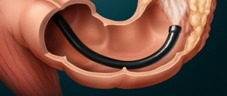
Irrigoscopy of the small intestine is a type of x-ray examination in which a barium contrast solution is injected into the patient's body. The use of contrast is necessary for the intestines to be visible on an x-ray (since they are not normally visible on x-rays). A barium solution is injected into the rectum, after which images are taken.
This analysis is used to find out whether the patient has disorders or pathologies in the structure of the intestine, whether there are neoplasms or other diseases of this organ.
The procedure is non-invasive - it does not require abdominal incisions - and not endoscopic (in a standard situation, no sensors are inserted into the human body to carry it out). It is painless and does not cause discomfort to the patient.
What does the study show?
Irrigoscopy is used to examine and assess the condition of different parts of the intestine. It allows you to determine:
- The presence or absence of disturbances in the operation of the valve (bauhinium valve) - it should open only in one direction
- Condition, location, diameter, elasticity and shape of the colon
- The quality of functioning of the intestinal sections
- Presence of gastric pathologies
- Presence of obstruction
- Qualitative condition of the inner lining of the intestine (presence of polyps, ulcers, ruptures, scars, cracks)
- Does the patient have any neoplasms (tumors) or diverticula (protrusion of the intestinal wall)
- Are there pathologies of interaction between the intestine and adjacent organs?
Stages of irrigography
Irrigoscopy of the large and small intestines takes place in two stages:
- General x-ray examination of the lower abdominal cavity
- Barium contrast examination
The first stage is used to diagnose the presence of surgical pathologies of the intestine. At this time, the patient lies on his back.
A barium solution is then injected into the patient's body through the rectum. After it has spread through the intestines, several pictures are taken. For this stage, the patient takes the following position: lying on his left side, pressing his legs to his stomach and placing his hands behind his back. After this procedure, the patient has a bowel movement, and doctors take another picture.
If there is a suspicion of cancer or another tumor, the doctor may do a double contrast study. In this case, after the previous stage, air is pumped into the rectum and then another series of pictures is taken.
When examining children using irrigoscopy, there are several nuances:
- If necessary, an ultrasound probe may be inserted into the intestine during the examination.
- If the procedure is prescribed for a small child, it will be performed under general anesthesia.
Indications for irrigography
This study is prescribed in the following cases:
- The patient has nonspecific ulcerative colitis
- There is a suspicion of cancer or another tumor
- The doctor suspects the appearance of polyps, ulcers or other disorders of the mucous membrane
- Possibility of having Crohn's disease (irreversible changes in the intestines)
- Possibility of obstruction
- There is a suspicion of a foreign body in the intestine
- Diagnosed with tuberculosis or Grishsprung's disease (congenital anomaly in organ development)
- Suspected injury or damage
Also, irrigoscopy of the large and small intestines is prescribed in the presence of symptoms such as:
- The appearance of bloody and mucous discharge in the stool
- Sudden weight loss for unknown reasons
- Permanent abdominal pain, discomfort in the anal area
- Regular bowel dysfunction (constipation, diarrhea)
It is worth undergoing a similar examination in the event of a decrease in hemoglobin levels, which occurred for unclear reasons. It is also prescribed if it is impossible to do a colonoscopy.
It is necessary to undergo irrigography for preventive purposes for people over fifty years of age whose family has a predisposition to colorectal cancer (one of the relatives suffered from this disease).
Contraindications
Irrigography cannot be used in such cases as:
- The patient has been diagnosed with cardiovascular disease
- The patient suffers from an infectious disease accompanied by fever
- The man is in a coma
- Woman pregnant
- The patient is in lactation period
- The patient has swelling or inflammation of the brain
- Exacerbation of ulcerative colitis diagnosed
- Allergy to contrast agent
- The patient's intestinal patency is extremely impaired
- There is injury or rupture of the intestinal wall
In the last two cases, the barium solution may enter the abdominal cavity, which will lead to serious complications in the patient's condition.
Preparing for the examination
Irrigoscopy of the large and small intestines requires simple preparation. But it is important to remember that the accuracy of the research results depends on its quality, so you must strictly follow the doctor’s instructions.
Preparatory procedures include two important stages:
- Compliance with diet rules
- Colon cleansing before the procedure
Two to three days before the examination, you need to start following a diet, namely, exclude foods such as:
- Vegetables (cabbage, carrots, beets)
- Legumes (corn, beans, peas, lentils)
- Porridge (barley, oatmeal, millet)
- Fruits (apples, apricots, bananas, oranges)
- Bakery products
- Dairy
- Sweet
- Fast food
- Fatty and smoked foods
Such products lead to excessive gas formation and take a long time to digest.
The last meal should be on the eve of the study, no later than 6 pm.
Also, the day before and in the morning of the test, it is necessary to do a cleansing enema or use drugs that stimulate the release of feces from the intestines.
Advantages of carrying out the procedure at MEDSI
- In the MEDSI network of clinics, the procedure for irrigoscopy of the large and small intestines is performed by experienced coloproctologists, oncologists and surgeons of the highest qualification categories, candidates of medical sciences
- Clinical centers have high-tech modern equipment that uses gentle X-ray radiation, but allows you to take the most accurate images
- Specialists will help you prepare correctly for the procedure to ensure the best possible result.
- To make an appointment for a consultation, you do not need to go to the reception desk, since you can easily make an appointment by calling 8 (495) 7-800-500
Which technique is preferable?
What's better
: X-ray examination of the intestines or colonoscopy - the doctor decides depending on the individual characteristics of the patient. Irrigoscopy with barium is often preferable, as it does not create discomfort for the patient.
But often it is impossible to do without endoscopic examination. This technique is the “gold standard” for diagnosing colon cancer; it allows detection of tumors at the earliest stages of development. X-ray methods do not provide such accuracy; their resolution is much lower.
Conditions for obtaining reliable diagnostic results
To avoid distorted results of hardware diagnostics of the stomach and intestines, patients are advised to follow a simple diet the day before. Alcohol should be discontinued one day before the test, tobacco and medications that can affect intestinal motility – 12 hours before.
to undergo fluoroscopy of the intestines on an empty stomach, after a three-day slag-free diet. There should be no metal objects in the clothes or on the patient.
The results can be distorted by:
- increased gas exchange;
- full stomach (metabolic disorders, in this case an enema is given a few hours before fluoroscopy of the stomach and intestines);
- metal on the patient (jewelry, crowns, prosthetic plates).
At the end of the study, barium suspension will be released for another 2-3 days. It can cause constipation or mechanical obstruction of the canal, which will aggravate the disease, if any. Therefore, if you experience serious discomfort (bloating, pain, nausea) during this period and (especially) after 72 hours, you should contact your doctor.
Advantages of carrying out X-rays of the chest cavity at the CELT clinic
We have our own diagnostic center with a full range of equipment necessary to conduct research in a wide variety of areas. Modern X-ray equipment not only makes the procedure safe, but also takes it to a new quality level. Our patients can count on:
- affordable prices;
- accurate diagnosis;
- the procedure is performed by radiologists with over 15 years of experience;
- use of effective safe means;
- use of modern gentle technologies.
You can find out our prices and make an appointment by calling in advance.
Sigmoidoscopy, colonoscopy and irrigoscopy: what is the difference?
Based on your medical history (symptoms, complaints) and examination, your doctor will determine which test is right for you. The methods are similar in implementation, but differ in information content.
Colonoscopy is considered by many to be an unpleasant procedure, but in our case it is performed with sedation (during medicinal sleep). The endoscopic method has its advantages; the images are of very high quality. In addition, during a colonoscopy it is possible to take a piece of tissue for a biopsy.
Our digital X-ray machine also produces clear images when performing irrigoscopy.
1 Digital X-ray unit ITALRAY CLINODIGIT COMPACT
2 Digital X-ray machine ITALRAY CLINODIGIT COMPACT
3 Digital X-ray machine ITALRAY CLINODIGIT COMPACT
Colonoscopy can also be a therapeutic procedure: with the help of a colonoscope, an endoscopist can excise tumors and coagulate blood vessels.
During sigmoidoscopy, the intestines are examined at a depth of no more than 20-30 cm. This simple procedure allows you to evaluate only the anus and rectum. Colonoscopy allows screening at a depth of about 160 cm.
1 We perform sigmoidoscopy at KARL STORZ
2 We perform sigmoidoscopy at KARL STORZ
3 We perform sigmoidoscopy at KARL STORZ
Irrigoscopy does not require anesthesia or sedation. However, a person is exposed to X-ray radiation, albeit in minimal doses. Preparation for irrigoscopy and colonoscopy is approximately the same: you need to go on a diet and cleanse the intestines. Before sigmoidoscopy, the intestines are also cleansed.
Please note that before irrigoscopy you should undergo a consultation and examination with a proctologist. The procedure has a number of side effects; it is necessary to determine how necessary irrigoscopy is for diagnosis.
Many people consider this procedure not the most pleasant. Contact good radiologists (such as those at MedicCity, our doctors have the highest qualification category) and choose a clinic that has the appropriate equipment. We conduct X-ray examinations using a modern digital device Italray Clinodigit Compact. It allows you to evaluate the study area in real time and obtain high-quality images.
For details, please contact our contact center. The phone number is listed in the header of the site.
The material was prepared with the participation of a specialist: