How do blood vessels bleed?
Bleeding can be mild, moderate or severe. His character may be:
- arterial - strong, jet;
- venous - blood loss occurs gradually;
- capillary - minor discharge due to damage to small vessels.
The problem itself can be stable/unstable, recurrent. Vessels of the mucosa, submucosal and intermuscular plexus, as well as those located outside the gastrointestinal tract, can bleed.
The expiration can last for several hours or days. In medicine there are:
- profuse blood loss - the patient loses more than 1 liter of blood in 1-3 hours and needs urgent medical attention;
- acute - less than 1 liter expires in 1-2 days, the patient’s vital signs are relatively stable;
- chronic - develops slowly, often over several weeks or even months, the intensity of symptoms gradually increases.
Diagnostic features
When a patient is admitted to the hospital, if he is conscious, the doctor clarifies the complaints, the presence of provoking factors, and examines the patient. Clinical data may suggest gastric or intestinal bleeding, but its exact source can only be determined with further examination. To do this, gastroscopy is performed using endoscopic equipment, during which esophageal varices, stomach ulcers, polyps or diverticula, and ruptures of the mucous membrane may be detected.
A clinical and biochemical blood test, determination of coagulability and platelet count are required, and stool is examined for the Gregersen reaction. If necessary, an ultrasound of the abdominal organs, diaphragm, chest, radiography of the stomach, angiography, radioisotope scanning, and MRI are performed.
The information obtained during the examination helps to carry out a differential diagnosis of bleeding in the stomach, that is, to exclude intestinal or pulmonary foci of blood loss. In addition, it is necessary to accurately identify pathologies leading to blood loss.
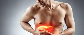
Why is there a problem?
Medicine knows about 200 causes of bleeding in the digestive tract. At the first symptoms, you should seek professional help from a doctor and under no circumstances self-medicate, as this will only worsen the condition.
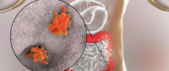
Ulcer
The most common reason. With an ulcer, the integrity of the mucous membrane of the organ is disrupted, and the main difference of the disease is deep tissue damage. The disease is chronic - with remission and exacerbations. On the mucous membrane of the stomach, esophagus or duodenum, areas of inflammation are formed, in which the protective function (mucus secretion) is reduced. Gradually, the mucosal tissues, including the walls of blood vessels, become thinner, which leads to their rupture.
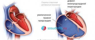
Phlebeurysm
The problem may occur in the esophagus or stomach due to increased pressure in the portal vein. The most common cause is cirrhosis of the liver. Rupture of a large vessel with varicose veins is extremely dangerous, since at this moment there is a copious outflow of blood. According to statistics, in 40% of cases it stops spontaneously. And the activity of bleeding depends on the degree of liver damage.
Colon diverticulosis
With this disease, the lining of the colon bulges, forming diverticula. The reasons for their formation in medicine are not completely clear; they are mainly associated with increased intraluminal pressure. Basically, the pathology is typical for adult patients over 50 years of age. In the acute course of the disease and rupture of intrawall blood vessels in the area of diverticula, intestinal bleeding occurs.
Tumors and polyps
They occur in the small and large intestines and are benign neoplasms that grow into the intestinal lumen. Most often, bleeding is minor and chronic. The danger lies in possible degeneration into malignant tumors.
Haemorrhoids
This is the formation of venous nodes around the rectum in the anal area. The main causes are thrombosis or tissue inflammation. The disease can be acute or chronic, and its common causes are a sedentary lifestyle, excessive exercise, and obesity. Bleeding (the color may be scarlet or dark) is minor and occurs most often after bowel movements.
More rarely, esophagitis, acute hemorrhagic gastropathy, erosive duodenitis, and Mallory-Weiss syndrome are found as causes of bleeding in the upper gastrointestinal tract. In the lower part, bleeding can be caused by tumors, vascular malformations, and various inflammations.
Determining the cause and assessing the intensity of bleeding
Often the probable cause of bleeding can be determined by history.
Important things to consider:
- symptoms of dyspepsia (especially at night);
- symptoms of stomach ulcer;
- side effects of medications taken, especially those that inhibit blood clotting, such as non-steroidal anti-inflammatory drugs;
- likelihood of alcohol abuse;
- the presence of hepatitis B or C - these may indicate liver cirrhosis and portal hypertension as possible causes of bleeding.
Who is at risk?
Basically, diseases that lead to bleeding are observed in adults. Moreover, according to statistics, men are 2 times more likely than women to be diagnosed with problems with the gastrointestinal tract - the stomach, duodenum. As we noted above, ulcerative pathologies hold first place in terms of the number of diseases. The peak age for diseases is 40-45 years.
However, the problem is not limited to adults. The diagnosis associated with ulcerative lesions of the gastrointestinal tract is often made to adolescents who uncontrollably consume junk food and drinks. Cases of the formation of intestinal polyps are also common.
Gastric and intestinal bleeding is increasingly being detected even in newborns. Basically, they are caused by intestinal volvulus. In 3-year-old children, leakage can be caused by the formation of a diaphragmatic hernia, as well as abnormalities in the development of the organs of the lower gastrointestinal tract.
Sigmoidoscopy (rectoscopy)
If necessary, an examination of the rectum is performed. Sigmoidoscopy (rectoscopy) is a method of endoscopic examination of the rectum and distal sigmoid colon by examining their internal surface using a sigmoidoscope inserted through the anus.
Sigmoidoscopy is the most accurate and reliable examination of the rectum and lower sigmoid colon. In the practice of a coloproctologist, sigmoidoscopy is an obligatory component of every proctological examination. The examination allows you to visually assess the internal surface of the rectum and distal third of the sigmoid colon to a level of 20-35 cm from the anus.
What symptoms to look out for
Patients with the diagnoses we listed above should be especially monitored for the appearance of alarming symptoms. If you are taking medications for the liver and gastrointestinal tract, carefully monitor your well-being. If you are concerned about the changes discussed below, consult your doctor. However, knowing these signs is useful for every person, since many diseases of the lower and upper gastrointestinal tract develop without obvious painful sensations. Often their first manifestation may be the symptoms of bleeding.
Weakness
This is the main sign of any prolonged bleeding. Weakness gradually increases, the patient's skin turns pale, he feels cold sweat, a ringing in the ears, and trembling of the limbs. The weakened state may last for several minutes, after which it passes and returns periodically. If blood is bleeding actively, fainting or semi-fainting and even shock are possible.
Vomit
This symptom accompanies severe blood loss - more than 0.5 liters. If the vomit is dark cherry in color, it is most likely coming from a vein near the esophagus. If unchanged blood is clearly visible in the vomit, the integrity of the artery in the esophagus is most likely compromised. If the patient vomits so-called “coffee grounds” of brown color, the problem lies in the gastric vessels. Only a doctor can accurately determine the nature, location and intensity of blood loss.
Chair
Traces of blood in the stool may appear within a few hours or 1-2 days after the integrity of the blood vessels is damaged. With significant problems with the stomach or duodenum, as well as blood loss in a volume of more than 0.5 liters, you can observe melena - loose stools that resemble tar in color and consistency. If the blood loss is smaller, which often happens, for example, with intestinal bleeding, then the stool remains formed, but its color darkens.
Please note that darkening of the stool can occur due to eating foods that contain dark coloring substances, for example, blueberries and cherries. Dark stools are not an absolute sign of the presence of blood in the stool and problems in the upper or lower gastrointestinal tract. The diagnosis can only be made by a qualified specialist.
Peroxidase test to confirm the presence of guaiacol
This is the most commonly used stool blood test. The test available is called Hemoccult. In this study, a stool sample is distributed into a guaiacol-impregnated cardboard box, which, if the stool contains heme, turns blue (heme causes a reaction similar to peroxidase). The more blood in the stool, the higher the likelihood of a positive result.
The Hemoccult test gives a positive result in 50% of cases when 10 ml of blood enters the gastrointestinal tract during the day. Under physiological conditions, 0.5-1.5 ml of blood is normally extravasated into the lumen of the gastrointestinal tract during the day.
The sensitivity of the first test is estimated to be approximately 30%. Performing 3 tests (which is the standard) increases the confidence to 92%. False negative results are especially common in people taking medications or foods high in vitamin C.
Relatively often, the test gives false positive results when consuming large amounts of red meat and foods high in peroxidase (radish, radish, horseradish). Taking iron supplements does not give false positive results in the study.
How to make a diagnosis
The doctor examines the patient, assessing his external condition, the shade of the skin and mucous membranes. Then he measures blood pressure - often it is low.
In the clinic, the patient undergoes a general blood test. Using it you can quickly get an idea of the level of hemoglobin and the volume of other blood cells. Additionally, the diagnosis is made by biochemical analysis, but it is usually prescribed several days after the onset of blood loss, since the chemical composition of the blood changes only over time.
The main diagnosis concerns the detection of the very cause of the violation of the integrity of blood vessels. To do this, doctors use the following hardware examinations.
- Endoscopy - examination of the esophagus, stomach, duodenum using a flexible tube with a miniature camera allows you to quickly detect a problem area;
- Contrast radiography - an effective method for detecting bleeding in the gastrointestinal tract involves injecting a safe contrast solution into the organ, followed by an X-ray;
- Magnetic resonance imaging is a modern method that allows you to obtain comprehensive information about the condition of all tissues of a particular organ of the gastrointestinal tract.
Physical examination
Physical examination
The gastroenterologist begins the examination from the head - carefully examining the nose, mouth and throat, since sometimes the cause of bleeding can be localized.
Next, they are identified with
symptoms of anemia and hypovolemia. The patient's heart rate and blood pressure are determined. A bad symptom is an acceleration of heart rate and a drop in blood pressure (>10 mmHg). Orthostatic hypotension involves rapid or severe blood loss.
Other signs that may indicate significant blood loss include:
- cool skin;
- oliguria;
- disturbances of consciousness.
Patients with anemia feel weak, dizzy, see spots before their eyes, have chest pain, and may faint. The severity of these symptoms depends on the amount of blood lost and the rate of bleeding.
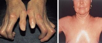
It is also important to carefully examine the patient's body for symptoms of chronic liver disease. A number of specific symptoms indicate a pathological condition of the liver:
- presence of spider veins on the skin of the breast
- gynecomastia;
- loss of axillary and pubic hair;
- yellowing of the skin;
- palmar erythema;
- enlarged spleen;
- ascites;
- swelling of the legs;
- hand tremors
Antisecretory therapy
Optimal conditions for the implementation of vascular-platelet and hemocoagulation components of hemostasis are created at pH > 4.0. Proton pump inhibitors and H2-histamine receptor blockers are used as antisecretory drugs.
Attention! It is not advisable to prescribe H2-histamine receptor blockers and proton pump inhibitors at the same time.
Medicines of both groups suppress the production of hydrochloric acid in the stomach and thereby create conditions for stable hemostasis of the bleeding vessel. But proton pump inhibitors show more consistent results in reducing gastric acidity and are significantly more effective in reducing the risk of recurrent bleeding. The antisecretory effect of proton pump inhibitors is dose-dependent. Therefore, the use of high doses of drugs is currently recommended, so the prescription regimens indicated below are not a typo by the author.
Patients are prescribed an intravenous infusion of one of the following proton pump inhibitors:
- Omeprazole (Losec) 80 mg IV as a loading dose, followed by 8 mg/hour.
- Pantoprazole (Controloc) 80 mg IV as a loading dose, followed by 8 mg/hour.
- Esomeprazole (Nexium) 80 mg IV as a loading dose, followed by 8 mg/hour.
The loading dose of the drug is administered in approximately half an hour. Intravenous administration of the drug is continued for 48-72 hours, using, depending on possibilities, a bolus or continuous route of administration. In the following days, they switch to oral administration of the drug at a daily dose of 40 mg (for all of the proton pump inhibitors listed in this paragraph). The approximate duration of the course is 4 weeks.
Attention. The administration of proton pump inhibitors should be started before endoscopic intervention, as this reduces the likelihood of recurrent bleeding.
In the absence of proton pump inhibitors, or patients intolerant of them, intravenous H2-histamine receptor blockers are prescribed:
- Ranitidine 50 mg IV every 6 hours or 50 mg IV followed by 6.25 mg/hour IV. After three days, 150-300 mg orally 2-3 times a day;
- Famotidine 20 mg IV drip every 12 hours. Orally for treatment use 10-20 mg 2 times/day or 40 mg 1 time/day.