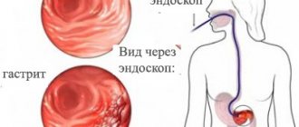
Very often, patients come to the appointment who, due to pain in the upper abdomen, heaviness in the epigastric region after eating, and belching, have undergone gastroduodenoscopy and made a conclusion, based on which the doctor has diagnosed “atrophic gastritis.” A person is worried about what to do and whether this gastritis can turn into stomach cancer?
Today we will try to answer the question of what methods will help confirm the presence of atrophic gastritis and how to treat such patients.
Among stomach diseases, the most common diagnosis made by practitioners is “chronic gastritis.”
Chronic gastritis occupies a central place among stomach diseases and is the most common disease of the gastrointestinal tract. It is believed that a third of the adult population suffers from chronic gastritis, but only 10-15% consult a doctor. It is important that chronic gastritis precedes or accompanies diseases of the stomach that are serious in course and prognosis - peptic ulcers, stomach cancer, as well as nearby organs of the digestive system.
Atrophic gastritis - what is it?
What do we mean by atrophic gastritis? It is based on atrophic processes in the gastric mucosa, inflammatory and dystrophic processes that lead to functional failure of the mucosa. Atrophy of the glands of the gastric mucosa consists of a decrease in the number of cells that produce enzymes (pepsins) and parietal cells that secrete hydrochloric acid, and cells that produce Castle factor.
Anatomy of the stomach
Conventionally, there are several sections in the stomach:
- the cardiac section is the most initial one, where food comes from the esophagus,
- fundus - the uppermost dome-shaped part of the stomach, located at the very top of the stomach and where there is always some air,
- the body of the stomach (fundus), where enzymes and hydrochloric acid are produced and food is located during digestion,
- the outlet section or pyloric section of the stomach (antrum), which ends with the pyloric sphincter, which limits the cavity of the stomach and separates it from the duodenum.
Atrophy of stomach cells can affect all sections of the stomach, and then we are talking about total damage to the organ or affect specific sections - fundus (damage to the body of the stomach), antrum (outlet section of the stomach), or selectively affect part of the cells of the stomach. The clinical manifestations of atrophy in different parts of the stomach will be different.
It is known that the stomach is a hollow organ where food enters from the oral cavity and where gastric digestion of food occurs: food deposition, mechanical and chemical processing of food and its evacuation from the stomach into the intestinal tract.
Cytology of the stomach
In the mucosa of each section there are various cells responsible for the digestion of food and participation in hematopoiesis:
- parietal (or parietal) and main cells of the body of the stomach, which produce hydrochloric acid,
- pepsinogens, which under the influence of hydrochloric acid are converted into the active enzyme pepsin, which digests food proteins,
- chymosin and lipase, which digest milk,
- Castle factor (gastromucoprotein), which is responsible for the participation of the stomach in hematopoiesis and vitamin B12 metabolism. Castle factor binds to vitamin B12 in protein foods in the presence of calcium and transports vitamin B12 to the intestines, where it is absorbed into the body. With a lack of production of Castle factor, B12-deficiency anemia develops (decreased blood hemoglobin).
Additional flocculi of the gastric mucosa form mucus in almost all parts of the stomach. This mucus performs a protective function against bile, medications that the patient takes, and hydrochloric acid.
In the outlet section of the stomach there are endocrine cells - G cells, which produce a stimulator for the formation of pepsinogens, hydrochloric acid, and are responsible for the state of the motor function of the stomach.
Atrophic gastritis - causes of development
The most common cause of atrophic gastritis at present is considered to be Helicobacter pylori infection. Superficial gastritis develops in the stomach and, with prolonged infection, becomes atrophic.
Stomach infection is associated with damage to the antrum of the stomach in the form of limited or total damage to this department - atrophic gastritis of the antrum and body of the stomach. The development of atrophic processes in the stomach can be the result of age-related changes in the mucosa, genetic characteristics caused by hypo- or atrophy of the gastric mucosa, drug-induced damage to the stomach, or long-term use of antisecretory drugs (PPIs).
Autoimmune atrophic gastritis, when the body begins to produce antibodies against its cells in the gastric mucosa, is quite rare.
First signs
The disease is accompanied by characteristic signs of a local and general nature:
- belching;
- nausea;
- regurgitation;
- a feeling of heaviness and pressure in the epigastric region after eating;
- unpleasant sensation in the mouth (especially in the morning);
- heartburn;
- burning in the epigastrium.
Immediately after eating, a dull pain occurs in the epigastric zone; Moreover, the pain sensation increases in a standing position and when walking. Intestinal dyspepsia is very often observed: rumbling, flatulence. With low acidity, diarrhea usually occurs, which worsens significantly after drinking milk or eating fatty foods.
How to confirm the diagnosis of “atrophic gastritis”
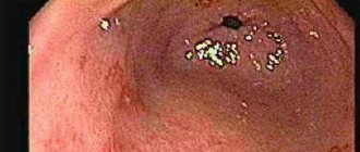
The diagnosis of chronic atrophic gastritis is morphological . If an endoscopic examination is carried out, then if atrophic processes in the stomach are suspected, biopsies of the gastric mucosa should be taken and a histological interpretation of the changes in it given.
The morphological diagnosis should take into account: damage to the stomach, the condition of the glands, the severity and depth of inflammation and changes in the mucosa (metaplasia, dysplasia). The clinical diagnosis of “chronic atrophic gastritis” without morphological confirmation does not make sense.
The most accurate diagnostic method is endoscopic examination with biopsy . However, due to the uneven distribution of atrophic processes in the gastric mucosa, histological studies can give false negative results. In addition, biopsy is an expensive and time-consuming method and cannot be performed on every patient, and endoscopic examination itself is a seriously invasive method of examination.
Currently, there is an alternative to biopsy of the gastric mucosa - a screening method of research called “serum biopsy”, which can objectively reflect the functional state of the gastric mucosa and its morphological basis using blood serum samples.
Serum biopsy or Gastropanel (or test panel ) provides a simple and reliable way to obtain great information about the structure and function of the gastric mucosa, with great sensitivity and specificity to identify patients with atrophic gastritis and those who need further examination - endoscopic examination of the stomach. If the test panel reveals atrophic gastritis, then endoscopic examination is mandatory with oncological alertness.
How is Gastropanel performed?
Blood sampling to determine markers of atrophic gastritis is carried out in the morning on an empty stomach and 20 minutes after a protein breakfast. Blood is collected into serum tubes, which are centrifuged for analysis.
Blood serum analysis determines markers of chronic atrophic gastritis. Markers of atrophy of the mucous membrane of the fundic and antral parts of the stomach are:
- Pepsinogen I, Pepsinogen II and their ratio,
- Gastrin-17 and Gastrin-17 stimulated after a protein breakfast,
- determination of antibodies to Helicobacter pylori Ig G.
The tests are based on enzyme immunoassay technology.
Besides,
- Antibodies to parietal cells of the body of the stomach are determined in autoimmune atrophic gastritis with a high risk of disorders associated with vitamin B12 deficiency, for which the level of vitamin B12 is determined.
- Homocysteine is identified as a risk factor for vascular and thromboembolic diseases.
- To clarify Helicobacter pylori infection, an additional test of the acute phase of infection is performed, Ig A and IgM antibodies are determined.
Which doctor should I contact?
If it is necessary to diagnose and treat atrophic gastritis, we invite patients from Moscow to the medical center in Khimki. Advantages of treatment in our department:
- Gastritis is diagnosed quickly, accurately with minimal loss of time and nerves of the patient. It is possible to perform gastroscopy in your sleep.
- After determining the exact form of the disease, the degree of its development and other features, gastroenterologists will prescribe a comprehensive course of effective treatment.
- Capabilities of modern endoscopic equipment
- At the request of the patient, gastroscopy is performed in a state of medicated sleep.
- Available: doctor’s appointment from 1500 rubles
- Convenient: open daily from 8:00 to 21:00
- Quickly: we will carry out all the diagnostics at the first appointment
- Complete: has all the necessary equipment
A few words about serum markers
We have a diagnostic algorithm for identifying stomach diseases, which is recommended for all patients who have pain or discomfort in the upper abdomen.
Pepsinogens
There are seven isoforms of pepsin precursors, five of which are designated as group Pepsinogen I of the main cells of the stomach body and Pepsinogen II, uniformly secreted by the glands of the entire stomach and duodenum. Pepsinogens formed in the stomach are absorbed into the blood and determination of their serum level is a generally accepted marker of atrophic gastritis “serological biopsy”.
A decrease in the level of Pepsinogen I indicates the severity of atrophic gastritis of the body of the stomach, and since the activation of Pepsinogen I into active pepsin occurs with the participation of hydrochloric acid, then the level of Pepsinogen I can roughly represent the level of stomach acidity. Pepsinogen II is produced in all parts of the stomach and in the duodenum. As the severity of atrophy increases, the ratio of serum levels of Pepsinogen I and Pepsinogen II decreases, which indicates the severity of atrophy and the spread of the process.
Gastrin
Gastrin-17, produced in the outlet section of the stomach after stimulation of cells by various factors (stomach distension, protein foods). In case of atrophy of the gastric antral mucosa, the secretion of Gastrin-17 decreases. To assess the presence and severity of the atrophic process in the stomach, it is necessary to conduct a protein stimulation test, the decrease of which shows the severity of atrophy; in the case of atrophy of the mucous membrane of the antrum of the stomach, the secretion of Gastrin-17 is proportionally reduced. In patients with severe atrophic gastritis in the antrum of the stomach, the risk of developing stomach cancer is 90 times higher than in people with normal gastric mucosa.
Homocysteine
Homocysteine is an early marker of cellular functional deficiency of B12, B6, folic acid due to the development of atrophic gastritis and other reasons - age, smoking, Helicobacter pylori infection, etc. With atrophic gastritis, the level of homocysteine increases in the blood and becomes toxic to the body. When donating blood for homocysteine, you should avoid protein foods, vitamins, and hormonal contraceptives 1 day before the test. This test can be an addition to the Gastropanel or an independent test for other diseases.
O.Ya. Babak, Doctor of Medical Sciences, Professor, Director of the Institute of Therapy named after. L.T. Small Academy of Medical Sciences of Ukraine, Kharkov
Atrophic gastritis is understood as a progressive inflammatory process of the gastric mucosa, characterized by the loss of gastric glands. The clinical and morphological feature of atrophic gastritis is a decrease in the number of specialized glandulocytes that provide the secretory function of the stomach, and their replacement with simpler cells, including those that produce mucus. Extensive atrophy of the gastric mucosa is usually associated with hyposecretion of hydrochloric acid and impaired pepsinogen production.
What is known today about atrophic gastritis? The most common etiological factors causing atrophic gastritis are Helicobacter Pylori (H. Pylori) infection and autoimmune gastritis. Moreover, the occurrence of the vast majority of atrophic gastritis is associated with H. pylori. The bacteria H. pylori, persisting on the gastric epithelium, cause chronic Helicobacter superficial gastritis. Long-existing superficial Helicobacter gastritis without appropriate treatment transforms into atrophic. Atrophic gastritis, as a rule, does not manifest itself clinically for a long time, therefore the diagnosis of chronic gastritis is rather morphological than clinical. The main method for diagnosing atrophic gastritis is endoscopic examination. During endoscopy, the esophagus, stomach, and duodenum are examined. With severe atrophy, the gastric mucosa has characteristic differences compared to that with, for example, superficial gastritis. The final diagnosis can be established by morphological analysis of biopsy samples of the gastric mucosa taken during endoscopy. Morphologically, atrophy is determined by a decrease in the number of functioning specialized cells of the stomach. It has been proven that in H. pylori-associated gastritis, atrophy processes more often occur when infected with certain strains (Cag A+ and Vac A+) of H. pylori. One of the morphological signs of atrophic gastritis is intestinal metaplasia, which has traditionally been considered as a precancerous change in the gastric mucosa. Other research methods - radiography of the stomach, ultrasound examination of the abdominal cavity and computed tomography - are not informative in terms of diagnosing atrophic gastritis.
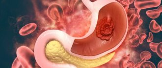
What should you be wary of with atrophic gastritis? What is the prognosis of the disease? Hypothetically, the presence of atrophic gastritis in its natural course can have two scenarios. The first is that long-term chronic gastritis leads to a significant decrease in the acid-forming function of the stomach, requiring replacement therapy, without which signs of impaired digestive function will be observed. The second option is that as a result of long-term chronic persistent inflammation in the gastric mucosa, characteristic of H. pylori-associated gastritis, a disruption of cellular renewal in the stomach occurs, which contributes to the emergence of target cells for the influence of carcinogenic substances on them, and subsequently to cellular mutations. As a result, the normal cellular epithelium of the stomach is replaced by metaplastic, dysplastic and neoplastic. According to the WHO definition, dysplasia is understood as cellular changes in which part of the epithelium is replaced by cells with varying degrees of atypia. In the international classification of epithelial neoplasia of the digestive tract (2000), dysplasia is neoplasia, in other words, a tumor. So, atrophic gastritis can transform into stomach cancer. The greatest danger in terms of cancer development is atrophic gastritis with reduced acid-forming function of the stomach (cancer incidence is up to 13%). Among the currently known molecular mechanisms underlying hereditary predisposition to gastric cancer are: induction of TGF-β1 expression, partial polymorphism of the IL-1 gene cluster (IL-1β). As a result of the development of atrophy of the gastric mucosa, its antitumor protection is reduced, and conditions are created for the active influence of carcinogens. When severe atrophy of the gastric body epithelium occurs, the risk of developing gastric cancer increases 5 times compared to that with non-atrophic gastritis. The bacteria H. pylori are classified as biological carcinogens in relation to gastric cancer. Most researchers believe that H. pylori is the main etiological factor in the development of chronic gastritis, which is an obligatory link in the cascade of processes leading to stomach cancer. Based on an analysis of the results of multicenter studies, the International Agency for Research on Cancer under the WHO in 1994 recommended that H. pylori infection be considered an absolute carcinogen for humans. Currently, gastric cancer is considered as the end result of a long-term multi-stage and multifactorial process, in which cellular changes in the gastric mucosa are caused by disturbances in the microenvironment. This process is called the Correa cascade (1995) after the author who described it. It includes chronic gastritis, intestinal metaplasia, dysplasia and cancer. H. pylori-associated gastric carcinogenesis is a multi-stage process characterized by the development of chronic gastritis - the first step in the evolutionary cascade. Subsequent changes lead to the formation of atrophy, small intestinal (types I and II) and large intestinal (type III) metaplasia and dysplasia of the gastric epithelium, ultimately leading to gastric adenocarcinoma. It is atrophic gastritis that occupies a middle position in the chain of the above changes on the path to stomach cancer.
How to avoid the transformation of atrophic gastritis into stomach cancer? The answer to this question consists of parts of equal importance: the earliest possible detection of precancerous changes, their adequate treatment and prevention (prevention) of the manifestation of the latter. When observing patients with chronic gastritis, it is important to catch the moment when atrophy of the gastric mucosa occurs and begins to progress, and it is advisable to do this in a simple informative and non-invasive way.
Timely detection of atrophy of the gastric mucosa is the first diagnostic stage in identifying the risk of gastric cancer.
Numerous studies in recent years have shown that areas of complete and incomplete intestinal metaplasia of the gastric mucosa cannot be regarded as a reliable marker of an increased risk of developing gastric cancer. Research shows that it is much more important to assess not the type of metaplasia, but its volume. Thus, with a large volume of metaplasia exceeding 20% of the surface of the gastric epithelium, real conditions are created for the development of dysplasia with the subsequent formation of gastric adenocarcinoma. Consequently, the risk of developing gastric cancer increases with severe atrophy of the gastric epithelium, characterized by extensive foci of intestinal metaplasia. How can one determine the area of such a lesion in practice? It should be remembered that these changes occur at the cellular level and cannot be recognized with conventional endoscopy. An accessible and effective way to diagnose metaplastic changes in the gastric mucosa is the method of chromogastroscopy - intravital staining of the gastric mucosa with a dye (usually methylene blue), carried out during an endoscopic examination. This technique is based on the absorption of dye by foci of intestinal metaplasia, which makes it possible to assess their size, perform a targeted biopsy for histological analysis of the mucosal biopsy, and identify possible dysplasia or metaplasia. However, the morphological diagnosis of atrophic gastritis is associated with a number of difficulties. The difficulty of diagnosing atrophy using the morphological method is due to the fact that in the early stages the process is never diffuse, therefore, the results of gastrobiopsy can contribute to over- and under-diagnosis. With inflammation, the microscopic picture may change and the manifestations of atrophic gastritis may be inadequately assessed due to the false conclusion about the loss of glands. The subjectivity of the methodology is also high. All this makes us look for other reliable ways to test atrophic changes in the gastric mucosa. A number of minimally invasive hematological tests (Biohit test panel) have been developed to avoid diagnostic errors and provide a comprehensive assessment of the condition of the gastric mucosa, the degree of its atrophy and loss of normal glands and cells in the antrum and body of the stomach. During an endoscopic examination, detection for the presence of H. pylori must be carried out. In this case, urease or histological research methods (from gastrobiopsy specimens) should be considered the most appropriate. Determination of the level of serum pepsinogen (S-PGI) or the ratio of pepsinogen I to pepsinogen II (PGI/PGII) is a non-endoscopic method for diagnosing atrophic gastritis with damage to the body of the stomach. As the degree of atrophy of the gastric mucosa (loss of normal acid-producing glands) increases, the levels of S-PGI and PGI/PGII gradually decrease. Determination of the level of gastrin in blood serum, mainly gastrin-17 (SG-17), can be used as an indicator of the morphological state of the mucous membrane of the gastric antrum. That is, a decrease in SG-17 is a biochemical marker of atrophic gastritis with damage to the antrum of the stomach (loss of antral G-cells). Decreased levels of SG-17 and S-PGI can be considered a result of progressive atrophic gastritis with loss of normal glands and cells of the mucous membrane of the body and antrum of the stomach. G-17 is almost completely synthesized and secreted by G-cells of the antrum of the stomach. These cells are components of normal antral glands; in the case of progression of atrophic gastritis, their number decreases against the background of damage to the antral glands and the appearance of intestinal metaplasia. In H. pylori-associated gastritis, there is a tendency for serological levels of G-17 and PGI to increase. Low intragastric acidity contributes to an increase in the serological level of G-17, and vice versa. Permanent, long-term hypo- or achlorhydria leads to extremely high levels of G-17 in the blood. This is especially often observed with low acidity (atrophic gastritis with damage to the body of the stomach) in combination with preserved mucous membrane of the antrum. This clinical picture is most typical for autoimmune atrophic gastritis. If there are concomitant signs of mucosal atrophy in the antrum (multifocal atrophic gastritis), then SG-17 does not increase and the test panel shows low levels of S-PGI and SG-17. The overall accuracy of the test panel in diagnosing atrophic gastritis is about 80% (when compared with the results of endoscopy and biopsy). This test panel is a minimally invasive alternative for the initial evaluation of patients with suspected gastric atrophy and dysplasia. It allows you to reliably identify patients with various forms of gastritis, determine the localization and etiology of the pathological process, assess the likelihood of developing stomach cancer and build further tactics for managing the patient. Considering the connection between the occurrence of atrophy of the gastric epithelium and intestinal metaplasia with H. pylori infection, the choice of treatment and prevention of further progression of the process becomes obvious. The method of choice is anti-Helicobacter therapy. In 2002, Japanese researchers convincingly proved the possibility of regression of metaplastic changes in the gastric mucosa after successful destruction of H. pylori bacteria. Using chromoscopy, they were able to establish that within five years after successful anti-Helicobacter therapy, the size of the foci of intestinal metaplasia decreased by almost 2 times in comparison with the initial ones. Subsequent studies confirmed the feasibility of this therapeutic approach. Currently, there is no doubt about the need for anti-Helicobacter therapy for patients with atrophic gastritis. Preliminary data from several multicenter studies monitoring H. pylori -associated gastric precancer and cancer support reversal of gastric mucosal inflammation and associated atrophy, intestinal metaplasia, and genetic instability. In this regard, ideally, patients with H. pylori-positive chronic atrophic gastritis should undergo eradication therapy, and if there is no effect, a study to identify markers of genetic instability and careful monitoring. This recommendation is reflected in the international recommendations for the diagnosis and treatment of diseases associated with H. pylori - Maastricht Consensus 3 (2005). To destroy H. pylori bacteria, as in the Maastricht Consensus 2 (2000), three- and four-component regimens of antibacterial drugs in combination with proton pump inhibitors (PPIs) in standard doses are recommended: PPI + clarithromycin + amoxicillin and PPI + tetracycline + metronidazole ( furazolidone) + colloidal bismuth. At the same time, it should be remembered that complete restoration of the structure of the mucous membrane in severe atrophy to normal requires a long time, and in some cases, apparently, this is not possible. In cases where pre-tumor processes do not undergo reverse development or progress, it is necessary to apply more radical methods of treatment, using the arsenal of modern endoscopic operations, up to resection of the gastric mucosa. The main goal of primary prevention of atrophic gastritis is timely and effective treatment of superficial Helicobacter gastritis. For this purpose, standard anti-Helicobacter therapy regimens are used, in accordance with the recommendations of the Maastricht Consensus 2 (2000) and 3 (2005). An important point is subsequent monitoring of the success of this therapy. Monitoring must be carried out using non-invasive methods (breath urease test or stool test). If eradication is unsuccessful, repeat courses of treatment. In addition, it has been proven that by adhering to a healthy diet, you can reduce the risk of cancer (progression of atrophy), which has been confirmed in studies conducted in a number of countries. It is recommended to avoid eating canned, pickled and smoked foods, stop smoking and drinking strong alcoholic drinks (especially in combination with fatty, fried, smoked and salty foods), and avoid overeating. It is necessary to control body weight, perform active physical activity, consume more fresh vegetables (including onions and garlic), fruits and natural juices, vitamins A, C, b-carotene, herbs, whole grains, and dairy products. In some developed countries of Europe and the USA, the introduction of a healthy lifestyle has led to a several-fold reduction in the incidence of stomach cancer; today this disease in these countries is considered rare, accounting for only 3% of malignant neoplasms. Monitoring - constant observation with periodic re-examination - is absolutely mandatory for patients with atrophic gastritis. So, at present, the need for special attention to atrophic gastritis is obvious. The integrated use of modern research methods - endoscopic, morphological, hematological (test panel) and others - contributes to its accurate diagnosis. The use of effective methods for the treatment and prevention of atrophic gastritis, the elimination of conditions that contribute to its development, present today a real opportunity to improve the prognosis of this disease and eliminate the risk of developing stomach cancer.
References 1. E.G. Burdina, E.M. Mayorova, E.V. Grigorieva, I.I. Timofeeva, O.N. Minushkin // Gastrin-17 and pepsinogen I in assessing the condition of the gastric mucosa // Russian Medical Journal, 2006, No. 2, p. 9-11. 2. H. Vaananen, M. Vauhkonen, T. Helske, I. Kaarijanen, M. Rasmussen, H. Tunturi-Hihnala, J. Koskenpato, M. Sotka, M. Turunen, R. Sandström, M. Ristikankare, A. Jussila, P. Sipponen // Non-endoscopic diagnosis of atrophic gastritis based on a blood test: correlation between the results of histological examination of the stomach and the levels of gastrin-17 and pepsinogen I in the serum // Clinical perspectives of gastroenterology, hepatology, 2003, No. 4, p. 26-32. 3. V.D. Pasechnikov, S.Z. Chukov, S.M. Kotelevets / Prevention of stomach cancer based on eradication therapy of pretumor diseases // Russian Journal of Gastroenterology, Hepatology, Coloproctology, 2003, No. 4, p. 11-19. 4. A.A. Sheptulin, V.A. Kyprianis / Diagnosis and treatment of Helicobacter pylori infection: main provisions of the Maastricht-3 conciliation meeting // Russian Journal of Gastroenterology, Hepatology, Coloproctology, 2006, No. 2, p. 88-91. 5. Kim N, Lim SH, Lee KH et al. Long-term effects of Helicobacter pylori eradication on intestinal metaplasia in patients with duodenal and behign gastric ulcers Dig. Dis. Sci. – 2000. – Vol. 45. – 1754-1762. 6. Malfertheiner P., Megraud F., O'Morain C. et al. Current concepts in the management of Helicobacter pylori infection – The Maastricht 2 Consensus Report // Aliment. Pharmacol. Ther. – 2002. – Vol. 16. – P. 167-180.
Advantages of Gastropanel for diagnosing “atrophic gastritis”
Thus, Gastropanel allows you to answer the following questions:
- does the patient have atrophic gastritis, in which part of the stomach are the changes localized,
- assess the risk of developing stomach cancer and peptic ulcers,
- does the patient suffer from gastritis caused by Helicobacter pylori,
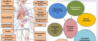
- identify autoimmune gastritis or atrophic gastritis with damage to the body of the stomach, which may determine the risk of developing vitamin B12 deficiency, which can be associated with many diseases (atherosclerosis, vascular damage to the brain and heart, depression, polyneuropathy, dementia, etc.)
What advantages of Gastropanel can be noted: it is a simple non-invasive serological test, high sensitivity, specificity, safety and convenience for the patient, quick results, and meets the principles of evidence-based medicine.
Symptoms
The more acidity is reduced, the more clearly the symptoms of gastritis with low acidity will appear:
- Signs of excessive bacterial growth include rumbling, loose stools, milk intolerance, and bloating. If gastritis is accompanied by frequent diarrhea, weight loss may occur and signs of mineral or vitamin deficiency may develop.
- Dystrophy - signs of deficiency of B vitamins, as well as C, E, D, protein deficiency (weight loss, “polished” or coated tongue with a thick white coating).
- Dyspepsia - loss of appetite, feeling of heaviness or fullness in the stomach, belching of rotten food, bad breath (cocosmia), unpleasant taste, nausea.
- Dull, aggravated after eating, without clear localization, aching pain that occurs due to stretching of the stomach.
- Development of anemia due to decreased absorption of iron and vitamin B12 (due to Castle factor).
Since inflammation of the stomach also affects other parts of the digestive system, hypoacid gastritis is often accompanied by diseases such as enterocolitis, cholecystitis, and pancreatitis.
What to do if you are diagnosed with atrophic gastritis
A few words about the principles of treatment of chronic atrophic gastritis. Treatment approaches are individual, it all depends on the severity of atrophic processes, the presence of Helicobacter, and vitamin B12 deficiency.
The problem will be dealt with by a doctor who will prescribe:
- treatment,
- repeated studies to monitor treatment,
- repeated consultations to assess the clinical manifestations of the disease and adjust treatment.
It is most difficult to eliminate autoimmune mechanisms of damage to the gastric mucosa; the question of prescribing hormonal drugs arises only when gastritis is accompanied by anemia. Helicobacter pylori eradication therapy has its own characteristics. First of all, the question arises about the acidity of gastric juice, and more often treatment is carried out without antisecretory drugs. The study of the acidity of gastric juice using the pH-metric method is now rarely carried out (usually daily monitoring in a hospital setting), but it is possible to orient the acidity according to the level of Pepsinogen I.
A high level of Pepsinogen and periodic heartburn in a patient may indicate preserved gastric secretion. In case of atrophic gastritis in a hypo or anacid state with damage to the body and outlet of the stomach, antisecretory drugs are excluded and Helicobacter pylori eradication is carried out with antibiotics. Effective eradication of infection improves processes in the gastric mucosa, and can be considered as a prevention of the development of gastric cancer.
Diagnostics
A gastroenterologist and endoscopist are involved in the diagnosis and treatment of gastritis with low acidity. The specialist examines the patient and conducts a number of examinations:
- morphological studies;
- X-ray of the stomach;
- esophagogastroduodenoscopy;
- endoscopic biopsy;
- gastroscopy;
- determination of pepsinogen level;
- gastric probing with intragastric pH-metry;
- examination of gastric juice;
- diagnosing pylori using stool ELISA, PCR testing, detection of antibodies in the blood and a breath test for this microorganism.
The purpose of these studies is not to confuse the disease with other pathologies and to make an accurate diagnosis. Based on the examination results, the gastroenterologist prescribes a treatment regimen. If required, the attending physician will refer you for a consultation with a nutritionist, who will create an appropriate diet. In some cases, to eliminate concomitant diseases, consultation with other specialists of a narrow focus (cardiologist, therapist) is required.
Nutrition for chronic atrophic gastritis
There are no strict dietary restrictions, but you need to know that some foods alkalize the stomach (mainly protein foods) and further reduce the acidity of gastric juice, and there are foods that stimulate it (vegetable and fruit juices, vegetables, fruits).
Most often, doctors prescribe diet 2, the purpose of which is to mechanically spare the gastric mucosa and preserve chemical irritants.
But medical experience shows that strict restrictions are not required. The necessary diet - eating in small portions with replacement therapy drugs allows for good gastric digestion, in which protein foods are also digested in the stomach.
Atrophic gastritis - treatment
Replacement therapy
Replacement therapy includes the use of hydrochloric acid preparations and gastric juice enzymes:
- Natural gastric juice is a preparation of animal origin, the composition corresponding to human gastric juice, contains hydrochloric acid and enzymes. Use 1-2 tablespoons (15 - 30 ml) during meals or after meals 2-3 times a day.
- Acidin-pepsin , containing betaine hydrochloride, which is hydrolyzed in the stomach to release free hydrochloric acid and the enzyme pepsin. Before use, tablets (500 mg) are dissolved in 50-100 ml of water and taken with meals. These medications are administered through a straw.
- Abomin - the drug contains the sum of proteolytic enzymes of the stomach and is obtained from the gastric mucosa of calves and lambs. Use 1 tablet (0.2 g each containing 50,000 units in 1 tablet) 2 - 3 times a day with meals for 1 - 3 months.
Stimulant therapy
Some drugs that stimulate the secretion of gastric juice:
- Mineral water (Essentuki N17, Narzan) is used warm 15-20 minutes before meals, the course is 2-3 weeks, twice a year.
- Rosehip decoction , cabbage, tomato, carrot, lemon juice, 50 ml each, diluted with boiled water 1:1 before meals, alternate.
- Medicinal mixtures: plantain, St. John's wort, wormwood, thyme in equal proportions.
- Plantaglucide is widely used - granules of plantain leaves to prepare a suspension, for which take 1/1 -1 teaspoon (½-1 g) per ¼ glass of warm water and apply 30 minutes before meals 2-3 times a day, course of treatment 3- 4 weeks, for preventive treatment 1-2 months. Plantaglucide activates gastric secretion, has an enveloping, regenerative, anti-inflammatory and antispasmodic effect.
- Collection of herbalist Mikhalchenko S.I.:
Ingredients: angelica, yarrow, dandelion, birch, trifoliate, plantain, caraway, burnet, licorice, fumifera, calendula, chamomile. Application: pour 1 dessert spoon of the mixture into 500 ml of boiling water, leave in a thermos by volume overnight, strain. Take 150 ml 3 times a day an hour before meals. Before use, warm up by diluting with hot water. Keep refrigerated. Reception: 10 days admission - 2 days break, 3-4 months.
During the period of relative remission of atrophic gastritis, the main drugs are replacement and stimulating therapy, which affect the secretory insufficiency of the stomach.
Diet and proper nutrition
If you have gastritis with low acidity, a strict diet is required. It should include:
- Cereals and pasta. Rice, buckwheat, oatmeal, and semolina are allowed.
- Soups - with low-fat chicken or fish broth, vegetable, vermicelli, cereal, with meatballs.
- Lean meat (chicken, beef, rabbit, turkey). Fish (pike perch, hake, pollock, pike).
- Eggs – no more than 2 pieces per day.
- Seasonings and sauces - vegetable oil, salt, herbs.
- Vegetables - beets, pumpkin, carrots, tomatoes, zucchini, cauliflower.
- Dairy products - cottage cheese, sour cream, mild cheese, milk, yogurt.
- Sweets - biscuits, marshmallows, marshmallows, marmalade.
- Fruits and berries – pureed (jelly, compotes, jelly, soufflé).
- Drinks – tea, rosehip infusion, weak coffee.
- Bakery products - yesterday's white bread, uneatable crackers.
Exclude from the diet:
- Pastries, sweets, fresh bread.
- Soups with mushrooms and fatty broths.
- Legumes, barley, corn and millet porridge.
- Large-grained and rough-skinned berries and dried fruits.
- Hot sauces, store-bought dressings, mayonnaise.
- Fatty meat and fish.
- Cucumbers, bell peppers, cabbage, turnips, radishes, mushrooms. Pickles and marinades are not allowed.
- Fast food, alcohol, carbonated drinks, packaged juices.
It is important that meals are fractional - 5-6 times a day. It is better to snack on vegetable or fruit purees or dairy products.
The diet for gastritis with low acidity is quite loyal - the patient will not suffer from hunger, but at the same time will not experience a feeling of overeating. If the proposed diet is followed for a long time, the symptoms of gastritis will appear less and less often and a long-term remission will occur.