Screening during pregnancy is the most important and highly informative method for the early detection of congenital malformations of the fetus. Screening during pregnancy allows you to assess the baby's development over time and identify possible pathologies of maternal and formed organs that can cause miscarriage. Ultrasound is included in the prenatal screening complex along with biochemical diagnostics, when the level of hormones in the blood is determined, which can also indicate the presence of certain pathologies.
According to the schedule approved by the Ministry of Health, routine ultrasound screenings are carried out three times during the entire gestational period, but in some cases there is a need for additional ultrasound examinations according to indications. Then ultrasound is performed on the patient as often as needed.
Timing of scheduled ultrasound screenings
The content of the article
When, from the first days after conception, pregnancy proceeds without pathologies or any complaints from the woman, ultrasound screening is done three times according to the following schedule:
- 10 weeks – 13 weeks 6 days.
- 18 – 22 weeks.
- 30 – 34 weeks.
Ultrasound in the first and second trimester is recommended to be planned strictly for the specified period of pregnancy. This is due to the fact that if gross malformations are detected in a timely manner in the fetus for up to 14 weeks, the woman is given an abortion for medical reasons. After this period, such manipulation is not carried out. If serious abnormalities in the development of the fetus are detected only at the second ultrasound - before 22 weeks, the woman is advised to have an abortion.
Requirements for the timing of the last - third planned ultrasound are discussed personally with the supervising doctor and can be postponed closer to childbirth if the woman is not worried about anything and other diagnostic techniques for dynamic monitoring of the condition of the pregnant woman and the fetus do not indicate possible problems in the development of the child.
Do I need to undergo a 3D/4D ultrasound?
The 3D/4D ultrasound method allows you to evaluate fetal parameters in three dimensions - length, depth, height - and in real time. This test is not mandatory and a woman can undergo it at her own request. The best time for this is 20-24 weeks, since it is already possible to clearly distinguish the facial features and gender of the child.
Basically, future parents are curious to look at the baby without waiting for the birth, because this way they can get a fairly clear and colorful first “photo” of the child. However, there are a number of cases when a doctor can refer an expectant mother to a 3D/4D ultrasound for medical reasons:
- multiple pregnancy;
- high risk of congenital pathologies - in this case, 3D/4D ultrasound is performed at 13-18 weeks;
- pregnancy occurs with complications;
- pregnancy occurred as a result of IVF, MESA, ICSI;
- The family had already given birth to children with severe genetic diseases.
The procedure takes about an hour. The doctor writes the result onto a flash card or CD and gives it to the parents.
Screening during pregnancy: Ultrasound in the early stages
Pregnant women are not recommended to have a routine ultrasound scan earlier than the 10th week of the gestational period. However, in some cases, an early visit to an ultrasound diagnostic specialist is justified, but this must be confirmed by an obstetrician-gynecologist and issued an appropriate referral describing the reason for the early ultrasound examination.
Early ultrasound is done in the following cases:
- if a woman’s previous pregnancies ended in spontaneous miscarriage;
- after the IVF procedure, when early monitoring of embryo development is required;
- if you have pain in the lower abdomen;
- if a woman is bothered by frequent headaches, dizziness and severe weakness;
- if you suspect a hydatidiform mole;
- when bloody discharge from the genitals appears - scarlet or brownish;
- if a woman does not have her next menstruation, but a home pregnancy test shows a negative result.
If there are disturbing symptoms, the patient will undergo a vaginal ultrasound examination even on the first day of a missed period. This technique allows you to determine an ectopic pregnancy already on the 12th day after fertilizing sexual intercourse.
The presence of an embryo in the uterine cavity can be determined already on the 5th day of delayed menstruation. However, in the absence of special indications for early ultrasound examination, it is better to postpone the first visit to an ultrasound diagnostician until 10-13 weeks of pregnancy.
Frequently asked questions by expectant mothers
When is 1st trimester screening done?
An ultrasound examination and biochemical blood test should be performed between the 11th week of pregnancy and the 13th week. Meeting deadlines is an important condition for the reliability of the results. If you do the examination earlier or later, you cannot vouch for the accuracy.
How is perinatal screening performed?
The expectant mother comes to the clinic, where she undergoes an ultrasound examination, and then blood is taken from a vein. After this, the patient goes home, nothing more is required from her. In terms of time, both procedures fit into one day.
How to prepare?
No special preparation is needed for an ultrasound. But since blood is usually given for analysis immediately after it, you need to come to the clinic with an empty stomach. The patient can drink as much as she wants. Before the procedure, the expectant mother is advised to have a good rest and try to be positive.
What risks does the survey reveal?
Blood tests and ultrasound screening reveal the likelihood of Down syndrome, Edwards syndrome, Patau syndrome, other chromosomal pathologies and hereditary diseases, defects of the central nervous system, and threats to a healthy pregnancy.
First trimester screening
The optimal period for its implementation is 11–13 obstetric weeks of gestational age. According to the recommendations of the Ministry of Health, such a study is carried out on all pregnant women to determine serious pathologies in the fetus, as well as to assess the condition of the maternal organs, including those formed during the period of gestation. The doctor evaluates the anatomy of the fetus, the number of fertilized eggs in the uterus (in the latter case, you can determine whether the twins will be identical or fraternal), can examine the heart and the vessels extending from it, listen to the heartbeat, and assess the mobility of the embryo.
The most important indicators, the assessment of which allows us to identify gross pathologies in the fetus, are:
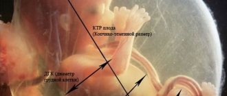
- coccyx - parietal size (KTR);
- collar space thickness (TNT);
- distance between the parietal tuberosities (BPD);
- Head circumference;
- heart rate (HR);
- size of the heart and large vessels;
- the structure of the cerebral hemispheres and their symmetry;
- volume of the fertilized egg (FE);
- distance from the frontal to the occipital bone;
- location of the stomach and heart in the fetus;
- bone lengths: femur, humerus, tibia, forearm.
The data obtained from ultrasound is compared with fetal fetometry norms and the risk of having a child with serious diseases is assessed:
PRENATAL SCREENING IN THE FIRST TRIMESTER OF PREGNANCY
Prenatal screening in the first trimester has great potential for identifying pathology in the early stages of pregnancy, when it is possible to exclude not only anatomical anomalies, but also to determine the risks of giving birth to a sick child. The optimal time for prenatal screening in the first trimester (measuring the thickness of the nuchal translucency, nasal bones, assessing blood flow in the ductus venosus, identifying tricuspid regurgitation, etc. is:
- gestation period 11+0 - 13+6 weeks, - the minimum value of the fetal CTE is 45 mm, - the maximum value of the fetus CTE does not exceed 84 mm. * KTP - coccygeal-parietal size of the fetus.
Ultrasound screening - at 11+0 -13+6 weeks it is possible to identify ultrasound markers (signs) of chromosomal pathology of the fetus - an increase in the thickness of the fetal nuchal space, absence of the nasal bone, pathological blood flow in the venous duct, the presence of tricuspid regurgitation (insufficiency of the tricuspid heart valve).
Neck thickness (TCT)
The nuchal translucency is an ultrasound manifestation of fluid accumulation under the skin in the dorsum of the fetal neck in the first trimester of pregnancy. The thickness of the nuchal space normally increases during pregnancy with an increase in the coccygeal-parietal size of the fetus.
| TVP is normal | Increase in TVP | Increase in TVP |
During the second trimester of pregnancy, the nuchal translucency usually disappears or, in rare cases, transforms into either cervical edema or cystic hygroma, with or without generalized fetal edema.
Nasal bones (NB)
The nasal bones are normally detected by ultrasound from the 10th week of pregnancy. In fetuses with chromosomal pathology, ossification processes occur later, so the absence of nasal bones or reduced size at this time may be a sign of chromosomal pathology of the fetus. During ultrasound examination at 11–13+6 weeks, the nasal bones of the fetus are not visualized in 60–70% of fetuses with trisomy 21 and in 2% of fetuses with a normal karyotype.
| NK is the norm | NK is the norm | Pathology - absence of NC |
Ductus venosus (DV)
The pathological shape of the blood flow velocity curves in the ductus venosus is observed in 80% of fetuses with trisomy 21 and in 5% of fetuses with a normal karyotype.
| No. 1 - VP - norm | No. 2 - VP - norm | No. 3 - VP - pathology |
Pathological blood flow - zero or reverse blood flow in the venous duct (Fig. No. 3 (peaks on - or - below the isoline). Zero and reverse values of blood flow in the VP during the phase of atrial contraction are a marker of CA. It is known that CA is often accompanied by congenital heart defects, which in early pregnancy can lead to changes in blood flow in the VP. Pathological blood flow in the VP is not common in fetuses with a normal karyotype.
Tricuspid valve (TV)
Tricuspid regurgitation is often observed in fetuses with CA in early pregnancy. CAs were found in 94.7% of fetuses with an enlarged nuchal space and isolated tricuspid regurgitation at 11-14 weeks of pregnancy. Cases of isolated tricuspid regurgitation have been described at 12-13 weeks of pregnancy in fetuses who were diagnosed with trisomy 21 during prenatal karyotyping. There are also reports of tricuspid regurgitation at 11-14 weeks in fetuses with trisomy 21 and in fetuses with trisomy 18, then, in fetuses with a normal karyotype - only in 0.7% of cases.
| No. 1 - TC - norm | No. 2 - TC - pathology | No. 3 - TC - pathology |
Tricuspid regurgitation Fig. No. 2 and No. 3 - pathology (below the isoline, additional signals are visible that indicate the reverse flow of blood from the right ventricle to the right atrium, with the valves closed). The detection of tricuspid regurgitation at any stage of pregnancy indicates the need for a detailed examination of the fetal heart to exclude cardiac pathology.
The frequency of occurrence of other markers of fetal chromosomal pathology, such as omphalocele, megacystis, single umbilical cord artery, etc., with certain chromosomal diseases is higher than with a normal fetal karyotype.
IMPORTANT
The main marker of CA (chromosomal abnormalities) in early pregnancy is the expansion of the fetal nuchal space. In cases where this marker is detected, prenatal karyotyping is a necessary component of prenatal examination in early pregnancy. Doppler technology and assessment of the fetal nasal bones should be considered important additional signs to increase the effectiveness of early prenatal diagnosis of CA, especially in cases of borderline or “controversial” dilatations of the nuchal space. In some cases, assessment of the fetal nasal bones and detection of pathological blood flow in the ductus venosus makes it possible to diagnose CA in the presence of normal nuchal translucency values. In cases where the nuchal translucency is expanded, additional detection of pathological blood flow in the ductus venosus and the absence or hypoplasia of the fetal nasal bones, the presence of tricuspid regurgitation indicate the need for prenatal karyotyping (invasive diagnostics). The current level of development of medical technologies makes it possible to examine pregnant women and identify among them a risk group for the presence of Down's disease (trisomy 21) and other chromosomal diseases in the fetus starting from the 11th week of pregnancy. The combination of ultrasound and biochemical examination within a period of 11 weeks - 13 weeks 6 days was recognized as the most effective.
BIOCHEMICAL SCREENING of the blood of a pregnant woman allows you to determine the concentration of proteins produced in the placenta: PAPP-A and beta-hCG. Each week of pregnancy has its own norms for the content of these proteins in the blood; with chromosomal pathology of the fetus, the ratio of proteins will change. With trisomy 21 in a fetus at 12 weeks of gestation, the concentration of β-hCG in the blood serum of a pregnant woman is increased (about 2 MoM) compared to normal values for this stage of pregnancy, while the concentration of PAPP-A is reduced (about 0.5 MoM). At the same time, the degree of difference in the concentration of β-hCG with trisomy 21 and with a normal karyotype increases with increasing gestational age, while the degree of difference in the concentration of PAPP-A decreases. These temporary changes in the levels of biochemical markers and their dependence on such an indicator as the weight of the pregnant woman should be taken into account when determining the methodology for calculating the individual risk for a particular patient. The combination of ultrasound and biochemical examination data can increase the detection efficiency of fetuses with Down syndrome to 85%. Currently, this examination method is the most effective way to prevent the birth of a child with Down syndrome or other chromosomal diseases.
To calculate the risk of chromosomal abnormalities, our Center uses a program
Astraia Software GmbH and ViewPoint GE Healthcare
Specialists involved in calculating the risk of chromosomal pathology of the fetus based on an assessment of its profile have undergone appropriate training and certification (FMF), confirming the level of quality of performing this type of ultrasound examination. More detailed information about specialists with an FMF certificate of competence can be obtained on the official FMF website. FMF Certificate
FMF - Fetal Medicine Foundation - (Fetal Medicine Foundation) fetalmedicine.org is a registered UK charity that provides training and certification for specialists in ultrasound screening in the first trimester of pregnancy. The training includes a theoretical course, practical training in ultrasound imaging standards and TVP measurements, and the provision of a training report consisting of ultrasound images of the examinations performed.
Risk assessment for trisomies
PRENATAL DIAGNOSIS
SCREENING first trimester. Comprehensive diagnostics at 11 weeks. - 13 weeks pregnancy. Results are ready on the same day!
Risk assessment is carried out:
- for trisomies 21, 18 and 13 at gestational age 11-13 weeks by combining maternal age + ultrasound data - nuchal translucency thickness, fetal heart rate + maternal blood tests - maternal serum ß-hCG and PAPP-A and risk calculation for syndromes the ASTRAYA program in order to exclude fetal pathology at the stage of intrauterine development.
An integrated approach to diagnosis makes it possible to detect fetuses at high risk of Down syndrome (trisomy 21); trisomy 18 (Edwards syndrome); trisomy 13 (Patau syndrome), identify developmental disorders of the internal organs of the fetus, exclude malformations of the brain, heart and other organs in the early stages. If a pathology is detected in the fetus, parents are provided with complete information about the nature and characteristics of the developmental defect, and the possibilities of modern medicine in terms of rehabilitation of the child. As a result, the family makes a decision about the fate of this child, carefully weighing its own possibilities.
Individual approach, interpretation of results, support at all stages of ultrasound diagnostics. The study protocol with comments (in any foreign language), if desired, can be sent by e-mail.
- NEW! Additionally, a risk assessment is carried out
- risk of preeclampsia; - risk of gestational diabetes mellitus; - risk of miscarriage at 11-24 weeks; - risk of stillbirth; - risk of restriction (delay) of fetal growth - risk of macrosomia (fetal weight at birth more than 4 kg); - risk of premature birth ( - risk of cervical incompetence ( FETAL MEDICINE
Fetal medicine is a modern branch of obstetrics that studies modern approaches to the prevention of fetal diseases, the diagnosis of congenital diseases, as well as the treatment of some threatening conditions directly in the womb. The preventive direction allows us to determine the risks of having children with congenital malformations through a comprehensive assessment of the results of biochemical and instrumental screenings. Expecting a baby brings the expectant mother not only joy, but also various fears. And the fear of the upcoming birth is not the strongest of them. According to statistics, the health of the unborn child becomes a source of constant concern. “How is he doing? Is everything okay with him? — these questions are answered by screening ultrasound studies of pregnant women, designed to identify possible pathologies at the stage of intrauterine development of the future person.
INVASIVE DIAGNOSTIC METHODS
1. Chorionic villus biopsy (10-12 weeks) obtaining results (several days); risk of possible complications 3-4% 2. Placentocentesis (13-18 weeks) obtaining results (several days): risk of possible complications 3-4% 3. Amniocentesis (17-22 weeks) obtaining results (several days): risk of possible complications 0 ,5%
Fetal fetometry norms by week in the first trimester
| Fetal fetometry norms by week in the first trimester | ||||||
| TVP in mm | BPR in mm | Heart rate beats per min. | PI in mm | CTE in mm | Nasal bone in mm | |
| 10 weeks | 1,5 – 2,2 | 14 | 161 – 179 | 44 | 33 – 41 | Only its presence is assessed without determining the size |
| 11 weeks | 1,6 – 2,4 | 17 | 153 – 177 | 51 | 42 – 50 | Only its presence is assessed without determining the size |
| 12 weeks | 1,6 – 2,5 | 20 | 150 – 174 | 57 | 51 – 59 | > 3 |
| 13 weeks | 1,7 – 2,7 | 26 | 147 – 171 | 63 | 62 – 73 | > 3 |
In this case, fetometry data will be valid only in the case when the coccyx - parietal size of the fetus is more than 45.85 mm.
NORMAL HCG IN BLOOD SERUM
| HCG levels during pregnancy | HCG norm, honey/ml |
| 1-2 week | 25- 300 |
| 2-3 week | 1500- 5000 |
| 3-4 week | 10000- 30000 |
| 4-5 week | 20000- 100000 |
| 5-6 week | 50000- 200000 |
| 6-7 week | 50000- 200000 |
| 7-8 week | 20000- 200000 |
| 8-9 week | 20000- 100000 |
| 9-10 week | 20000- 95000 |
| 11-12 week | 20000- 90000 |
| 13-14 week | 15000- 60000 |
| 15-25 week | 10000- 35000 |
| 26-37 weeks | 10000-60000 |
PAPP-A (PAPP-A) is a protein that modulates the immune response of the maternal body and is one of the factors ensuring the functioning of the placenta.
The analysis is carried out in the first trimester of pregnancy.
A decrease in PAPP-A levels indicates the likelihood of:
- fetal chromosomal abnormalities;
- Down syndrome, Edwards syndrome, Corneille de Lange syndrome;
- Threats of miscarriage or termination of pregnancy.
Fetal pathologies detected during early screenings
After assessing the pregnancy screening data obtained on ultrasound, the doctor compares them with the results of biochemical screening. Such diagnostics allows early detection of the following serious pathologies of fetal development:
- abnormal development of the neural tube;
- Down syndrome is a common genetic disorder;
- Cornelia de Lange syndrome is a genetic anomaly with malformations of internal organs and mental retardation in the future;
- Edwards syndrome is a genetic disease associated with the abnormal location of internal organs, the battle of the umbilical arteries, and the absence of nasal bones;
- Patau syndrome is a disease manifested by severe damage to internal organs during fetal development;
- Smith-Opitz syndrome is a genetic disease associated with dysmetabolic disorders in the body, as a result of which such children are diagnosed with multiple malformations of organs, autism and other serious diseases;
- triploidy is a disease in which the fetus is formed with a triple set of chromosomes, which is manifested by multiple malformations;
- Omphalocele is an anomaly in the location of internal organs: they develop in the hernial sac.
During the first screening during pregnancy, the following indicators are also determined, on the basis of which the risk of spontaneous abortion is assessed:
- Condition of the internal os of the cervix: open/closed, shortened cervix, etc.
- Absence or presence of uterine hyperactivity (tone).
- Thickness of the placenta and its location.
- Number of vessels in the umbilical cord.
- Amount of water
When is pregnancy screening necessary?
Undergoing routine screening in the first trimester is a recommended procedure that every pregnant woman should undergo to maintain her health and prevent the birth of a child with gross malformations, which are often incompatible with life. But there is a risk group for whom screening of pregnant women is a mandatory event, the completion of which must be strictly controlled by the supervising doctor. This group includes:
- women aged 35 years and older;
- smoking pregnant women;
- patients who had a history of spontaneous abortion;
- those who have prerequisites for spontaneous abortion;
- women who have children with developmental abnormalities or previous studies during previous pregnancies have established the risk of such pathologies;
- patients who had a history of regressing or frozen pregnancy;
- those who have had infectious diseases in the early stages of pregnancy;
- women who took medications prohibited or not recommended for use during the gestational period;
- patients suffering from drug addiction or alcoholism;
- those who are closely related to the father of the child in the womb;
- women who have serious hereditary diseases in their family (or their husband’s family).
As a rule, such patients undergo ultrasound more than three times during pregnancy to dynamically monitor the condition of the fetus. Screening during pregnancy in this case is strictly required.
Why is it recommended to choose a paid clinic?
Paid clinics are equipped with modern equipment for conducting research. In government institutions you need to take a number of things with you (diaper, shoe covers, towel, etc.), but in paid clinics all this is usually provided.
It is worth considering that this procedure is performed not only for pregnant women. When performing a free ultrasound, there are queues; as a rule, the procedure is carried out by appointment, and people sign up for the queue a month or two in advance, but in a paid clinic there are rarely queues. When selecting a clinic, you should pay attention to the availability of a license, work experience and qualifications of specialists. When you contact the Diamed clinic, you will be provided with all the necessary documents. Come to us!
Second trimester screening
The pregnant woman is sent for a second planned ultrasound at 18–22 weeks. This study will definitively confirm or refute the presence of congenital malformations, even those that may not be detected during the first screening. Prenatal determination of their presence becomes the reason for prescribing additional diagnostic procedures that will finally determine whether such a pregnancy needs to be prolonged. Otherwise, at up to 22 weeks, it is still possible to terminate the pregnancy for medical reasons without causing artificial labor.
It is important to do a maternal assessment at this stage of pregnancy and determine whether the woman needs corrective therapy, which is usually aimed at maintaining the pregnancy. Screening during pregnancy allows you to assess the condition of the internal os of the cervix. The cervix must remain closed until the last weeks of pregnancy, otherwise the patient is given special sutures or pessaries to prevent spontaneous miscarriage.
A diagnostician examines the condition of the placenta. The most important characteristics are:
- location of the placenta;
- its thickness;
- its degree of maturity.
The data obtained is used for preliminary planning of the method of delivery, identifying the likelihood of fetoplacental insufficiency, assessing the development of maternal organs and the ability to fully carry a pregnancy to term.
In the second trimester, the amount of amniotic fluid is of great importance. By calculating the amniotic fluid index using ultrasound, it is possible to determine polyhydramnios or oligohydramnios, which can negatively affect the development of the fetus.
Fetometry in the second trimester
The second ultrasound screening of pregnant women also involves conducting detailed fetometry of the fetus and assessing the dynamics of fetal development. To do this, a competent diagnostician first studies the results of the first screening carried out at 10–13 weeks of pregnancy, after which he conducts an ultrasound examination of the fetus to determine the most important parameters of the fetus:
- Head circumference;
- abdominal circumference;
- size and location of internal organs;
- thigh length;
- biparietal size, etc.
Based on the ultrasound results obtained, the doctor makes a conclusion about the exact duration of pregnancy and determines whether the fetus has intrauterine growth retardation (IUGR). If there is a suspicion of IUGR, ultrasound diagnostics are performed on the patient every 2 weeks.
The second ultrasound screening is the most anticipated for most parents, as it allows one to determine the gender of the unborn child with great certainty. This fact comes under the control of a doctor only in the case when the future parents have a serious hereditary disease in their family that is transmitted only to a certain gender, for example, hemophilia in boys.
Last trimester screening - third trimester requires special attention
Ultrasound diagnostics is carried out at a period of 30–34 weeks and its main function is to assess the location of the placenta, its function, study the condition of the fetus, its internal organs, as well as the degree of readiness of the maternal organs for childbirth. The final screening will show which route of delivery is safest for the baby and woman, how soon the birth will occur and whether additional prenatal preparation is needed.
The following indicators are subject to assessment:
- fetal presentation: transverse or pelvic requires cesarean section;
- gestational age, which corresponds to the size of the fetus;
- the thickness of the placenta, its structure, localization, degree of maturity - deviation from the norm of one of the indicators can cause oxygen starvation of the developing fetus;
- condition of the umbilical cord: ultrasound allows you to determine the entanglement and choose the appropriate delivery tactics in a particular case;
- the length of the cervix, the degree of its opening: if there is an opening or a short length of the cervix, the woman is indicated for corrective therapy;
- purity and quantity of amniotic fluid;
- in case of multiple pregnancy, the difference in the size of the fetuses is assessed.
At the third ultrasound, detailed fetometry of the fetus is also performed: the length of the limbs, the volume of the abdomen, chest, head are determined, the heart rate and other indicators are determined. According to the data obtained, the gestational age is determined, which is usually 1 to 2 weeks different from the obstetric period. This is associated with spasmodic intrauterine development of the fetus and cannot be a reason to suspect IUGR.
At the third ultrasound, the baby’s weight is determined, from which its approximate birth weight can be determined. The sex of the fetus is determined with great certainty if it does not cross its legs during the study.
The correct structure of the internal organs of the fetus is subject to assessment: brain, lungs, heart, abdominal organs, etc., and its motor activity is also assessed. The third ultrasound can reveal:
- developmental anomalies that are compatible with life, but may require urgent correction - medicinal or surgical - immediately after birth;
- oxygen starvation of the fetus, which is well determined by its motor activity and muscle relaxation; During the ultrasound, the child must move at least 2 times;
- fetal presentation: if it is pelvic, an external rotation is made through the abdominal wall.
References
- Tsibizova, V.I., Govorov, I.E., Pervunina, T.M. and others. Prenatal screening of the first trimester in multiple pregnancies. Part I: comparative analysis of serum proteins PPAP-A and β-hCG during pregnancy occurring spontaneously or as a result of in vitro fertilization. Obstetrics, gynecology and reproduction, 2021. - No. 1. - P. 25-33.
- Normal pregnancy. Clinical recommendations. Society of Obstetricians and Gynecologists of Russia, 2021. - 80 p.
- Makarov, I.O., Yudina, E.V., Borovkova, E.I. etc. Prediction of adverse pregnancy outcomes based on biochemical screening in the first trimester. Obstetrics, gynecology and reproduction, 2011. - No. 1. - P. 18-21.
- Poon, L., McIntyre, H., Hyett, J. et al. The First trimester of pregnancy — A window of opportunity for prediction and prevention of pregnancy complications and future life. Diabetes research and clinical practice, 2021. - Vol. 145. - P. 20-30.
Ultrasound with Doppler in the third trimester
According to indications, an additional Doppler ultrasound may be prescribed - a study that allows you to assess the blood supply to the fetus. During Doppler, the speed of blood flow in the umbilical cord, vessels of the uterus, middle cerebral artery, and fetal aorta is studied. The study allows you to determine whether the fetus has enough oxygen and nutrients for full development.
Indications for prescribing Doppler ultrasound:
- umbilical cord entanglement;
- Rhesus conflict pregnancy;
- fetoplacental insufficiency;
- history of pregnancy pathologies;
- diabetes;
- gestosis;
- multiple pregnancy;
- unsatisfactory cardiogram result;
- small increase in the height of the uterine fundus;
- if the size of the fetus determined on a previous ultrasound does not correspond to the actual gestational age.
Doppler ultrasound may also be prescribed in the second trimester if there is a suspicion of fetal hypoxia.
What does biochemical screening examine?
- Free subunit of human chorionic hormone (hCG)
- PAPP-A is pregnancy-associated plasma protein A.
The hCG hormone is produced by the cells of the embryonic membrane (chorion). It is thanks to the hCG analysis that pregnancy can be determined already on the 6-10th day after fertilization. The level of this hormone increases in the first trimester and reaches its maximum by the 10-12th week. Then it gradually decreases and remains constant during the second half of pregnancy.
The hCG hormone is made up of two units (alpha and beta). Of these, beta is a unique one, which is used in diagnostics.
If beta-hCG levels are elevated, this may indicate:
- multiple births (the hCG rate increases in proportion to the number of fetuses);
- Down syndrome and some other pathologies;
- toxicosis;
- diabetes mellitus in the expectant mother;
- incorrectly determined gestational age.
If the beta-hCG level is low, this may indicate:
- presence of ectopic pregnancy;
- non-developing pregnancy or threat of spontaneous abortion;
- delayed development of the unborn baby;
- placental insufficiency;
- fetal death (in the second and third trimester of pregnancy).
Additional ultrasounds
Sometimes a doctor can refer a pregnant woman for ultrasound diagnostics not three, but more times during the entire period of bearing a child. This need arises in cases where, based on the results of tests or other methods for monitoring the condition of a pregnant woman, abnormalities have been identified or the woman has had disturbing symptoms.
Additional ultrasound - unscheduled screening - is prescribed regardless of the stage of pregnancy in the following cases:
- if a pregnant woman has pain in the lower abdomen, heavy vaginal discharge of unknown origin, especially with an unpleasant odor, weakness or dizziness;
- when a woman does not notice fetal movements even after 20 weeks;
- if a pregnant woman has suffered bacterial or viral infections at any stage of pregnancy;
- in the case when labor activity began before 36 weeks; the study can be carried out even during contractions;
- on the eve of the expected date of birth, if the third ultrasound determined a dangerous presentation of the fetus;
- before birth to determine the final location of the fetus and choose delivery tactics.
How to prepare for screening
Only abdominal ultrasound in the first trimester requires preparation. The vaginal diagnostic method and ultrasound in the second and last trimesters are carried out without preparatory measures.
When conducting screening in the first trimester, placing the sensor in the lower abdomen, the following rules must be observed:
- The bladder must be full: for this you need to drink 500-700 ml of water 40 minutes before the examination or do not urinate for 3-4 hours before the ultrasound;
- 2 days before screening, it is recommended to follow a diet that is aimed at reducing gas formation in the intestines; To do this, you need to exclude gas-forming foods: fresh fruits, vegetables, legumes, fermented milk products, etc.
You should not refuse an ultrasound scan if your doctor insists on it. An ultrasound examination can be performed as many times as necessary to adequately assess the condition of the fetus and dynamically monitor its development. In some cases, pregnant women visit an ultrasound diagnostic room every 2 weeks.
There is nothing dangerous in this, since ultrasound is only sound waves that are reflected from the tissues of the fetus and the woman’s organs, and therefore cannot affect the condition of the child and mother. It is much more important to promptly identify gross malformations or conditions that can be corrected if treatment is started in a timely manner.