Early recognition makes it possible to completely remove the lesion.
Thus, alveococcosis of the liver remains a surgically dependent disease. Radical liver resection for alveococcosis can completely cure most patients and gives good long-term results. When using modern research methods, there is no problem of making a correct diagnosis, but the lack of a screening system leads to the detection of the disease in late stages. As a consequence, at the time of diagnosis, in most cases, the possibility of radical surgery is excluded. Methods of local destruction and liver transplantation are used in limited observations due to the specificity of the parasitic disease.
Publications in the media
Regarding the treatment of this disease, you can contact the Department of X-ray surgical methods of diagnosis and treatment of the Clinic of Faculty Surgery named after. N.N. Burdenko
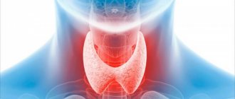
Alveococcosis is a zoonotic helminthic invasion that occurs as a volumetric process in the liver. In some cases, infiltrating growth and development of metastases in the lungs, brain, heart and bones are possible.
Etiology. The causative agent is the flatworm (cestode) Echinococcus multilocularis, type Platyhelminthes.
Epidemiology. The natural reservoir and definitive hosts are dogs (the main definitive host in Russia), arctic foxes, foxes, wolves, coyotes, and cats. Humans become infected by eating the liver of infected intermediate hosts. The eggs come out with feces, and active crawling of the segment is possible (they can crawl on the ground, leaving eggs behind), which leads to contamination of the fur, the environment (soil, water) and objects. Intermediate hosts (muskrats, voles) swallow segments or oncospheres (eggs in a special shell), which penetrate through the bloodstream into the liver, where the hatched larvae form a parasitic node. Most often, a person becomes infected when cutting carcasses or skins of infected animals, or by not following the rules of personal hygiene and keeping animals (especially dogs). Less common cases of infection are observed when eating wild berries and herbs contaminated with animal feces. In the Russian Federation, alveococcosis is widespread mainly in the Republic of Sakha (Yakutia), Krasnoyarsk, Altai and Khabarovsk territories, in Tomsk, Omsk and other regions. The disease is also reported sporadically in Tatarstan and Bashkortostan. Clinical picture. Compared to echinococcosis, it is more malignant. Characteristic is a gradual increase in symptoms. The main target organ is the liver. An enlarged and hardened liver (“iron” liver), pain and a feeling of heaviness in the right hypochondrium, decreased appetite, weight loss, jaundice and (rarely) ascites are observed.
Diagnostics • OAC: pronounced eosinophilia, increased ESR • Serological and immunological methods (RNGA, ELISA, RSK, latex agglutination reaction with Ag from the fluid of echinococcal blisters) give positive results in 60–90% of cases. A skin allergy test (Cazzoni reaction) is also used; it is most informative for liver echinococcosis • X-ray methods: cysts in the liver (against the background of pneumoperitoneum) or in the lungs look like rounded shadows with clear contours; rings of calcification are often found around cysts in the liver • Ultrasound, computed tomography, angiography.
Treatment. Surgical removal of parasitic nodes. Before and after surgery, mebendazole is prescribed in courses of 30 days in increasing daily doses from 200 to 600 mg or more.
Complications • Portal hypertension • Budd–Chiari syndrome • Perihepatitis • Spreading and metastasis to internal organs (gallbladder, anterior abdominal wall, diaphragm, pericardium, lungs, brain).
Synonyms • Alveolar echinococcosis • Multilocular echinococcosis.
ICD-10 • B67.5 Liver invasion caused by Echinococcus multilocularis • B67.6 Invasion of other sites and multiple echinococcosis caused by Echinococcus multilocularis • B67.7 Invasion caused by Echinococcus multilocularis, unspecified
Stages of the disease
During alveococcosis there are several stages:
| Asymptomatic | Can last up to 10 years. The disease is discovered as an incidental diagnostic finding during the examination of a patient for another reason. |
| Uncomplicated | The pathological process is localized in the liver, that is, the location of the primary tumor. Patients complain of digestive disorders. |
| Complicated | It is characterized by the presence of metastatic tumors and significant dysfunction of a number of internal organs. |
What awaits the patient?
The prognosis depends on the extent of the disease and the involvement of organs other than the liver. In the early stage, success rates for timely surgery are high. Patients who have undergone node removal are registered with a dispensary for life for regular examination. Such observation helps not to miss relapses and complications after treatment.
A person can live for a long time with a slowly growing parasitic “tumor”, not knowing about it until the terminal (last) stage appears. Without medical care, about 10 years after infection, death may occur from liver failure or damage to other organs by the parasite.
In general, the prognosis of alveococcosis is considered unfavorable due to the difficulty in removing the parasite from the body.
Treatment
Radical surgery for alveococcosis can be performed in approximately 15% of patients. More than 80% of patients are admitted to surgical departments when radical removal of parasitic nodes is impossible due to the prevalence of the process.
A large alveococcal node excised from the right lobe of the liver along with the gall bladder spread out on it (visible from above). The knot is cut
The node can be excised within healthy tissue - segmentectomy, liver lobectomy (see Hemihepatectomy), enucleated or partially resected and partially enucleated (resection - enucleation - A. N. Velikoretsky). If there are two or more nodes, and the patient’s general condition does not allow them to be removed simultaneously, the operation is performed in two or even three stages. Often the gallbladder is spread out on a parasitic tumor; in these cases, it is removed along with the alveococcal node (Fig.). If radical surgery is impossible (for example, when it grows into the inferior vena cava), and the parasitic tumor, having reached a large size, mechanically compresses neighboring organs, palliative resection is indicated, which is performed in order to delay the compression of the extrahepatic bile ducts by the growing tumor. The remaining unremoved areas of the alvesococcal node are subjected to local action of antiparasitic agents: injections at several points of 10-15 ml of trppaflavin solution (1:1000) or 3-4 ml of tepal (thymol ester of palmitic acid). In addition, treatment is carried out with steroid drugs and vitamins. The use of chemotherapy is being studied.
The technique of palliative resections developed by I. L. Bregadze does not present any particular difficulties, since the majority of parasitic nodes do not bleed. Bleeding from single large vessels is stopped by biological plugs from the omentum. After opening the abdominal cavity, a piece of the greater omentum is excised, from which biological tampons are immediately made. Pieces of omentum 0.5×0.5 cm are powdered with dry thrombin and fixed with two knots to the center of the ligature. At the moment of bleeding from a vessel gaping in the dense stroma, a sharply curved needle with a ligature with a biological swab threaded into its eye is inserted into its lumen. By tightening the ligature, it is possible to clog the lumen of the vessel and stop the bleeding. If there are decay cavities in the center of unremovable parasitic nodes, the cavities are drained, followed by systematic washing with antiparasitic drugs, and in case of infection - with antibiotics. In case of occlusive jaundice caused by germination of the porta hepatis, various bile drainage operations are performed.
Life cycle of a parasite
An adult has 4 suckers on its head and a corolla with hooks for best attachment. With the help of these devices, helminths are absorbed in large numbers in the small intestine of the main host: most often these are foxes.
- Each individual has up to 400 eggs in the mature segments of the body, inside which larvae sit. Together with feces, they end up in the soil, water bodies and on the surface of plants. Interestingly, adult worms can crawl out of the anus on their own, after which they burst, scattering hundreds of their eggs, including onto the animal’s fur.
- Rodents, consuming contaminated water and grass, swallow oncospheres, which almost immediately come out of their shells. With the help of their hooks, they drill into the intestines and head towards the liver. Settling in the tissues, the larvae transform into vesicles about 2 mm in size, resulting in the formation of a node at this location.
- Then the alveolar cyst with the larva goes out into the external environment and again enters the intestine of the main host, which becomes infected through eaten rodents.
Causes of infection
Humans are considered an accidental intermediate host, which is generally not typical for the natural cycle of alveococcus. However, cases of infection occur in some areas, and even widespread outbreaks are observed. The main causes of infection, as a result of which alveococcosis of the liver develops, are identified
- drinking contaminated water from natural reservoirs;
- eating wild berries, fruits, herbs, mushrooms without prior washing or cooking;
- infection during hunting during cutting of hunted carcasses of animals carrying the parasite;
- work related to the removal or dressing of skins from wild animals;
- contact with animals of zoo workers and fur farms.
How can you get infected?
People become infected by ingesting parasite eggs or failing to comply with hygiene rules. There is a high risk of infection among hunters and anyone who has anything to do with animals. In rare cases, you can become infected from domestic animals, but when cutting up a fox carcass you need to be especially careful.
The disease does not spread from person to person because the parasite inside cannot reach maturity and cannot produce eggs. People do not infect each other with alveococcosis; this is impossible.
It is possible to become infected by eating unwashed berries and herbs that have been contaminated with the feces of various wild animals. In rare cases, eggs enter the human body through inhalation of dust.
Treatment of alveococcosis at the EUGENE clinic.
Our clinic offers you an innovative drug treatment method for alveococcosis. Our chief physician and part-time director of the clinic, Evgeniy Li, developed this technique in order to find a way to effectively get rid of parasites without the use of surgery.
And for ten years now we have been successfully treating patients with these problems. During this time, we have cured a lot of people who now live full lives and are periodically observed in our clinic for the purpose of prevention.
Get rid of alveococcosis with the EUGENE clinic and breathe deeply again!
Pathogen
The larva of the alveococcus tapeworm Echinococcus multilocularis is the cause of a parasitic disease; this parasite is from the family Echinococcus. Experts classify it as a species of Alveococcus, which is why it is called alveococcus. This parasite is popularly called the fox tapeworm, due to the fact that foxes are often its final hosts.
- The parasite in the larval stage looks like a small bubble with a diameter of 1-20 mm; this nodule causes the pathogen alveococcosis in both humans and animals. These larvae are dangerous because they reproduce vegetatively; they can bud and, with a prolonged illness, a conglomerate of very small bubbles (vesicles) is formed in the patient’s body. As a result of reproduction, they adhere tightly to each other, and sometimes grow together. The bubbles are filled with a viscous liquid of a yellowish or dark color; they contain the heads of parasites (scolex).
- In the normal life cycle of the parasite, such small bubbles are preferred by the organisms of small infected animals, these are rodents, which most often feed on foxes and other canines. Next, the larvae turn into adult worms, living in the small intestine, they reproduce. For the development of alveococcus, humans are a dead end because the larva will not be swallowed by the definitive host.
As an adult, the parasite reaches a length of up to 4 mm. This helminth is a species of flatworm, class cestodes (tapeworms), order Tapeworm (Cyclophyllidea). But it differs from its well-known counterparts (bovine or pork tapeworm) in that it is content with only a maximum of five segments, and does not have thousands, like others.
Alveococcosis in the liver
Diagnosis of alveococcosis
Diagnosis of echinococcosis and alveococcosis is difficult; at the first signs, doctors often diagnose liver damage (for example, cirrhosis or hepatitis). A stool test does not show the presence of parasites; a blood test reveals a high ESR and then a diagnosis of cancer is made, while the true cause of the disease - helminthic infestation - remains unidentified.
NPK Optisalt has developed an innovative device, Optisalt IridoScreen, which makes it possible to establish the parasitic and toxic load on organs and identify the deficiency of vital microelements in the iris of the eyes. It has been proven that each organ has its own projection on the iris, where it transmits information about its condition through nerve impulses.
How does a doctor detect an infection?
It is not so easy to suspect alveococcosis. The first “bell” on the way to a correct diagnosis may be living or traveling to a region unfavorable for the spread of this parasite, even if it was a long time ago (up to 10–15 years ago).
Thus, diagnostic measures for suspected alveolar echinococcosis begin with collecting an anamnesis, which, among other things, takes into account:
- contacts with parasite carriers;
- features of the profession;
- cases of consumption of forest products, as well as water from natural sources.
Next, the doctor performs an examination: he pays special attention to the abdominal cavity. After this, palpation is used, that is, examination by feeling with the hands: sometimes, if the parasite cysts are close to the surface of the liver, during palpation the doctor may feel an unusual density and tuberosity of the liver, in addition, its enlargement can be noted.
Diagnostic procedures are required to help clarify the presence of the disease. Laboratory methods:
- A detailed blood test, since in those infected, ESR and eosinophils often increase, and hemoglobin decreases.
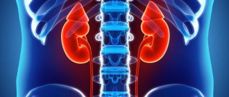
- A general urine test can reveal signs of kidney damage - proteinuria (increased protein) and hematuria (presence of blood).
- A biochemical blood test, which shows that the concentration of AST, ALT and bilirubin, and sometimes gamma-GT and alkaline phosphatase, increases.
- Serological tests (ELISA) to determine the presence of antibodies to the parasite.
Be sure to use methods of visualization of the affected organ:
- Ultrasound allows you to detect specific cysts and estimate their size.
- Computed tomography or MRI of the abdominal cavity (usually with contrast), chest, MRI of the brain - to clarify damage to internal organs.
- Laparoscopy for unclear diagnosis. Suitable for assessing nodes available for viewing, as well as when they grow outside the organ.
Such a complete examination helps to differentiate the disease from other liver lesions (that is, to distinguish from them): echinococcosis, liver cancer, polycystic disease, cirrhosis and hemangioma.
Consequences
The most common complication of alveococcosis is obstructive jaundice, which occurs due to compression of the bile ducts. Others include:
- aspiration pneumonia;
- chronic grosserulonephritis;
- renal failure;
- dilation of the blood vessels of the stomach and intestines with bleeding;
- fevers accompanied by severe chills and heavy sweats;
- breakthroughs of larvocysts into the cavity with the occurrence of peritonitis;
- abscesses due to bacterial infections;
- fistulas in the liver, lungs and pleura;
- germination of the parasitic node through the omentum and diaphragm into the mediastinum, lungs and heart.
Malignant alveococcosis with metastasis to the brain is particularly severe.
Symptoms of alveococcosis
For a long time (sometimes for many years), the symptoms of alveolar echinococcosis may not appear; the long latent period is explained by the slow growth of the cyst (larvocyst). The first signs of the disease are usually associated with dysfunction of the liver: it may be enlarged in size and lumpy to the touch. Pain in the right hypochondrium may appear, fatigue may increase, general weakness may develop, and appetite may decrease.
The growth of larvocyst causes disruption of blood flow in organs; toxins released by the parasite lead to tissue necrosis and acute allergic reactions. With the development of alveococcosis of the liver, symptoms such as:
- Increased pain in the liver (become permanent), liver dystrophy develops, and cirrhosis appears;
- The functioning of the gastrointestinal tract is disrupted (a feeling of heaviness after eating, diarrhea or, conversely, constipation);
- The appearance of skin itching (caused by both liver dysfunction and acute allergic reactions to worm toxins);
- Edema, varicose veins of the esophagus, accumulation of fluid in the abdominal cavity (if larvocysts have damaged large vessels);
- Disruption of the functioning of organs and tissues affected by metastases: if these are the kidneys, then problems with urination begin, if the tumor grows in the brain, symptoms of schizophrenia may appear, and so on.
In the last stages of alveococcosis of the liver (with the formation of a large number of metastases), the patient sharply loses weight, experiences severe pain, the functioning of almost all organ systems is disrupted - clinically the disease manifests itself as cancer. Alveococcosis causes serious immunity disorders and often causes complications such as abscesses of internal organs, purulent cholangitis, and amyloidosis.
Feeding at the expense of the host organism, alveococcus absorbs vital microelements that are not reproduced in the human body, but come from the external environment. Their deficiency provokes the development of severe endocrine disorders and has a detrimental effect on the functioning of the nervous system and the entire body as a whole.