Ultrasound examination of organs and tissues is carried out using ultrasonic vibrations.
In the process of passing between the boundaries of tissues of different thicknesses, sound reflection does not occur in the same way. The sensor, which performs the function of receiving the reflected echo signal, converts them into graphic symbols, which are subsequently recorded on the device’s monitor and, if desired, printed on special photographic paper.
The huge advantage of this method is that it has absolutely no contraindications; it can be performed as many times as necessary to monitor the course of the disease or diagnose. Even if necessary, it is allowed to do an ultrasound a couple of times a day.
Difficulties in conducting the study or it may not be informative due to the presence of dense scars in the patient after surgery, excess body weight, and flatulence.
History of ultrasound
Ultrasonic vibrations were discovered by Italian scientist Lazzaro Spallanzani back in 1794. While watching the bat, he wondered how it was possible to navigate in space at night while flying. It turned out that this is carried out using high-frequency sound vibrations, inaudible to humans. But animals, particularly bats, perceive them very well. The principles of echolocation are also used by marine animals - killer whales, dolphins, whales, etc. Over time, they became known as ultrasonic waves.
In 1942, for the first time, the German doctor Theodor Dussick and his brother, scientist and physicist Friedrich Dussick, tried to use ultrasound to diagnose a tumor in a patient’s brain. The first medical ultrasound device was created in 1949 by the American scientist Douglas Haury.
Doppler Christian Anders made a great contribution to the development of ultrasound diagnostics. While working on his scientific work “On the collometric characteristics of the study of double stars and some other stars in the sky,” he noticed that the frequency of the signal reflected from an object depends on its speed and direction of movement. This property is called the Doppler effect, which is widely used in Dopplerography to examine blood flow parameters.
Diagnostics
In the first weeks of life, it is very difficult to diagnose congenital hypothyroidism and this is possible only in 10-15% of cases, so doctors need to focus mainly on the existing symptoms, which were voiced in the article above. If symptoms exist in the first months and years of life, a number of studies are carried out that will be aimed at identifying the disease:
- routine examinations by a pediatrician, and before the child reaches the age of one month – by a neonatologist;
- taking a general and biochemical blood test to determine the concentration of thyroid hormones, cholesterol and other important indicators;
- monitoring the child’s pulse and blood pressure, since with hypothyroidism in children these indicators will be reduced;
- conducting an ECG, which may show bradycardia and other abnormalities;
- ultrasound examination of the thyroid gland, which can reveal both some deviations from the norm and the complete absence of the thyroid gland;
- in some cases, a CT or MRI of the baby’s head may be prescribed.
What is the thyroid gland?
The thyroid gland, or “thyroid” for short, is a gland that is part of the endocrine system (a system for regulating the functioning of internal organs with the help of hormones that secrete endocrine cells into the blood) that stores iodine and secretes hormones containing iodine.
Regulates metabolism, cell growth and the growth of the entire organism in general. The thyroid gland is located in the neck in front of the trachea under the larynx. It looks like a butterfly located on the surface of the cartilage. If there are changes in the functioning of the thyroid gland, the patient will feel unwell (this may indicate an incipient disease), and later various complications may arise. The method of ultrasound examination of the thyroid gland will help to effectively identify incipient diseases.
If the thyroid gland does not function properly, the following pathologies may appear:
- hyperthyroidism – excessive activity of the gland;
- hypothyroidism – low activity of the gland, characterized by a reduced level of hormones produced;
- cancer;
- cyst;
- benign formations;
- thyroiditis is an inflammatory disease of the gland;
Why is it important to get checked periodically?
Hormones secreted by the thyroid gland regulate many processes in the body:
- Metabolic rate;
- Appetite;
- Activity of the brain and nervous system;
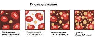
- The breakdown of glucose and its absorption by cells;
- Absorption of nutrients;
- Breakdown of fats;
- Cholesterol levels;
- Strength and frequency of heartbeat and breathing;
- Oxygen consumption by cells;
- Body temperature;
- The work of the mammary and reproductive glands;
- much more.
A malfunction of the thyroid gland can have serious consequences for the entire body.
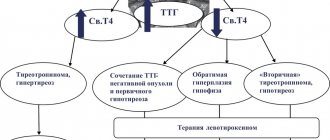
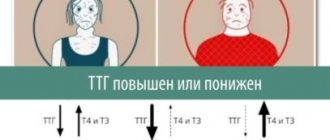
Disturbances in the functioning of the thyroid gland have two extremes: an increase in the organ - hyperthyroidism, and a decrease - hypothyroidism.
With hypothyroidism, metabolism slows down (pulse, appetite, blood pressure decrease, and weakness appears). In an adult, hypothyroidism can cause diabetes, hypertension, kidney disease and cardiovascular disease.
Hyperthyroidism, on the contrary, causes increased metabolism (rapid heart rate, sleep disturbances, heart palpitations, irritability, etc.).
Ultrasound of the thyroid gland
, an ultrasound diagnosis of the thyroid gland should be performed . This procedure is invasive, fairly cheap, fast and provides accurate results.
Ultrasound is a study of the body using ultrasonic waves. The echo signals reflected from the internal organs, after certain transformations, create on the monitor screen an image of a section of the gland in different shades of gray.
A specialist can determine the condition of the organ: geometric dimensions, state of borders, lymph nodes, blood vessels. Ultrasound determines the acoustic density of an organ, called echogenicity.
May be:
- normal;
- reduced;
- increased;
- echonegativity.
The condition of the organ being studied is determined by the types of echogenicity.
Reduced (hypoechogenicity) is characteristic of liquid formations (dark gray areas on the screen). Perhaps it is a cyst, liquid formations, vascular formations and cancer in 5% of cases. A doctor cannot always reliably determine the nature of these formations, so other diagnostics, such as a biopsy, will be required.
Increased (hyperechogenicity) indicates a lack of iodine (endemic goiter), damage to the organ by poison (toxic goiter), autoimmune thyroiditis, subacute thyroiditis, oncology, areas of sclerosis. Visualized on the monitor as white spots. Hyperechogenicity may occur in a healthy gland, or it may indicate serious pathologies, therefore, to obtain a reliable diagnosis, it is advisable to conduct additional research.
Echonegativity (anechoicity) is displayed as black areas. Perhaps it is a cyst, pseudocyst, colloid cyst, adenoma. It is also recommended to undergo other examinations to make a more accurate diagnosis.
With normal echogenicity, the silhouette of the gland is smooth and clear. A benign tumor has smooth edges, while a malignant tumor has curved edges.
Since the frequency of ultrasound is not high, there is no harmful effect on the body.
Symptoms of hypothyroidism in children
Typically, children have mild symptoms of the disease and without proper diagnosis it is not possible to make a diagnosis. However, some symptoms may indicate that such a diagnosis is necessary:
- such children have a birth weight greater than the average;
- severe swelling of the baby’s limbs and face;
- the child has a low, bass voice, wheezing when crying;
- wheezing may be heard during normal breathing;
- such children are lethargic and sleep a lot;
- yellowness of the skin may occur;
- Almost half of children with congenital hypothyroidism are born late; postmaturity is also a reason for additional diagnostics.
In addition to the symptoms presented, a cause for concern may be: the child later than usual began to roll over on his own, sit up, hold his head up, and other aspects of normal physical development. In the photo you can see the characteristic appearance of a baby with congenital hypothyroidism.
How to prepare for an ultrasound
The method is accessible and simple, does not require special preparatory operations. You can eat absolutely any food product. But there are a number of restrictions:
1. Regardless of the feeling of nausea, older people are not recommended to eat before the procedure. Age-related changes may have an effect.
2. Women are recommended to undergo examination on days 7-9 of the menstrual cycle to avoid inaccurate results.
3. Immediately before the procedure, you need to free your neck from elements that interfere: jewelry, collars, etc.
4. Preparation for an ultrasound examination of the thyroid gland in children is no different from the procedure for adults. The child must be psychologically prepared for the procedure so that he is not afraid of either the doctor or what he will do.
Doctors recommend that women undergo testing both during pregnancy and at the planning stage. If it is impossible to get pregnant, gland pathology may be one of the possible causes.
After the child has suffered stress, it is also worth going to the clinic so that an endocrinologist can perform an ultrasound of the thyroid gland.
Stages of the disease
Thyroid hypoplasia has 2 degrees of development - mild and complicated. With a mild degree, the disease practically does not manifest itself in any way, symptoms do not appear, and the glands do not change their size.
The second degree of the disease is characterized by noticeable manifestations of the main signs of the disease, while the thyroid gland decreases in size. In the absence of proper treatment, the pathology leads to severe and irreversible consequences, including cretinism and the occurrence of myxedema.
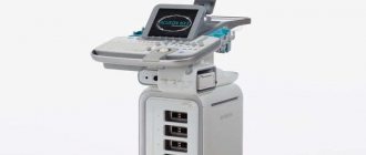
How is the procedure carried out?
The procedure is quite simple.
The patient lies down on a special couch with his head thrown back, a soft pillow or cushion is placed at the level of the shoulder girdle. A gel that conducts ultrasound well is applied to the neck in the area of the thyroid gland. An ultrasound transducer is applied to the neck and ultrasonic waves are emitted. The sensor is a transceiver that emits and receives a reflected echo signal. This information is processed by a computer and the result is displayed on the monitor. During the procedure, the patient does not experience any discomfort; sometimes there may be slight discomfort due to incorrect body position.
Consequences of the disease
The consequences of insufficient thyroid hormone production can be catastrophic for a child without the necessary treatment.
Consequences of congenital hypothyroidism:
- delay in physical, emotional and intellectual development;
- since thyroid hormones affect the functioning of many organs, we can talk about emerging pathologies also from the heart, nervous system and psyche;
- in advanced cases, myxedema occurs (insufficiency of thyroid hormones in tissues and other organs), which leads to the patient’s coma.
Dear patients, in our medical center you can do an ultrasound of the thyroid gland.
Interpretation of ultrasound results
The diagnostician describes the results obtained in the study protocol. Typically, the time to prepare a written report does not exceed 15 minutes.
Determination of exact geometric dimensions
The location of the gland will normally be typical or low, the shape will be classic, the contour will have a clear outline. It consists of two lobes with an isthmus that connects them. Sometimes a pyramidal lobe may be present.
Minor (less than one centimeter) tissue growths may be visible. If during the period of intrauterine growth the gland develops with pathology, the tissue may not be divided into two sides, but may move to one side. This is called aplasia of one lobe.
In the case of a completely undeveloped gland, they speak of complete aplasia. The length of the lobe should be from 4 to 6 cm, width from 1.3 to 1.8 cm, thickness from 1.5 to 1.8 cm; the shares are normally the same; the lintel has a thickness of 4 to 8 cm.
The volume of a healthy gland depends on the patient’s weight and is:
- weight 50 kg, gland volume 15.5 cm³
- weight 50-60 kg, volume 18.7 cm³
- weight 60-70 kg, volume 22 cm³
- weight 70-80 kg, volume 25 cm³
- weight 80-90 kg, volume 28.4 cm³
- 100 kg or more, volume 32 cm³
Non-compliance with standard sizes will indicate possible pathologies.
Definition of structure
The echogenicity of a healthy thyroid gland, without any peculiarities, the structure of the jelly tissue is fine-mesh, homogeneous, echogenic granularity is 1 mm or less. Fibrous and connective tissues are not detected. In inflammatory processes, the structure is heterogeneous.
Focal education. There should be no new growths. If the formation is present, it must be classified. If the size is up to 10 mm, then it is a focal formation, more than 10 mm, then it is a node.
Blood flow analysis. The parameters of blood flow, its nature and density, and parameters of the lymph nodes are determined. Normal nodes have clear boundaries, the width is approximately 2 times less than the length.
Contours. Clear contours are the norm. Fuzzy outlines indicate an inflammatory process or tumor.
Focal formations. Assessed for the presence of nodes, cysts, and calcinitis.
Echogenicity is the grayscale image of the tissue being examined and its tone on a monitor.
Parameters of visible lymph nodes (if present), their structure, size, structure. By their presence, the onset of tumor formation can be diagnosed.
The structure of the salivary gland and the level of response to ultrasound.
The size and structure of the soft tissues of the neck and larynx. Those areas that are located near the thyroid gland are examined.
Women over 35 years of age are recommended to have their thyroid gland examined at least once a year. This category of people is at risk for thyroid diseases.
The human neck is quite complex; it contains many nerve trunks and large vessels, the esophagus, trachea, many lymph nodes, and other glands. Therefore, the specialist performing the ultrasound procedure must be highly qualified.
The doctor conducting the research records the data obtained in a special protocol, which describes the geometric parameters of the lobes and isthmus, calculates the volume of the gland, and writes a conclusion.
The document evaluates the position and contours, tissue structures, analyzes the parathyroid glands and lymph nodes, and the images taken are also attached to the protocol. In case of normal indicators, a corresponding entry is made in the document. This takes no more than 10 minutes.
Symptoms of hypoplasia
The main signs of thyroid hypoplasia develop gradually over many years. Such signs of the disease include:
- deterioration in the general condition of the skin, nails and hair - the skin becomes dry and flaky, hair becomes brittle and increased loss, nails become thin and weak;
- appetite, in most cases, is completely absent - at the same time, body weight rapidly increases;
- general weakness, increased fatigue, constant drowsiness;
- memory impairment, decreased body temperature;
- constant tremor in the muscles;
- sexual desire is practically absent;
- menstrual flow becomes extremely long and heavy.
The main echo sign in the process of diagnosing the disease is determining the exact size of the thyroid gland and its compliance with age standards. The dimensions of the organ are determined during the ultrasound examination.
In children, the symptoms of thyroid hypoplasia look somewhat different and can be expressed as follows: a teenager has constant drowsiness, decreased appetite, increased weakness and fatigue. The child may simply refuse to eat and have constant digestive problems - constipation or diarrhea.
Forecast
If the pathology is detected early, the consequences of congenital hypothyroidism will not be detrimental to the patient’s health, but are assessed by doctors as favorable.
With proper therapy, bringing the main thyroid hormones to normal levels, we can say that nothing will hinder the child’s development (taking into account the fact that hormone replacement therapy will be lifelong).
If congenital hypothyroidism is detected not in the first months of life, but at 3-6, then the chances of complete normalization of the condition are significantly reduced, even with proper treatment. At the same time, it is possible to achieve the required level of psychophysical development of the child, but there is a high probability of lagging behind in intellectual development.