- Ultrasound machine
Ultrasound machine
SAMSUNG SONOACE R7
Class:
Expert (installation year 2019)
What is included in the cost of ultrasound:
Ultrasound diagnostics, interpretation of images, written diagnostic report
What is important to know about making an ultrasound appointment?
Do I need to make an appointment: ultrasound diagnostics are carried out by appointment
Appointment schedule: you can make an appointment 24 hours a day
Form of payment for diagnostics: cash, credit cards
Doctor's referral: not required
Ultrasound screening during pregnancy: we do not provide
Minimum patient age: 18 years.
Content:
- Why do you need to do an ultrasound of the thyroid gland?
- How to do an ultrasound of the thyroid gland correctly?
- Ultrasound of the thyroid gland: interpretation
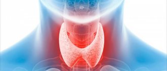
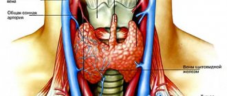
What is thyroid ultrasound?
The tissues of the human body have different structures, different densities and, accordingly, will reflect ultrasonic waves differently. Ultrasound diagnostics are based on this physical phenomenon. The diagnostic apparatus is equipped with sensors of various frequencies that supply and receive the return signal. During the examination, the scanner sends a signal of ultrasonic waves into the soft tissues of the neck. Some of the waves, reflected, return to the sensor, and some continue on their way. All data on the behavior of waves is sent to the device’s processor, and based on them, the computer constructs an image that is sent to the monitor. The computer of a modern ultrasound machine is capable of making an accurate calculation of tissue thickness, distance from other nearby organs or vessels, and tissue density. During the examination, the diagnostician has the ability to change the frequency and intensity of sound waves using a control panel, which is located just below the monitor, thereby obtaining the necessary information.
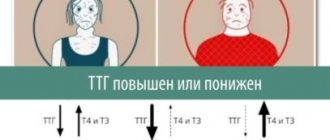
Consequences of deviation from the norm
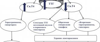
The normal functioning of almost all body systems depends on the condition of the thyroid gland. The production of TSH by the pituitary gland is directly related to the production of T4 and T3. If the concentration of T4 and T3 is low, TSH in men increases. With low levels of T4 and T3, the concentration of TSH in the blood will decrease due to deterioration in synthesis.
If the analysis shows increased production of TSH with reduced or normal concentrations of T3, T4, this condition is called hypothyroidism. If thyrotropin is low, this is a signal of hyperthyroidism.
Any deviations in the production of thyroid-stimulating hormones from the norm indicate changes occurring in the pituitary gland, thyroid gland or other organs. Men are usually prescribed a comprehensive analysis to study the amount of TSH, T3, T4 in the blood, which helps to establish the cause of simply feeling unwell or the appearance of various diseases.
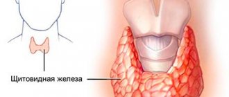
Why do you need to do an ultrasound of the thyroid gland?
Ultrasound of the thyroid gland allows you to timely detect the disease in the early stages of its development or prevent it. Using this type of diagnostics you can determine:
- size and structure of the organ, size of the lobes;
- benign and malignant tumors, sites of metastases;
- cysts and fibrous tissue, inflammation;
- local lymphatic drainage;
- anomalies and malformations.
An ultrasound of the thyroid gland with interpretation of the results will allow us to determine the true condition of the patient and the nature of his complaints. Otherwise, with a lack of hormones in the body, you can get, for example, a violation of the natural bone density and osteoporosis. An excess of hormones leads to increased blood pressure, increased cholesterol, poor daily well-being and new problems with the functioning of other organs.
Free triiodothyronine (free T3)
Thyroid hormone stimulates the exchange and absorption of oxygen by tissues (more active than T4).
Produced by follicular cells of the thyroid gland under the control of TSH (thyroid-stimulating hormone). In peripheral tissues it is formed during deiodination of T4. Free T3 is the active part of total T3, accounting for 0.2 - 0.5%.
T3 is more active than T4, but is found in the blood in lower concentrations. Increases heat production and oxygen consumption by all body tissues, with the exception of brain tissue, spleen and testicles. Stimulates the synthesis of vitamin A in the liver. Reduces the concentration of cholesterol and triglycerides in the blood, accelerates protein metabolism. Increases calcium excretion in urine, activates bone turnover, but to a greater extent, bone resorption. It has a positive chrono- and inotropic effect on the heart. Stimulates reticular formation and cortical processes in the central nervous system.
By 11–15 years, the concentration of free T3 reaches adult levels. In men and women over 65 years of age, there is a decrease in free T3 in serum and plasma. During pregnancy, T3 decreases from the first to the third trimester. One week after delivery, serum free T3 levels return to normal. Women have lower concentrations of free T3 than men by an average of 5 - 10%. Free T3 is characterized by seasonal fluctuations: the maximum level of free T3 occurs from September to February, the minimum in the summer.
Limits of determination:
1.5 pmol/l - 46.1 pmol/l.
Nodular goiter on ultrasound of the thyroid gland
Using an ultrasound of the thyroid gland, the doctor will be able to quickly see all the signs of nodular or diffuse goiter. Nodular goiter is a limited compaction in the tissues of the thyroid gland, which can be detected even by palpation. This is a dense node, which on ultrasound of the gland is significantly different from healthy tissue of the organ. Diffuse goiter is a disease associated with an increase in the volume of the thyroid gland and the hormones it produces. The increase in volume in women can reach more than 19 ml, and in men - more than 25 ml. The cause of the formation and development of the disease is autoantibodies that cause active stimulation of the thyroid gland. Signs of diffuse goiter include:
- irritability;
- increased excitability;
- sudden weight loss with good appetite;
- rapid heartbeat (attacks);
- slight increase in body temperature.
Ultrasound diagnostics for diffuse goiter reveals an increase in the size of the scale insect with a constant homogeneous structure.
Hyperthyroidism on thyroid ultrasound
Hyperthyroidism is a disease of the thyroid gland associated with excessive production of the hormones triiodothyronine and thyroxine. There are primary, secondary and tertiary hyperthyroidism (pathology of the thyroid gland, pituitary gland and hypothalamus, respectively). It is difficult to make this diagnosis based only on ultrasound results. In addition to the scan results, the patient needs to donate blood for hormones. Severe hyperthyroidism can actively affect the functioning of the brain and heart, thereby threatening the patient’s life.
Hypothyroidism on thyroid ultrasound
Hypothyroidism is a disease associated with decreased activity of the thyroid gland, low production of hormones, and a decrease in the volume and size of the thyroid gland. The main causes of hypothyroidism are: surgery on the thyroid gland, Hashimoto's goiter (replacement of healthy gland tissue with connective or fibrous tissue). With this disease, an ultrasound scan of the thyroid gland may reveal some diffuse changes.
Thyroiditis on ultrasound of the thyroid gland
Thyroiditis is inflammation in the tissues of the thyroid gland. Symptoms of the disease may include pain in the neck and head, a slight increase in body temperature and an increase in the size of the gland, although not in all cases. When carrying out ultrasound diagnostics in case of thyroiditis, small cavities with liquid (pus), swelling and an increase in the volume of the organ are detected. In the case of autoimmune thyroiditis, changes in the structure and size of the organ (decrease or increase) are detected.
Previous NextCyst on ultrasound of the thyroid gland
A thyroid cyst is a cavity filled with fluid. The cyst can be either congenital or acquired. It can be detected by ultrasound examination. In the case of an inflammatory process or suppuration of cysts, the patient may notice a sore throat and a slight increase in body temperature. Ultrasound of the thyroid gland notes the formation of cysts in the form of round, clear cavities with fluid inside. The rest of the organ tissue remains healthy. In some cases, in order to exclude malignant formations, fluid is taken from these cavities (puncture) directly during ultrasound diagnostics using a needle, or the patient is sent for further examination using MRI of the thyroid gland with contrast.
Tumor on ultrasound of the thyroid gland
In addition to cysts, benign or malignant tumors can form in the thyroid tissue. Ultrasound of the thyroid gland diagnoses tumors in the form of limited (for benign tumors) or invasive compactions. In most cases, when a tumor is suspected, special attention is paid to the lymph nodes. In some situations, evidence of cancer is enlarged lymph nodes and associated diseases - lymphadenitis. However, there is no direct relationship between cancer and lymph nodes, that is, lymphadenitis does not always become a determining factor in cancer. If an ultrasound of the thyroid gland shows signs of a tumor, the patient is necessarily referred for either a biopsy or an MRI of the soft tissues of the neck and lymph nodes with contrast.
Interpretation of ultrasound of the thyroid gland
To obtain comprehensive information that reflects the condition of the thyroid gland, the diagnostician during the ultrasound procedure evaluates and enters into the protocol the following parameters:
- Location . The typical location of the gland is considered standard when it is within the anatomical norm. If there is a pathology, a record of its aberrant location is entered into the protocol, when the thyroid gland may be located behind the sternum, in the area of the root of the tongue, etc.
- Size . Determining this parameter allows one to judge the presence or absence of hypo- or hyperplasia of the thyroid gland.
- Structure . Normally, the thyroid gland consists of two symmetrical lobes, which are connected by a small isthmus. In the case of abnormal intrauterine development of the gland or its pathological acquired modification, tissue outgrowths, an additional lobe, and bifurcation may be absent.
- Form . Normally, the thyroid gland is flat, but may be slightly elongated. If it is greatly increased in volume, swollen, with thyroid tissue, diffuse goiter, viral or bacterial pathology is suspected.
- Outlines . The clear contours of the thyroid gland are taken as normal. Otherwise, if its contours are not clearly visualized, one can judge the presence of a tumor or inflammatory focus in the gland.
- Structure . If there is no pathological process in the gland, its structure is homogeneous and has a specific granularity.
- Echogenicity . Visually, this parameter can be defined as a gradation of gray shades over the entire area of the study area. Normally, the echogenicity of thyroid tissue should be visible in light gray shades.
- Focal formations . During ultrasound diagnostics, the doctor must examine the gland for the presence of pathological formations - nodes, calcifications, cysts or tumors.
- Blood supply to tissues . Impaired blood supply to the thyroid gland can lead to the death of its tissue. Normally, the multi-colored signals visualized on the ultrasound monitor screen should be uniform over the entire area of the thyroid gland. An increase in color and “pulsation” of shades indicates an increase in blood flow, which may be associated with an inflammatory process inside the gland.
- The structure of the cervical lymph nodes . To determine their condition, the following indicators are determined: structure, size, presence of pathological inclusions, cystic transformation, presence of the “gate” of the lymph node, assessment of blood flow.