Ultrasound of the thyroid gland - (Thyroid Ultraschall) is the simplest and most highly informative diagnostic method, which provides a detailed assessment of the condition of the gland itself, as well as the anatomical formations adjacent to it - blood vessels and muscles. This is possible using modern ultrasonic sensors with additional capabilities. In particular, Doppler ultrasound allows you to examine the blood supply to the thyroid gland and nearby lymph nodes.
Make an appointment, ultrasound or tests
PRICE OF THYROID ULTRASOUND 1000 rub.
Indications for thyroid ultrasound
The content of the article
Before scheduling a procedure, please read the important information regarding indications and contraindications. A classic ultrasound examination of the thyroid gland is recommended to be done regularly as part of an annual medical examination, regardless of the patient’s age and the absence of characteristic symptoms. If the following symptoms appear, it is necessary to make an appointment with an endocrinologist as soon as possible and undergo an ultrasound examination of the thyroid gland:
- visible increase in the volume of the neck, the appearance of a compacted formation;
- redness of the neck area near the location of the gland;
- hoarseness not associated with colds;
- difficulty breathing, discomfort during swallowing;
- increase in body temperature within 37-37.5 degrees;
- the appearance of swelling;
- weakness, apathy, drowsiness, severe irritability;
- sweating, feeling hot or, conversely, cold;
- trembling of limbs;
- sudden change in body weight;
- cardiopalmus;
- baldness.
These symptoms may be an indicator of a malfunction of the thyroid gland, and therefore require examination by ultrasound. An endocrinologist may refer you for an ultrasound of the thyroid gland if palpation reveals lumps or the patient complains of pain when palpating the gland. A direct indication for ultrasound diagnostics is unsatisfactory results of hormone tests.
In addition, the following groups of people should sign up for a thyroid ultrasound :
- Pregnant women or those who are planning to conceive a child in the near future.
- People living in iodine-deficient areas.
- Patients diagnosed with obesity.
- For those taking hormone-containing medications.
Ultrasound of the thyroid gland is also prescribed to patients to monitor the treatment. In some cases, the frequency of such a procedure reaches several times a week, which is necessary for dynamic monitoring of the gland’s reaction to drug correction.
Ultrasound examination is completely safe for humans, has no contraindications, and if a pregnant woman undergoes it, it does not have any effect on the fetus. Therefore, ultrasound can be done as many times as needed.
How to prepare for a thyroid ultrasound
This gland is absolutely accessible for research due to its convenient location: ultrasonic waves easily reach the thyroid gland and are fully reflected from its tissues. Therefore, this procedure does not require special preparation. But there are a couple of recommendations that should be considered when planning an ultrasound of this gland:
- Patients who take medications that affect cardiac output and blood pressure should stop taking them the day before ultrasound diagnostics.
- Do not drink alcohol during the day.
- Elderly people are advised to come for the procedure on an empty stomach. This recommendation is associated with the appearance of a gag reflex at this age when pressing on the neck area.
When coming for an ultrasound, you can take a personal towel with you to wipe off any remaining gel from your neck after the procedure.
How to do an ultrasound of the thyroid gland
The procedure uses a linear sensor with a high scanning frequency. With its help, you can obtain a high-quality image that shows the structure of the organ and focal changes. During the procedure, the size of the gland, its structure, configuration, and blood flow condition are determined. In parallel, the parathyroid glands and regional lymph nodes are examined.
No preparation is required for diagnosis, since nothing interferes with the examination of the organ. Ultrasound diagnosis of thyroid pathologies is an accessible, non-traumatic and painless diagnostic method. Complications after ultrasound are not observed. During the study, the patient is not administered drugs, and the organs are not irradiated, but the diagnostic efficiency is high. There are no contraindications to the ultrasound procedure. It can be done many times even during pregnancy.
The examination is carried out in a lying position. A special gel is applied to the patient’s neck to increase the conductivity of ultrasound. The results are displayed on the screen. Modern ultrasound machines determine not only the presence, but also the size of thyroid nodules, which is important for choosing treatment tactics.
The patient is given an image and a description, based on which the endocrinologist will prescribe treatment for the identified pathologies. To make a final diagnosis, the patient needs to undergo blood and urine tests and consult with a therapist and oncologist.
When to get tested
The frequency of visits to the ultrasound room is determined individually for each patient, depending on his age, state of health, presence of ailments, conditions and lifestyle, and so on.
- Patients under 50 years of age are recommended to undergo testing once every 5 years as a preventative measure.
- For people over 50 - one study every 2-3 years.
- For women - additionally when planning pregnancy.
An examination may be indicated urgently in the following cases:
- The doctor discovered that the patient had problems with the endocrine system.
- It is necessary to monitor the treatment process or evaluate the results of therapy.
- There is a risk of recurrent thyroid dysfunction.
- Nodules or lumps have appeared in the neck area.
- The thyroid gland has noticeably increased in size.
- The patient suddenly lost or gained weight.
- The patient has an unstable emotional state - nervousness, apathy, and so on.
Interpretation of ultrasound of the thyroid gland
To obtain comprehensive information that reflects the condition of the thyroid gland, the diagnostician during the ultrasound procedure evaluates and enters into the protocol the following parameters:
- Location . The typical location of the gland is considered standard when it is within the anatomical norm. If there is a pathology, a record of its aberrant location is entered into the protocol, when the thyroid gland may be located behind the sternum, in the area of the root of the tongue, etc.
- Size . Determining this parameter allows one to judge the presence or absence of hypo- or hyperplasia of the thyroid gland.
- Structure . Normally, the thyroid gland consists of two symmetrical lobes, which are connected by a small isthmus. In the case of abnormal intrauterine development of the gland or its pathological acquired modification, tissue outgrowths, an additional lobe, and bifurcation may be absent.
- Form . Normally, the thyroid gland is flat, but may be slightly elongated. If it is greatly increased in volume, swollen, with thyroid tissue, diffuse goiter, viral or bacterial pathology is suspected.
- Outlines . The clear contours of the thyroid gland are taken as normal. Otherwise, if its contours are not clearly visualized, one can judge the presence of a tumor or inflammatory focus in the gland.
- Structure . If there is no pathological process in the gland, its structure is homogeneous and has a specific granularity.
- Echogenicity . Visually, this parameter can be defined as a gradation of gray shades over the entire area of the study area. Normally, the echogenicity of thyroid tissue should be visible in light gray shades.
- Focal formations . During ultrasound diagnostics, the doctor must examine the gland for the presence of pathological formations - nodes, calcifications, cysts or tumors.
- Blood supply to tissues . Impaired blood supply to the thyroid gland can lead to the death of its tissue. Normally, the multi-colored signals visualized on the ultrasound monitor screen should be uniform over the entire area of the thyroid gland. An increase in color and “pulsation” of shades indicates an increase in blood flow, which may be associated with an inflammatory process inside the gland.
- The structure of the cervical lymph nodes . To determine their condition, the following indicators are determined: structure, size, presence of pathological inclusions, cystic transformation, presence of the “gate” of the lymph node, assessment of blood flow.
How much does a thyroid ultrasound cost?
The cost of the procedure depends on the medical center where the study is carried out and on the quality of the device used to perform the procedure. In St. Petersburg, the price of an ultrasound scan of the thyroid gland varies from 1000 rubles to 2000 rubles. At the Diana Clinic, you can undergo a study on a new device for 1 thousand rubles.
ADDITIONAL RESEARCH
Is it possible to do ultrasound for pregnant women and children?
Ultrasound examinations are safe for patients, so they can be performed on pregnant women and children.
- The child can undergo the procedure while sitting next to mom or dad. Ultrasound is prescribed for babies under severe stress and nervous shock.
- Women should have an ultrasound scan at the planning stage of pregnancy and at the time of its onset. All organs during this period must work correctly to ensure full development of the fetus. Violations can affect the baby's health.
- Women who cannot get pregnant should also undergo testing, since the problem may lie specifically in the thyroid gland.
Interpretation of ultrasound of the thyroid gland: norm and deviations
To understand whether a pathological process is present in the thyroid gland, the following points must be carefully assessed:
Structure, contours
The homogeneous structure of the gland is considered standard; its granularity is clearly visible. Normal variants include a moderately heterogeneous structure in patients in whom laboratory tests have revealed an increase in the titer of antibodies to thyroid peroxidase and thyroglobulin. If the gland is visualized as a “honeycomb,” it means that there is an abnormal degeneration of its tissues. The contours of the gland should be clear. If they are blurred and difficult to read, it means that an inflammatory process or tumor is developing in the gland. If the structural outlines of the thyroid gland are poorly visualized, there is reason to believe that malignant tumors have grown into nearby organs.
Main settings
To determine the presence of edema, tumor or thyroid tissue, the sizes of the lobes must be assessed: the length should not go beyond the range of 2.5-4 cm, width - 1.5-2 cm, and the range of 1-1.5 cm is taken as the norm for the thickness of the gland During the study, the volume of the thyroid gland is determined. Its norms may vary based on the patient’s weight:
| Patient weight (kg) | Maximum gland volume (cm cube) |
| Up to 55 | 15,5 |
| 56-65 | 18,5 |
| 66-75 | 22 |
| 76-85 | 25 |
| 86-95 | 28,5 |
| More than 96 | 32 |
In case of non-compliance with the standards, a record is made in the study protocol - a diffuse increase, which may indicate the presence of bacterial or viral damage to the gland, diffuse goiter, an oncological process, etc.
Echogenicity
The density of the echostructure may vary depending on the age and gender of the patient, but should be visualized in light gray tones. Inflammatory foci are visualized in a dark gray color, closer to black. Black echogenicity is a sign of a malignant process in the thyroid tissue. The following echogenicity characteristics may be included in the study protocol:
- isoechoic area – a sign of the absence of pathological processes in the tissues of the gland;
- hyperechogenicity - light areas that indicate the absence of diffuse changes in the area under study;
- hypoechogenicity - the presence of dark areas in the examined area, which indicate the presence of inflammation;
- anechogenic zone - black areas indicating the presence of cysts or tumors in the thyroid gland.
Focal changes
The study protocol must indicate the absence or presence of focal formations - neoplasms of a benign or oncological nature. If cysts or nodes are identified, the protocol indicates a focal lesion with a description of their location and nature.
Small cyst-shaped inclusions up to 4 mm are considered an acceptable variant of the norm.
Benign neoplasms are visible on ultrasound as clearly isolated objects, while a cancerous tumor has blurred boundaries and is black in color.
Cysts are fluid-filled blisters and can be congenital or acquired. If the doctor diagnoses the presence of nodes, then they were formed from the growing tissue of the gland itself. The presence of calcifications in the thyroid gland suggests that these formations consist of potassium salts, and therefore can contribute to the death of thyroid cells and their further degeneration into a tumor.
Blood supply to tissues
The intensity of this parameter is determined using Doppler ultrasound. Normally, single signals of pulsating blood flow can be seen on the surface of the thyroid gland. When its speed increases, one can judge the inflammatory process or tumor inside the gland.
Regional lymph nodes
The norm is smooth, clearly visualized contours, the absence of any neoplasms, cysts, the presence of a “gate” of the lymph node, moderate blood flow, length greater than width. Any deviation from the norm indicates a developing pathology.
The protocol has a conclusion, which indicates all the information about the study performed and the presence or absence of identified pathologies. Based on the ultrasound examination, the endocrinologist can clarify the diagnosis and select the appropriate treatment.
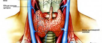
The thyroid gland and its normal size
To determine the correct norms of the thyroid gland, you need to know what it is.
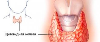
The gland is presented in the form of two lobes - left and right, an isthmus, as well as an additional lobe (pyramidal), which is not always expressed. The normal weight of the thyroid gland can range from 25 to 40 grams. In different patients, its shape, size and thickness can vary significantly.
In normal condition, the size of the gland lobes is usually 4*2*2 cm, and the thickness of the isthmus between them usually varies in size 4-5 mm. The total volume of the unchanged gland is an individual parameter for each patient, but in men it should not exceed 25 ml, and in women it should not exceed 18 ml. The structure of the organ, if there are no changes, should be homogeneous.
In the case of the development of diseases of the thyroid gland, as well as the appearance of neoplasms in its tissue, the size of the lobes can greatly increase, and their volume will also change, which can significantly affect the organs surrounding the gland.
What diseases can be detected by ultrasound of the thyroid gland?
Based on the results of ultrasound diagnostics, an endocrinologist can diagnose the following diseases:
- Nodular goiter : a disease in which compactions are clearly palpable in the area of the gland. Ultrasound reveals an increased density of formations bordering unchanged tissues.
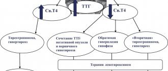
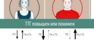
- Diffuse toxic goiter : a pathology in which the diffuse toxic state of the thyroid gland is within normal limits, but the volume of the gland is increased, and laboratory diagnostics show increased synthesis of thyroid-stimulating hormone (TSH) and T3.
- Hashimogo's thyroiditis : an inflammatory disease of an autoimmune nature, in which part of the thyroid tissue dies, therefore the function of the gland is impaired.
- Hypothyroidism : an endocrine disease characterized by the presence of diffuse degenerative changes in the thyroid tissues. In this case, the gland is small in size compared to the norm; thyroid hormones are produced in insufficient quantities.
- Thyroiditis : a disease in which inflammation develops in the gland. The main symptoms of this condition are an increase in the volume of the neck, pain in the thyroid gland, swelling of adjacent tissues.
- Cyst : a formation that resembles a bubble with fluid inside. On ultrasound it is visible in the form of formations with clear contours.
- Follicular adenoma : a nodular formation of a benign nature that may contain colloid.
- Papillary adenoma : a benign formation with a cystic structure and papillary growths inside.
- Cancerous tumor : a malignant neoplasm that is a mass accumulation of cancer cells.
In some cases, to clarify the diagnosis, additional tests for thyroid hormones are required, which reflect the balance of the production of T3 and TSH hormones.
Advantages of the method - ultrasound examination of the thyroid gland
If there is a suspicion of a malfunction in the endocrine system, the doctor will refer the patient for an ultrasound scan of the thyroid gland. This study is informative enough to determine the presence of pathologies in the area under study, while being absolutely safe for humans. MRI and CT are indicated only in cases where it is necessary to detail the structure of the pathological formation inside the gland.
The main advantages of ultrasound examination of the thyroid gland include:
- lack of special training;
- simplicity and speed of the procedure;
- absence of ionizing radiation;
- harmlessness, including for pregnant women and the fetus;
- no risk of infection.
Such diagnostics have no contraindications, so they can be performed on children of any age, pregnant and lactating women and the elderly.
If you find an error, please select a piece of text and press Ctrl+Enter
Why do you need to do the research?
The thyroid gland (TG) is one of the largest endocrine glands in the body. The primary tasks of the organ are storing iodine reserves and producing thyroid hormones.
In turn, thyroid hormones perform the following functions:
- affect metabolism, heat exchange, and the level of other hormones in the body;
- participate in the formation and maintenance of immunity;
- regulate the functioning of the cardiovascular, reproductive system, gastrointestinal tract, brain;
- activate protein synthesis, cell and tissue regeneration processes.
In addition, parafollicular cells of the thyroid gland produce calcitonin, a hormone that regulates the level of calcium and phosphorus in the blood and promotes the deposition of calcium in bone tissue.
Based on the multifunctionality of the thyroid gland, we can conclude that there is not a single internal organ that is not affected by it. Therefore, any failure in its operation can lead to serious consequences. It is possible to diagnose organ diseases in a timely manner using ultrasound.
Clinical endocrinology uses thyroid ultrasound as a primary screening method. The undoubted advantages of the technique include:
- non-invasive;
- availability;
- absolute safety for health;
- high information content.
There are several ultrasound examination methods:
- Classic ultrasound. Provides information about the structure, location, and size of the organ.
- Dopplerography. It is used to study blood flow and assess the condition of blood vessels.
In addition, ultrasound is used for differential diagnosis, assessing the effectiveness of treatment of endocrine diseases, and as an additional technique for fine-needle biopsy of the thyroid gland.