What is a hypermobile joint
The joint allows movement, but it is limited by the tendons and ligaments surrounding the joint. When the ligaments work normally, they allow, for example, bending the arm at the elbow. With hypermobility, a person can bend his arm in the other direction. This already indicates weak ligaments.
Hypermobility can be congenital or acquired. Thus, athletes involved in certain sports, ballet and circus performers also have excessively mobile joints, but this is usually the result of hard training. Although, on the other hand, congenital hypermobility of the joints is often the reason for choosing these particular professions.
In fact, there are quite a lot of people with hypermobile joints, according to some data – about 15%. But not everyone is diagnosed with this syndrome, since a person does not perceive this peculiarity as a pathology and does not seek medical help.
By the way
With the syndrome of “loose” joints, the issue of professional guidance arises especially acutely. Work associated with physical overexertion, long walking or long standing is dangerous for the patient. In these cases, the weak, stretched connective tissue part of the joint (ligaments, capsule) cannot withstand the load, and it “looses,” which contributes to the development of arthrosis. Despite the natural amazing flexibility, which seems to naturally push a person to practice choreography, you should categorically not become a dancer or ballerina with this syndrome - otherwise you may remain disabled.
What are the causes of hypermobility?
It is believed that this condition of the periarticular tissues is caused by excessive extensibility of collagen, a connective tissue protein that forms the basis of ligaments and tendons.
In some cases, hypermobility is a symptom of genetically determined connective tissue diseases, such as Ehlers-Danlos syndrome, Marfan syndrome. But such diseases are rare. More often the syndrome is associated with connective tissue weakness. Typically, this condition is inherited through the female line.
Notes
- Disease ontology database (English) - 2021.
- Monarch Disease Ontology release 2018-06-29sonu - 2018-06-29 - 2018.
- V. B. Simonenko et al., Connective tissue dysplasia (Hereditary collagenopathies), “Clinical Medicine”, 2006, N 6, 62-68.
- JH Kirk, BM Ansell, EG Bywaters, The hypermobility syndrome, Ann. Rheum. Dis., 1967, 26, 425-427.
- A. G. Belenky, Hypermobility syndrome - a systemic non-inflammatory disease of connective tissue, Department of Rheumatology RMAPO, Moscow
- Hypermobility syndrome: clinical manifestations, differential diagnosis, approaches to therapy N. G. Pravdyuk, N. A. Shostak, Department of Faculty Therapy named after. A. I. Nesterova, Russian State Medical University, Moscow
- M. M. Bikbov, V. K. Surkova, K. Kh. Oganisyan.
Keratoconus as a manifestation of connective tissue dysplasia (Russian).
Ophthalmology
(March 31, 2015). Date accessed: August 8, 2021. - Connective tissue dysplasia: main clinical syndromes, diagnosis, treatment. G. I. Nechaeva, V. M. Yakovlev, V. P. Konev, I. V. Druk, S. L. Morozov
- Attending physician
What are the dangers of hypermobile joints?
It would seem that there is nothing wrong with the fact that the joints can easily bend in different directions. But the fact is that the consequences of such hyper-mobility can be quite sad.
- People with joint hypermobility often suffer injuries such as dislocations and sprains, especially ankle sprains.
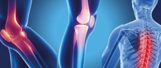
- After minor injuries and even ordinary physical activity, the joints begin to ache. The ankle and knee joints are the most vulnerable.
- Often an inflammatory process develops in the joint membrane - bursitis, synovitis, etc.
- People with hypermobile joints develop osteoarthritis in their young years.
- They suffer from back pain.
- A common consequence of the syndrome is scoliosis (curvature of the spine).
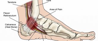
- Another consequence of hypermobility of the joints is flat feet.
Hypermobile type of Ehlers-Danlos syndrome.
One type of Ehlers-Danlos syndrome is hypermobile . It is distinguished from other types by significantly pronounced hypermobility of the joints, often accompanied by chronic pain and numerous dislocations. And since the probable cause of hypermobility is in the genes responsible for collagen synthesis, with this type, as with other types of EDS, the skin, internal organs and body systems suffer. Among the complaints of patients with the hypermobile type of EDS: instability in the joints, chronic and debilitating pain in the joints and ligaments, easy bruising, chronic fatigue, and pulse fluctuations.
To diagnose the hypermobile type of Ehlers-Danlos syndrome, the so-called Villefranche criteria are used.
Benign hypermobility syndrome and hypermobility type of Ehlers-Danlos syndrome - the same thing or different diseases?
Most often, doctors consider benign joint hypermobility syndrome and the hypermobile type of Ehlers-Danlos syndrome as separate diseases. However, in 2014, an article was published [3], refuting this opinion and proving that these are the same disease .
Often the same person at different ages fits different diagnoses. In childhood, he can be diagnosed with the hypermobile type of EDS based on pronounced hypermobility and elastic skin, and in adulthood, when all possible complications and symptoms have already appeared, he is diagnosed with hypermobility syndrome based on the Brighton scale.
The study [3] examined the medical records of 23 Italian families, each of which had patients with hypermobility. It turned out that in almost 90% of families there were several diagnoses within the same family. That is, for example, a parent was diagnosed with ADHS (in the article it is called joint hypermobility syndrome), and the child was diagnosed with a hypermobile type of EDS.
From the article it becomes quite obvious that both of these diagnoses have the same genetic cause. That is, this is, in fact, one and the same disease, or at least two facets of one disease. And while the diagnostic criteria are not developed as well as we would like, the doctor usually makes a diagnosis at his own discretion. The situation is aggravated by the fact that there is no genetic analysis to diagnose this syndrome.
- Simpson MR: Benign Joint Hypermobility Syndrome Evaluation, Diagnosis, and Management. JAOA 2006.
- Simmonds JV, Keer RJ: Hypermobility and the hypermobility syndrome. Manual Therapy 2007.
- Castori M, Dordoni C, Valiante M, Sperduti I, Ritelli M, Morlino S, Chiarelli N, Celletti C, Venturini M, Camerota F, Calzavara-Pinton P, Grammatico P, Colombi M. 2014. Nosology and inheritance pattern(s) of joint hypermobility syndrome and Ehlers-Danlos syndrome, hypermobility type: A study of intrafamilial and interfamilial variability in 23 Italian pedigrees. Am J Med Genet Part A 9999A:1–11.
How are hypermobile joints identified?
To diagnose hypermobility syndrome, the Beighton scale is used, based on assessing the mobility of various joints. The patient is asked to perform several movements, the results of which are awarded points.
1. Bend the little finger on your hand back with the help of your other hand (passive movement). The same is repeated with the other hand.
If you manage to bend your finger more than 90 degrees, 1 point is awarded for each hand.
2. Bend your thumb (also with the help of your other hand) and try to touch the area under the wrist. The same thing with the other hand. When touching - 1 point for each hand.
3. Reextend your arm at the elbow joint. The same thing with the other hand. For extension of more than 10 degrees – 1 point for each arm.
4. Re-extend the leg at the knee joint. When extension is more than 10 degrees - 1 point for each leg.
5. Standing with your knees fully straightened, bend over and touch your palms to the floor - 1 point.
In total you can get 9 points. Joint hypermobility is confirmed if a person scores 4 points or more.
Of course, this approach is very simplified and does not take into account either the age or gender of the patient, however, as a first approximation, this method allows us to talk about the presence of hypermobility syndrome.
Diagnostics
The first person to suspect this disease is the person himself. There are a number of criteria by which one can judge the presence or absence of the syndrome.
The first criterion is the Beighton assessment. It takes into account signs of joint hypermobility in the five most affected areas. The execution method is simple:
- Hypermobility of the little finger.
The little finger must be passively (i.e., with the second hand) extended all the way. If the angle with the back of the hand is less than 90º, then this is confirmation of increased flexibility.
- The second criterion confirming the presence of hypermobility of the finger joints is checking the flexibility of the thumb.
Thumb hypermobility is tested using passive flexion. If the finger reaches the forearm, then the criterion is considered positive.
- Hypermobility of the elbow joint.
The flexibility of the elbow joint is checked at its maximum extension. Normally, the angle is up to 10º.
- Hypermobility of the knee joint.
The flexibility of the knee joint is determined similarly to the elbow joint, the norm is also up to 10º
- Hypermobility of the spine.
To test the elasticity of the spine, you need to bend. If the palms touch the floor, hypermobility also occurs here.
Beighton tests are carried out in order, the correct technique must be taken into account
All criteria for hypermobility are divided into major and minor.
The big ones include:
- Score 4 or more on the Beighton test.
- The presence of arthralgia for more than 3 months in 4 or more joints.
Minor criteria:
- The presence of dislocations in two or more joints or recurrent dislocations in the same one.
- Beighton test from 1 to 3 points.
- Arthralgia for less than 3 months or in less than 4 joints.
- Presence of varicose veins.
- Hernias of any localization.
- Myopia.
- Thin, very elastic skin with stretch marks and atrophic scars.
- Bursitis, tendonitis, tenosynovitis in two or more joints.
The diagnosis is considered confirmed if there are 2 major criteria, 1 major and 3 minor, 4 minor.
Remember: the Beighton test must be performed in a calm state, without preliminary warm-up or stretching.
We also recommend studying the topic of this material:
Why does rheumatism develop in children and how to recognize it in time?
Rheumatism in a child is dangerous due to its consequences: if treatment is not started on time, heart disease develops, and the baby may remain disabled. With this disease they suffer greatly...
What to do
There is no cure for hypermobile joint syndrome. If there are no severe hereditary pathologies, then treatment is not required. You may have to treat the consequences of hypermobility: injuries, osteoarthritis, back pain. To prevent this from happening, you should change your lifestyle:
- eliminate heavy stress on joints;
- try to avoid injuries;
- exclude traumatic sports, perhaps change profession;
- strengthen muscles with isometric exercises, which involve working the muscles and minimal movements in the joints.
- correct flat feet using orthopedic insoles.
If joint pain occurs, you can use orthoses - special devices for unloading the joints.
Joint hypermobility syndrome
Prepared by: MedWeb
Joint hypermobility syndrome
Joint hypermobility syndrome
Joint hypermobility syndrome (JMS) is an excessive range of motion in the joints in the absence of signs of another rheumatic disease.
joints, cracking in joints, flat feet
Causes
VHMS is based on a hereditary feature of the structure of the main protein of connective tissue, collagen, which leads to greater than normal extensibility. The average normal degree of joint mobility varies significantly among different age, gender, and ethnic groups. It is known that representatives of eastern peoples have much greater joint flexibility than Europeans. In particular, when examining healthy people in Moscow aged 16–20 years, more than half of girls, and more than a quarter of boys, demonstrated a degree of hypermobility above average.
Signs
The main signs of VMS are joint pain for no apparent reason for three months or more. Most often, at any age, pain is observed in the knee joints. They are caused by increased sensitivity to load (supporting joints) and moderate orthopedic anomalies (impaired development of the hip joints, longitudinal and transverse flatfoot), which are often found in people with FHMS.
Prevalence
The prevalence of FHMS is difficult to estimate. According to a survey of practicing rheumatologists in the UK, each of them sees 25–50 patients with this disease per year. When analyzing the reasons for hospitalization in large rheumatology clinics, the diagnosis of FHMS was made in 2–5.7% of cases. However, these data are only the tip of the iceberg, since many do not receive the correct diagnosis or become patients of operating orthopedists with complications.
Clinical manifestations
The clinical picture of VHMS is diverse and includes both articular and extra-articular manifestations. A frequent fact in the patient’s life history is his participation in areas of activity where joint hypermobility has a certain advantage (dancing, ballet, acrobatics). However, at the same time, there is also a special sensitivity to physical activity and a tendency to frequent injuries (sprains, subluxations of the ankle joints). Often, in a family history (and when examining relatives), it is possible to establish the presence of joint hypermobility in blood relatives (mainly on the female side), often with similar problems.
SGMS in high school students
Although in 3/4 of cases the onset of clinical manifestations of VHMS occurs during high school age, they can occur at any period of life. As a rule, this is due to a violation of the balance between the load and the body’s capabilities that developed in the previous period (increase in body weight, change in motor stereotype).
FHMS at a young age
For young people with FHMS, pain in several joints is most common. Factors that provoke such pain can be psycho-emotional stress, unfavorable weather conditions, the phase of the menstrual cycle, a previous acute respiratory disease, or pregnancy.
Signs of FHMS and accompanying conditions
Joint subluxations (mainly ankles and knees) are typical in young patients with FHMS. Many of them learn about their predisposition to injuries and take preventive measures on their own (wear high-laced shoes, avoid traumatic situations).
Recurrent effusion (the appearance of excess fluid) in a joint (usually the knee) as a manifestation of VHMS is not common; its appearance, as a rule, is directly related to injury. The effusion is non-inflammatory in nature and undergoes rapid regression. Subsequently, these patients may experience persistent pain in the injured knee joint due to post-traumatic meniscus disease.
Crunching (“clicking”) of joints without pain is often the only manifestation of hypermobility and, in the absence of pain, is not a sign of pathology. It is associated with uneven sliding of the tendons relative to the bony protrusions in the joint area and does not require treatment.
Back pain, often combined with scoliosis and vertebral displacement, occurs in patients with VMS at any age.
Symptomatic flatfoot (longitudinal, transverse and combined) may be the only manifestation of FHMS, especially in young women, for a long time. A characteristic symptom is “tired legs” at the end of the day, the inability to wear high-heeled shoes or none at all. At an older age, orthopedic complications develop: flat feet - corns, hammertoe deformity, secondary arthrosis of the ankle joints.
Periarticular lesions (inflammation of tendons, joint capsule, etc.) occur in patients with FH over 40 years of age significantly more often than in the general population. As a rule, they occur in response to unusual (unusual) load or minimal trauma.
Often, patients over 30 years of age show several signs of non-inflammatory connective tissue damage. In this case, the same patient has pain in several joints, in the back, damage to soft tissues, and already complicated flat feet. In some cases, such a symptom complex in patients with FHMS in the older age group (40–50 years) is clinically and radiologically indistinguishable from polyosteoarthrosis. It is known that joint hypermobility is a risk factor for the development of early osteoarthritis.
Extra-articular signs
When examining patients with possible FHMS, attention is often drawn to extra-articular signs of systemic connective tissue damage - thinness, extensibility of the skin, atrophic scars, easy formation of subcutaneous hemorrhages with normal blood clotting, tall stature, thinness, elongation and thinning of the fingers, underdevelopment of the upper jaw, impaired normal row of teeth, sandal gap between the first and second toes, chest deformities, winged neck, etc. Detection of these signs provides significant assistance in making the correct diagnosis.
Ultrasound and nuclear magnetic resonance examination of the soft tissues of the joint area in some cases can detect signs of periarticular lesions that are not detected during examination. X-ray examination is important to establish signs of secondary and early osteoarthritis.
Non-drug therapy
Non-drug methods and lifestyle optimization play a decisive role in treatment. The latter involves ensuring a balance between physical activity and the patient’s tolerance threshold. It is necessary to exclude loads and situations that cause pain and discomfort in the joints, to minimize the possibility of injuries, which includes professional guidance and the exclusion of team sports.
Elastic orthoses
For persistent pain in one or more joints, elastic orthoses (knee pads, etc.) are used to artificially limit the range of motion and relieve pain. If orthopedic deviations (flat feet) are identified, their correction is indicated. The shape and rigidity of corrective insoles are determined individually: at a young age, hard insoles are effective, at an older age, soft polyurethane insoles are effective. In some cases, it is possible to completely relieve pain in the knee and ankle joints only by correcting longitudinal flat feet.
Rehabilitation
For back pain (related and not related to scoliosis), it is recommended to include the patient in an appropriate rehabilitation program (use of reclinators in childhood, strengthening of paravertebral muscles).
Strengthening the muscles surrounding the painful joint ensures optimization of local biomechanics and, as a result, the disappearance of pain. Gymnastics for HSMS includes isometric exercises, during which the muscles are tense, but the range of motion in the joints is minimal. Depending on the location of the pain syndrome, it is recommended to strengthen the muscles of the hips, shoulder girdle, back, etc. Swimming is useful, in which muscle tension occurs with passive resistance from the environment. You should not carry your bag on one shoulder.
Drug therapy
For joint pain, drug therapy is indicated. Taking analgesics (paracetamol, tramadol) has some effect. Intra-articular administration of glucocorticoids in the absence of signs of secondary inflammation of the articular membrane is ineffective.
Treatment of VMS in older patients with clinical and radiological signs of polyosteoarthrosis essentially differs little from the generally accepted therapy of osteoarthritis, including the use of drugs that strengthen cartilage (chondroprotectors).
For periarticular lesions, treatment tactics are practically no different from those in ordinary patients. Eliminate a possible provoking factor (uncomfortable shoes, excessive load, etc.). Drug therapy in moderate cases is ointments containing non-steroidal anti-inflammatory drugs, in the form of applications or compresses; in more persistent cases, local administration of glucocorticoids.
For severe orthopedic pathology (recurrent subluxations, vertebral displacement, meniscal pathology), surgical treatment is indicated.
Andrey BELENKY, rheumatologist, Ph.D. honey. sciences