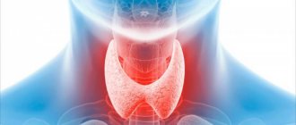
We have long known that Russia is an endemic area for thyroid disease. It is difficult to find a person whose friend or relative would not complain about problems with the thyroid gland. To be fair, it should be noted that the culture of preventive research in Russia has recently increased. There are especially long lines at the doors of ultrasound specialists. However, simply undergoing an ultrasound examination of the thyroid gland is not enough. Equally important is the correct reading of the examination result.
General information
Diffuse changes in the thyroid gland are structural disorders of the parenchyma, which are usually detected by ultrasound examination and are expressed in the form of enlargement of the organ, hypo-, iso- or hyperechogenicity (change in density, its unevenness), loss of tissue homogeneity as a result of destruction, lymphogenous infiltration, sclerotic processes, etc. In this case, transformations are mixed, diffuse-nodular, euthyroid, hypothyroid and cause various pathological functional states.
Approximately 30-50% of the world's inhabitants have problems with the thyroid gland and they can lead to the development of goiter (struma) , thyroiditis and tumors, and can also be accompanied by hyperthyroidism ( thyrotoxicosis ) or hypothyroidism ( myxedema ).
Which doctor should I contact?
In order for the treatment to be successful, it is necessary to initially contact a qualified specialist who will conduct a consultation and help with deciphering the diagnosis. I am such a highly qualified specialist in the field of endocrine health, Georgy Nikitich Romanov. I am a candidate of medical sciences, I have been practicing medicine for almost a quarter of a century, both in public and private hospitals. institutions, I have extensive experience in treating patients with thyroid nodules.
In addition to face-to-face appointments at the clinic, I conduct paid online consultations, which anyone can attend and ask questions of concern. To contact me and make an appointment, you can write to me via email on one of the social networks or messenger: Viber, Telegram, Instagram, WhatsApp, Skype, VKontakte.
Pathogenesis
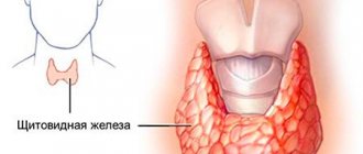
Normally, the structure of the thyroid parenchyma is characterized by homogeneity, the presence of clearly defined anatomical markers and easily differentiated adjacent tissues.
Structure of the thyroid gland
With diffuse changes in the parenchyma of the thyroid gland, the greatest clinical significance is affecting functional cells - follicles-thyrocytes containing iodine . As a result of proliferation, an enlargement of the organ occurs, which can even be visually noticeable and look like a goiter. If the amount of iodine in the blood is reduced and the level of thyrotropin , then diffuse hyperplasia of the gland first occurs, and then nodular forms of goiter. In addition, disorders may be associated with dysgenesis , aplasia , organ hypoplasia , focal degeneration, necrobiosis , sclerosis , hyperplasia and colloid involution.
Thyroid goiter
At first, moderate changes do not cause structural disturbances, so it is likely that it will function normally and maintain the level of thyroid hormones , but with more pronounced lesions of the thyroid gland, in addition to enlargement, structural and functional disturbances occur, which are usually caused by serious diseases such as Graves' disease or thyrotoxic goiter .
Classification
Diffuse changes are characterized by uniform damage to the entire thyroid gland and they can be congenital or acquired, leading to hypothyroidism or, on the contrary, thyrotoxicosis .
Diffuse focal changes in the thyroid gland
Diffuse-focal transformations are subject to the same mechanisms of pathogenesis, but differ in focality, that is, they develop in specific zones of the organ. Typically, these transformations are preceded by inflammation, while the organ works as usual and does not malfunction, but still in this case it is better to regularly consult a doctor.
Diffuse nodular changes in the thyroid gland
Diffuse nodular or focal changes are called changes that lead to the formation of nodular structures. However, their size should not exceed 1 cm. If colloidal content is found inside the formations and it is encapsulated, then they speak of diffuse cystic changes.
Most often, diffuse nodular formations are benign - in approximately 95% of cases, but there is a risk of their malignancy and becoming malignant. Very often, pathological changes do not cause symptoms, which significantly worsens the patient’s prognosis.
If a single node develops in an organ, the compaction is characterized by rapid growth, has a hard consistency and can cause changes in the lymphatic system. When we talk about multiple nodular formations, we talk about the development of diffuse goiter, which can be endemic, mixed and toxic. For example, as a result of chronic iodine deficiency, gland tissue grows and its functionality changes. The mechanism is based on the adaptation of the thyroid gland to iodine deficiency by increasing the amount of iodine uptake from the bloodstream and subsequent synthesis and secretion of triiodothyronine (T3). Maintenance of the process is caused by increased secretion of thyroid-stimulating hormone, which has goitrogenic properties. Despite the large volumes of the gland, with a reduced intrathyroidal iodine concentration, it is not able to maintain the optimal level of thyroid hormones, which ultimately results in hypothyroidism .
Diffuse changes in the thyroid gland like AIT
The most demonstrative example of diffuse changes in the thyroid gland is autoimmune thyroiditis , or otherwise Hashimoto's thyroiditis . Familial disease refers to genetically determined defects in the immune response, causing T-lymphocyte aggression against one’s own thyrocytes. Chronically progressive lymphoid infiltration leads to gradual destruction of the parenchyma. As a result, patients may develop lymphogenous goiter and primary hypothyroidism , which may further cause an increase in symptoms (decreased immunity , decreased performance, growth retardation, cretinism, gastrointestinal disorders, problems in sexual life) and require correction.
Diffuse changes of the AIT type may be associated with taking interferon , as well as hepatitis C and blood diseases.
Causes
Diffuse changes in the thyroid gland can be promoted by:
- poor ecology and radioactive contamination;
- hormonal disorders;
- mental trauma and nervous strain;
- deficiency or excess of iodine;
- congenital autoimmune disorders;
- infectious diseases and inflammatory processes;
- injuries and operations on the thyroid gland;
- increased body weight;
- viral and other chronic infectious processes, for example, carious teeth, inflammation of the sinuses;
- nutritional errors and lack of amino acids, minerals and vitamins in the diet.
Diagnostics
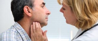
If there are suspicions of nodules in the thyroid gland, then you need to consult an endocrinologist, since this is entirely his field of activity. The doctor will collect anamnesis, ask the necessary questions about well-being, nutrition, diseases and heredity, and also palpate both lobes of the thyroid gland in the neck.
The main diagnostic procedures for identifying nodes and describing their nature are:
- ultrasound diagnostics, in which you can evaluate the structure of tissues and detect formations;
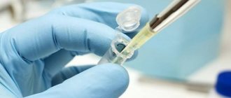
- fine-needle aspiration biopsy - taking the material of the nodes themselves for analysis to determine whether they are malignant or benign;
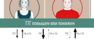
- hormone analysis - an analysis is performed for the main types of hormones TSH, T4 and T3, and the doctor may also prescribe additional blood tests.
The listed studies are aimed at identifying pathology and clarifying its nature in order to prescribe appropriate treatment.
Symptoms
General symptoms of pathological changes in the structure of the thyroid gland manifest themselves in the form of increased fatigue and muscle weakness, and also occur:
- decreased performance and concentration;
- depressed state, and a tendency to depression ;
- memory deteriorates;
- nail plates and hair become more brittle and dry;
- hot flashes may be followed by feelings of extreme cold;
- aggressiveness increases;
- an inexplicable and sudden increase in body weight occurs;
- abnormal frequency of colds or other illnesses.
If diffuse changes have led to the development of a goiter or nodes of quite impressive size, then typical complaints may be:
- feeling like there is a lump stuck in the throat;
- causeless cough and fever;
- difficulty breathing and swallowing;
- headaches and neck discomfort.
When diffuse changes provoke hyperthyroidism , a person may experience tachycardia , palpitations , frequent hot flashes, exophthalmos (bulging of the eyes), increased excitability, emotional lability, irritability and aggressiveness.
What does an ultrasound of the thyroid gland show?
The content of the article
Ultrasound examination of the thyroid gland allows you to obtain accurate information about its condition. First of all, attention is paid to the location, shape, contour and, of course, the size of the gland.
In terms of overall size and volume, the gland may be normal, enlarged (hyperplastic), or reduced (hypoplastic). There are also cases of asymmetry of the gland, absence of part of the lobe (hemiagenesis) and complete absence of the gland (agenesis-aplasia).
Thyroid volume levels vary depending on age and gender. The upper limit for women is 18 cm3, for men – 25 cm3. As children get older, the glands grow.
Tests and diagnostics
To detect diffuse changes in the thyroid parenchyma, ultrasound examination is sufficient. Usually, echography is resorted to if it is not possible to detect diffuse changes in the thyroid gland, but tests reveal a hormonal imbalance and palpation reveals lumps. However, most often, it is part of a routine health examination.
In some cases, to clarify the type and location of lesions, MRI, bronchoscopy or laryngoscopy, fine-needle aspiration biopsy, as well as tests to determine the titer of antibodies to thyroid tissue may be prescribed.
Signs of diffuse changes in the thyroid gland
A diffuse increase in the echogenicity of the gland, detected on ultrasound, is usually associated with the development of intraparenchymal strands of connective tissue, which creates a false impression of lobulation of the thyroid gland; multiple hypoechoic foci of approximately 1-6 mm can be identified inside it.
The main echographic signs are an enlargement of the organ and the detection of nodes or clusters of nodes on its surface. Echo signs are usually expressed in the form of diffuse heterogeneity of structure, echo density, the presence of hypoechoic inclusions in the tissues of the gland and increased vascularization.
Ultrasound of the thyroid gland: normal
Many patients who plan to have an ultrasound of the thyroid gland would like to understand for themselves what all these words written in the conclusion of the study mean. We will try to explain some of the most important terms used by doctors during ultrasound, as well as their meaning in terms of determining normality and pathology.
The most important ultrasound characteristics of thyroid tissue are:
- contours of the gland;
- gland tissue structure;
- echogenicity of gland tissue;
- presence or absence of focal changes (nodules, cysts);
- blood supply to gland tissue.
The ultrasound condition of the cervical lymph nodes surrounding the thyroid gland is also necessarily described.
Outlines of the thyroid gland
can be clear or unclear. Normally, the contours of the thyroid gland should be clear. The contours become fuzzy (blurred) with the development of inflammation, as well as with the occurrence of malignant tumors of the thyroid gland that grow into the surrounding muscles and adipose tissue.
Fabric structure
can be homogeneous or heterogeneous.
The thyroid gland normally
has a characteristic grainy tissue, which, with some skill, is difficult to confuse with anything else.
Inflammatory diseases of the thyroid gland
, developing as a result of aggression of the immune system (autoimmune thyroiditis, diffuse toxic goiter) are accompanied by the appearance of heterogeneity of thyroid tissue - sometimes like a “honeycomb”, sometimes doctors describe it as “moth-eaten tissue”, but always in the tissue with heterogeneity The structure contains more and less light areas, the tone of which is clearly different. It happens that doctors describe a pronounced heterogeneous structure of the gland, when the difference in the tone of the thyroid gland is large, or a moderately heterogeneous structure of the thyroid gland - this is often found in healthy people who have an increased titer of antibodies to thyroid peroxidase or thyroglobulin.
Echogenicity of thyroid tissue
– this is the same “tone” that is visible on the screen. It should be remembered that the image on the screen of the ultrasound machine is formed by a computer, which analyzes the reflected ultrasound rays coming from the internal organ and, based on this analysis, presents the image in grayscale to the operator. Echogenicity is the tone of gray that the computer represents thyroid tissue. Normally, the echogenicity of the gland tissue is equal to the echogenicity of the parotid salivary gland. With the development of inflammatory diseases, the echogenicity of the thyroid gland is most often reduced, but in the later stages of this process it can even be increased. A pronounced decrease in echogenicity is indicated when the tone of the gland becomes darker than the tone of the surrounding muscles (i.e., almost black) - such changes should always alert the doctor performing an ultrasound of the thyroid gland. The norm of echogenicity may vary slightly, but usually the echogenicity of the thyroid gland is higher than the echogenicity of the muscles, vessels, and esophagus (i.e., the gland looks lighter on the ultrasound machine screen).
Focal changes (nodes)
The thyroid gland does not normally contain. Acceptable are cystic formations up to 3-4 mm in size, which look uniformly black on the screen (i.e., anechoic - without echogenicity) - endocrinologists often call such formations enlarged follicles, accumulations of a hormone-containing gel - colloid. All formations over 4 mm in size that differ in echogenicity from the surrounding thyroid tissue are usually called nodes. Nodes can be:
- isoechoic, i.e. equal in echogenicity to the surrounding thyroid tissue;
- hyperechoic, exceeding the echogenicity of the surrounding thyroid tissue (i.e. lighter);
- hypoechoic, having less echogenicity than the surrounding tissue (i.e. darker);
- anechoic, i.e. completely, completely black (this color is typical for fluid formations, cysts).
A thyroid nodule is always not the norm. The thyroid gland should normally be homogeneous, without nodes. However, modern high-class ultrasound machines used at the North-Western Endocrinology Center make it possible to detect nodes as small as 1 mm. Endocrinologists who perform ultrasound at the endocrinology center understand that it is unreasonable to call every formation in the thyroid tissue measuring 1, 2 or 3 mm a node, since after this, from a formal point of view, it is necessary to establish a diagnosis of “Nodular goiter”. A patient with such a diagnosis then has a lot of problems when visiting other specialists who do not have sufficient knowledge in the field of endocrinology, who, instead of treating, for example, high blood pressure or cardiac arrhythmia, will tell the patient: “Well, what do you want - you have It's a goiter! First, go to an endocrinologist, have him write you a paper saying it’s not the thyroid gland’s fault, and then come to me.” As a result, the patient is forced to make unnecessary trips to the doctor, wasting time, nerves and money. That is why doctors must treat small focal formations very carefully - of course, certain examinations are necessary, but usually no treatment is required as a result.
For each thyroid nodule, the physician performing the ultrasound should describe:
- contours (clear, fuzzy);
- the presence or absence of a dark rim along the periphery of the node (halo rim);
- echogenicity of the node;
- the presence of micro- or macrocalcifications (i.e. calcium deposits that do not have an acoustic shadow = microcalcifications, or have an acoustic shadow = macrocalcifications);
- the presence or absence of cystic transformation of the node (i.e. the appearance of cysts inside the node);
- linear dimensions (it is advisable to describe three linear dimensions of the node, since this makes it possible to calculate the volume of the node and subsequently, with repeated ultrasound, reliably determine the dynamics of its change).
Blood supply to tissue
determined by conducting a Doppler study, which reveals the intensity of blood flow that the thyroid gland has. The norm is the presence of several color signals on the surface of the thyroid tissue. When the thyroid gland is inflamed, blood flow increases, and the entire gland seems to be “bursting with fire” on the ultrasound machine screen. Western researchers even came up with the poetic name thyroid inferno (“thyroid hell”) for this type of blood flow, comparing this picture with the tongues of hellish flames on medieval paintings.
Regional lymph nodes of the neck
Normally, an ultrasound scan of the thyroid gland should not look enlarged. Lymph nodes must have clear, even contours, the length of the lymph node must be at least 2 times the width of the lymph node (the so-called Solbiati index), the gate must be clearly visible in the structure of the lymph node - the place where the lymphatic vessel enters the lymph node. The tissue of the lymph node should not have increased blood flow and, especially, cysts - both of these signs are very alarming and often indicate a malignant lesion of the lymph node.
Ultrasound normal thyroid gland
- this is the standard that every endocrinologist must clearly know, and with which he must compare the image visible on the screen of the ultrasound machine. Of course, it is difficult to give a complete picture of all ultrasound aspects of a normal thyroid gland in a short article. If you doubt whether what was revealed during an ultrasound examination of your thyroid gland in a clinic or general medical center is normal, the most reasonable tactic would be to consult an endocrinologist or an endocrinologist surgeon at the North-Western Endocrinology Center, who will independently perform An ultrasound of the thyroid gland will compare what you see with what is described on your form and explain to you what needs to be done in the future. You will be surprised, but each of our specialists knows how often the “threatening” changes on ultrasound described somewhere, in the end, when carefully examined by experienced specialists using high-quality equipment, turn out to be just another variant of the norm...
Diet for diffuse changes in the thyroid gland
Diet for thyroid disease
- Efficacy: therapeutic effect after a month
- Timing: constantly
- Cost of food: 1600-1700 rubles per week
In most cases, thyroid problems are caused by iodine deficiency, so it is recommended to include in the diet:
- seaweed salad;
- buckwheat, oatmeal and wheat porridge;
- iodized salt;
- seafood, including fish and caviar;
- feijoa, cranberries, persimmons and apples.
Important! If an increased level of thyroid hormones in the blood has been established, then these products should be discarded.