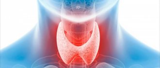
Nodules in the thyroid gland
The thyroid gland is one of the organs of the endocrine system; it is located in the neck, has two lobes and an isthmus. This organ produces important hormones that allow the body to function properly and are responsible for metabolism.
The main nutrient of the thyroid gland is iodine - its deficiency leads to deviations and the development of pathologies, which are best detected in time to prevent serious consequences.
ICD 10 code for thyroid diseases: E00-E07. Depending on the classification, a certain type of treatment is provided.
The nodule in the thyroid gland has a round shape and is formed due to the excessive process of cell division of the gland itself.
In practice, a person may have one or several nodes in any lobe or both. Most of these formations are benign, however, there are also malignant ones that require serious intervention.
To determine malignancy, thyroid nodules undergo a biopsy procedure. According to statistics on biopsies, 80-90% reveal the benign nature of the material. The photo below shows what a person’s neck looks like with the usual size of the left and right lobes of the thyroid gland, and what it can look like with a significant increase in the size of the lobes.
Why are nodules in the thyroid gland dangerous?
As the thyroid tissue increases, it can begin to produce additional amounts of hormones, which creates an imbalance in the body, metabolic processes are disrupted, and other disorders and diseases develop against this background. Also, strong tissue proliferation at some point can worsen a person’s quality of life; symptoms such as sudden weight loss may appear, which is associated with dysfunction of metabolic processes, excessive sweating, depression of the nervous system, etc.
Causes of nodules on the thyroid gland
What to do if a nodule appears in the thyroid gland is decided by the doctor based on its nature and causes.
Unfortunately, today there is no confirmed data on why this process occurs. But everyone is inclined to believe that this is due to a lack of iodine in the body and a hereditary predisposition. In addition to the fact that a person may receive little iodine from food, he may be exposed to a large flow of radioactive radiation. Radiation is also known to promote the formation of nodules and even cancer.
Symptoms of an enlarged thyroid gland and causes
A change in the volume of the thyroid gland indicates both the presence of pathological processes in the organ and a disruption of the functioning of the entire endocrine system as a whole. Therefore, when making the correct diagnosis, it is worth taking into account the entire symptom complex of the disease.
- An increase in the size of the thyroid gland is facilitated by unfavorable factors that have a negative effect on the entire body or hereditary predisposition - these are uncontrollable causes; the doctor can only eliminate the consequences.
- The appearance of this pathology can be caused by diseases of the pituitary gland, an imbalance of the hormonal system, a lack of iodine, vitamin A, cobalt, zinc and other trace elements. In this case, by finding out the exact cause, you can effectively help the patient.
Signs of an enlarged thyroid gland are observed in diseases such as hypothyroidism, hyperthyroidism and thyrotoxicosis. The appearance of benign or malignant neoplasms is also one of the causes of gland pathology.
Symptoms
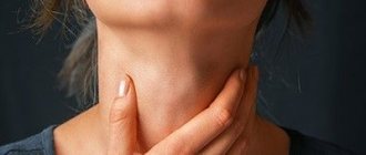
Thyroid nodules in women and men are usually not accompanied by symptoms, but are detected during routine ultrasound. Sometimes, however, with large sizes, a person may notice uncharacteristic swelling in the neck area or may notice discomfort in the neck area when turning the head and swallowing.
A large nodule (more than 1 cm) can be palpated by the doctor, and then its size and nature can be clarified using ultrasound. Small nodules may not be felt, so it is important to do an ultrasound yourself once a year to monitor the condition of the thyroid gland.
Sometimes patients confuse discomfort, the feeling of a lump in the throat, some attacks of suffocation, etc. with the formation of nodules in the thyroid gland, however, most often this is associated with improper functioning of other organs, such as the gastrointestinal tract.
The first signs and symptoms of thyroid goiter
The vast majority of patients do not have any symptoms if hormone production is normal. Enlargement of the lower third of the neck is possible with diffuse or multinodular goiter; individual nodules up to a centimeter do not change the configuration of the neck.
Compression of the trachea is possible if the trachea is very large or if the main mass of glandular tissue is located behind the sternum, as well as an increase in the diameter of the cervical vessels and a change in voice to the point of hoarseness.
Inadequate production of hormones does not correlate with the size of the gland and nodes; the symptoms of thyrotoxicosis or hypothyroidism are not obvious and nonspecific, but are manifested by impaired functioning of target organs: the cardiovascular system, nervous, subcutaneous fat, skin and muscles.
Initially, the patient complains of palpitations, nervousness, difficulty sleeping and concentration, weight loss or gain. A cardiac examination that does not detect cardiac pathology is performed, and only a blood test reveals abnormal hormone levels.
Very rarely, the classic thyrotoxic triad occurs: palpitations with trembling and exophthalmos - bulging eyes. An elderly patient may be hospitalized with thrombosis and heart rhythm disturbances with a visually undetectable pathology, but releasing too many hormones.
It is very rare that an adenoma develops that produces excess thyroid hormones; almost 80% of thyrotoxicosis is caused by a diffuse increase.
Diagnostics
If there are suspicions of nodules in the thyroid gland, then you need to consult an endocrinologist, since this is entirely his field of activity. The doctor will collect anamnesis, ask the necessary questions about well-being, nutrition, diseases and heredity, and also palpate both lobes of the thyroid gland in the neck.
The main diagnostic procedures for identifying nodes and describing their nature are:
- ultrasound diagnostics, in which you can evaluate the structure of tissues and detect formations;
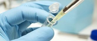
- fine-needle aspiration biopsy - taking the material of the nodes themselves for analysis to determine whether they are malignant or benign;
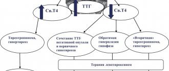
- hormone analysis - an analysis is performed for the main types of hormones TSH, T4 and T3, and the doctor may also prescribe additional blood tests.
The listed studies are aimed at identifying pathology and clarifying its nature in order to prescribe appropriate treatment.
Magnetic resonance imaging (MRI) in St. Petersburg
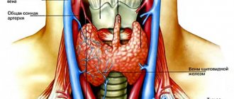
The thyroid gland consists of two lobes connected by an isthmus. The isthmus is located between the 2nd and 4th tracheal cartilages. The size of the lobe is approximately 4.0 x 1.5 x 2.0 cm (length-width-depth). The lower pole reaches the 5-6 tracheal ring. From above, the lobe reaches the thyroid plate. The gland is covered with bundles of the hyoid muscle. 40% of people have a pyramidal lobule located in the area of the upper pole of the isthmus. The gland weighs 15-25 g. The gland is supplied with blood from the superior and inferior thyroid arteries and its own artery, which arises from the aortic arch or innominate artery. The outflow of blood goes through the upper, middle and lower thyroid veins.
Clinical manifestations of pathologies consist of hypo- or hyperthyroidism. Hypothyroidism is associated with impaired hormone production, mainly in various types of thyroiditis. Hyperthyroidism is associated with various types of thyrotoxicosis.
ETIOLOGICAL CLASSIFICATION OF THYROID DISEASES
- I. Autoimmune thyropathies.
- Graves' disease.
1.1. Isolated thyropathy.
1.2. Thyropathies with extrathyroidal manifestations.
- Autoimmune thyroiditis.
2.1. Chronic autoimmune thyroiditis.
2.2. Transient autoimmune thyroiditis.
2.2.1. Painless (“silent”) autoimmune thyroiditis.
2.2.2. Postpartum autoimmune thyroiditis.
2.2.3. Cytokinin-inducing autoimmune thyroiditis
- II. Colloidal, proliferating goiter to varying degrees. Diffuse goiter.
- Nodular and multinodular goiter.
2.1. Nodular and multinodular goiter without functional autonomy.
2.2. Nodular and multinodular goiter with functional autonomy.
III. Infectious thyropathies.
- Subadult thyroiditis.
- Acute purulent thyroiditis.
- Specific thyroiditis.
- IV. Tumors Benign.
- Malignant.
- V. Congenital (hereditary) thyropathies.
- VI. Diseases of the thyroid gland in the pathology of other organs and systems.
Nodules in the thyroid gland occur in 4-7% of the adult population. These can be colloid nodes, adenoma, focal hyperplasia, cysts, metastases, thyroid tumor.
Diagnostic methods: radionuclide, ultrasound, and MRI .
Iodine-123 scintigraphy shows “hot” and “cold” nodes. Iodine-131 and technetium preparations are also used. “hot” nodes are usually adenomas, “cold” nodes can be malignant in 1-4% when studied with iodine and up to 30% when studied with technetium.
Ultrasound perfectly shows the anatomy of the gland and hypoechoic nodes, as well as cysts. A disadvantage is poor visualization of lymphadenopathy and surrounding tissue. Ultrasound is the most accurate in terms of diagnosing cysts. However, complicated cysts may turn out to be carcinoma. For accurate diagnosis, ultrasound is supplemented with fine-needle biopsy.
Tomographic studies better reflect the anatomy and are possible in thin sections. Since the administration of iodine contrast is undesirable, MRI is preferable to CT. MRI excellently demonstrates extension toward the mediastinum. It is believed that MRI reliably shows nodes up to 4 mm.
MRI. Thyroid cancer with invasion of the recurrent nerve. T1-dependent and T2-dependent axial tomograms.
The most common pathologies of the thyroid gland include nodular hyperplasia. On ultrasound, nodules are detected in 30% of subjects aged 19-50 years, and in 60% or more of those over 60 years of age. Only 1 in 20 nodes turns out to be malignant.
CT and MRI for tumors reflect signs of malignancy: infiltrative growth and vascular invasion. Carcinoma is contrasted on CT and MRI .
CT. Reformation into the coronal plane. Lymph nodes of the neck with calcifications.
Staging of thyroid cancer is carried out according to the TNM system:
T-tumor
- TX – Primary tumor cannot be assessed
- T0 – No evidence of tumor
- T1a - Tumor ≤1 cm, limited to the thyroid gland
- T1b – Tumor >1 cm but ≤2 cm in greatest dimension, limited to the thyroid gland
- T2 – Tumor >2 cm but ≤4 cm in greatest dimension, limited to the thyroid gland
- T3 – Tumor >4 cm in greatest dimension, limited to the thyroid gland or any tumor with minimal extension beyond the thyroid gland
- T4 - Extensive disease T4a - Moderately extensive disease - tumor of any size extending beyond the capsule into the subcutaneous soft tissues, larynx, trachea, esophagus, or recurrent nerve
- T4b – A very common disease - the tumor invades the paravertebral fascia or engulfs the carotid artery or mediastinal vessels
- cT4a – Intrathyroid anaplastic carcinoma
N: Lymph nodes
- NX – Regional lymph nodes cannot be assessed
- N0 – No metastases to regional lymph nodes
- N1 – There are metastases to regional lymph nodes N1a – Metastases up to level VI (pretracheal, paratracheal, and preglottic)
- N1b – Metastases to unilateral, bilateral and contralateral cervical (Levels I, II, III, IV, or V) or retropharyngeal or upper mediastinal lymph nodes (Level VII)
M: Metastasis
- MX – Distant metastases cannot be assessed
- M0 – No distant metastases
- M1 – There are distant metastases
In addition to the TNM system adopted by WHO, there is also a simplified TIRADS , adopted by radiology diagnosticians in some countries (for example, the USA). The system involves a visual assessment of the condition of the thyroid gland, for example, using ultrasound.
ULTRASONIC DIAGNOSTICS OF THYROID DISEASES
- Autoimmune thyropathies
Graves' disease (Diffuse toxic goiter, Graves' disease).
It is characterized by hyperplasia of the thyroid gland with an increase in its function. Echographically, unevenness or scalloping of the contours of the gland is determined. A decrease in echogenicity is not a specific sign of hyperthyroidism, since it can occur due to increased vascularization of the parenchyma or other processes leading to an increase in the hydrophilicity of the thyroid tissue (lymphocytic infiltration, reactive changes in the thyroid gland against the background of adenovirus infection). Hypervascularization of the thyroid gland using Doppler echo was first described by Rails in 1988 and called “Thyroid inferno” - “fire” in the thyroid gland. The uniformity of blood flow indicators in all regional vessels of the thyroid gland, as well as in the region of the lobes, in patients with Graves' disease confirms the well-known opinion about the uniform functional activity of the entire parenchyma of the thyroid gland. Against the background of hyperproduction of thyroid hormones, recanalization of the previously collapsed vessels of the gland parenchyma occurs.
Autoimmune thyroiditis.
This group includes acute, subacute and chronic inflammatory diseases of the thyroid gland, which are based on autoimmune, cytotoxic processes, with simultaneously occurring processes of degeneration and tissue regeneration. The echographic picture largely depends on the functional state of the gland. The gland can be either enlarged, normal or reduced in size, its contours are often uneven. The echo density of the gland is often reduced, the echo structure is heterogeneous due to multiple hypoechoic areas of various shapes and sizes, mostly without clear contours, as well as hyperechoic strands that give the gland a lobular structure. Large foci of low, or less often increased, echogenicity may resemble nodes, however, with ultrasound, the vascular pattern in the “node” zone does not differ from neighboring areas. Parenchymal blood flow is characterized by an increase in the total number of visualized vessels. In this case, a direct proportional relationship is found between the number of visualized vessels and the activity of chronic autoimmune thyroiditis (the higher the number of vessels, the higher the activity of the pathological process), the uniform distribution of vessels throughout the entire volume of the organ, the increase in speed indicators and the resistance index.
Autoimmune thyroiditis ( Hashimoto's goiter).
The disease is characterized by an increase in quantitative indicators equally in all regional vascular collectors with normal values of qualitative parameters. In the active stage, it is practically indistinguishable echographically from Graves' disease. Detection of hypoechoic pretracheal lymph nodes with a diameter of 4-8 mm is an indirect sign of thyroiditis activity. Clinical diagnosis is based on the detection of an increase in the titer of antibodies to thyroglobulin.
Ultrasound. Hashimoto's thyroiditis.
Chronic thyroiditis.
In the process of treating thyroiditis with medications, it is not always possible to achieve normalization of the structure of the thyroid tissue. At one of the stages of improvement of the patient’s general condition, the echographic picture seems to be frozen, and remission is defined as subclinical. When echography, a gland of normal, enlarged, and even sometimes reduced size is visualized, with alternating areas of reduced, medium, and increased echogenicity.
Ultrasound. Chronic autoimmune thyroiditis.
Fibrous-invasive thyroiditis (Riedel's goiter).
A very rare form of thyroiditis, characterized by focal or diffuse enlargement of the gland, most often one lobe and the isthmus change. As a result of the replacement of the parenchyma with hyalinized fibrous tissue, on echograms the iron appears heterogeneous due to the presence of iso.- and hyperechoic areas.
Ultrasound. Fibrous-invasive thyroiditis.
Atrophic thyroiditis.
The thyroid gland is reduced in size, its echostructure becomes heterogeneous, and is poorly differentiated from the surrounding muscle structures. Ultrasound angiography shows reduced vascularization of the thyroid gland.
Ultrasound. Atrophic thyroiditis.
Hypertrophic (nodular) thyroiditis.
The thyroid gland is enlarged due to intraparenchymal nodes. With ultrasound, one can see the displacement of parenchymal vessels by nodes to the periphery.
Ultrasound. Hypertrophic thyroiditis.
Colloidal, proliferating goiter to varying degrees
Diffuse goiter.
Characterized by an increase in the volume of the thyroid gland by more than a third. The following degrees of enlargement of the thyroid gland are distinguished:
grade 0 - the gland is not visible and not palpable;
grade 1 - the gland is not visible, but the isthmus is palpable and visible during swallowing movements;
degree 2 - clearly visible and palpable during swallowing, but the shape of the neck is not changed;
grade 3 - the gland changes the contour of the neck, giving it a thick appearance;
degree 4 - the enlarged gland reaches enormous sizes, which is often accompanied by compression of the esophagus and trachea with impaired swallowing and breathing.
The echostructure of the gland is medium- and coarse-grained, with areas of cystic degeneration in the form of anechoic and hypoechoic zones. With ultrasound, a slight increase in the number and size of the vessels of the gland is noticeable, which is associated with an increase in its volume.
Nodular goiter.
With nodular goiter, the thyroid gland is enlarged in size due to single or multiple nodes. The contours of the gland are clear, often even, but can also be uneven if the nodes are localized at the edges. The structure of the nodes is characterized by pronounced polymorphism - from clearly visualized formations of reduced or increased echogenicity to confluent isoechoic foci, barely distinguishable from the surrounding parenchyma. With ultrasound, three-dimensional reconstruction of the vessels of colloid nodes allows one to clearly determine the course of vascular structures, assess the vascularization of the node, both in the periphery and in the center, and also identify the vessels supplying the node. Colloidal formations are often characterized by extranodular blood flow.
Adenomas, when scanned in B-mode, do not have specific ultrasound signs that allow them to be distinguished from other nodular lesions. They can be visualized as single or multiple hyper-, hypo- or isoechoic formations of round or oval shape, with clear, even contours, homogeneous or heterogeneous structure (cystic hemorrhagic degeneration is observed in 1/3 of patients), with hypo- or hyperechoic perinodular capsule along the periphery. Sometimes single or multiple calcifications are visualized in adenomas. The presence of a hypoechoic rim along the periphery is primarily due to the vascular component, which is confirmed by ultrasound. Extranodular blood flow and/or a distal pseudoenhancement effect suggests a benign process.
Colloid nodes are the most common finding during surgical interventions and do not have pathognomonic ultrasound signs. In the case of multinodular colloid goiter, the structure of the gland can be represented by nodes of all echographic types. Ultrasound significantly improves the accuracy of diagnosing nodular formations. Thus, when nodes form in the gland tissue, there is a disturbance in the course of blood vessels, their displacement to the periphery with the subsequent formation of a vascular rim, where a high blood flow velocity is recorded - more than 40 cm/s. This is probably due to compression of the vessels by the nodular tissue. In relation to nodular formations, three types of vascular pattern can be distinguished: avascular, extranodular, mixed. In nodes less than 0.4 cm, vessels and blood flow are usually not detected (avascular type). The most typical image of a hyperplastic process is isoechoic formations with a hypoechoic rim of varying thickness (“halo”) along the periphery - hypervascularization around the node. Three-dimensional reconstruction of vessels in the nodes makes it possible to determine their course. More often, extranodular blood flow is detected in colloid formations. Nodules with extranodular blood flow are divided into hypovascular and hypervascular. Hypervascular (“hot”) nodes are characterized by: a significant increase in the number of visualized vessels, their concentration along the periphery of the nodes. Hypovascular (“cold”) nodes are characterized by an unchanged number of visualized vessels or their slight increase compared to the norm, extranodular blood flow. Sometimes in colloid formations a mixed type of vascular pattern is determined, which is characterized by the presence of extra- and intranodular blood flow.
Ultrasound. Colloidal nodes.
Ultrasound. Colloidal node.
Three-dimensional reconstruction of vessels makes it possible to differentiate the type of vascularization (normal or increased) due to the severity of the vascular pattern and the presence of dilated vessels, both along the periphery and inside the node. A mixed type of blood flow is characteristic of both non-toxic and toxic thyroid adenomas (autonomous adenoma, Plummer's disease). In most cases, they have the appearance of clearly defined hyperechoic formations with a perinodular rim 1-3 mm thick.
III . Infectious thyropathies
Subacute thyroiditis.
Subacute granulomatous thyroiditis (De Carvain's thyroiditis) is a rare disease. The occurrence of the disease is associated with a viral infection, but the causative agent has not been clearly identified. It is believed that subacute thyroiditis can be induced by the Coxsack virus, adenoviruses, endemic mumps virus, etc. Hereditary factors contribute to the development of De Carvin's thyroiditis. Genetic predisposition to this disease is associated with functional disorders of the histocompatibility system.
Most often occurs in women aged 40-50 years. It is characterized by degenerative-dystrophic changes in the follicles with simultaneous proliferation of the stroma. As a result of these processes, peculiar granulomas are formed, which contain giant cells and are immured among fibrous connective tissue. Diagnosis of the disease is difficult, which is explained by the vague symptoms and rarity of the disease.
Early signs of De Carvin's thyroiditis are: discomfort when swallowing, general malaise, pain in the thyroid gland. The stage of development of clinical symptoms is characterized by intense pain in the neck radiating to the lower jaw and occipital region. The pain intensifies when turning the head, chewing, and swallowing. Intense sweating, palpitations, insomnia, and arthralgia are noted.
On palpation, the thyroid gland may be moderately enlarged, dense, and painful. Regional lymph nodes are usually not enlarged. In most patients, symptoms of thyrotoxicosis are observed in the initial period of the disease.
In recognizing De Carvin's thyroiditis, laboratory data acquire a certain significance. A general blood test reveals an increase in ESR and lymphocytosis in patients. Nonspecific markers of an acute inflammatory reaction appear in the blood serum. An increase in the level of thyroxine and triiodothyronine in the blood is also characteristic. Some patients experience a sharp increase in thyroglobulin in the blood. The described changes are not pathognomonic for subacute thyroiditis. The final diagnosis is usually formulated by cytological examination of aspirates from thyroid tissue.
Ultrasound examination reveals an increase in the size and volume of the thyroid gland with a diffuse decrease in the echogenicity of the parenchyma. In a number of patients, loci of a distinct decrease in acoustic density are visualized.
Painless subacute thyroiditis.
It usually occurs sporadically or after pregnancy. Its peculiarity is the absence of pain. Painless subacute thyroiditis is manifested by symptoms of thyrotoxicosis in 30-40% of patients and hypothyroidism in 25-40%. A characteristic feature of this disease, accompanied by symptoms of thyrotoxicosis, is the relatively low level of accumulation of I¹³¹ by the thyroid gland during a scintigraphic study, which is the main criterion for differential diagnosis.
Ultrasound examination reveals an increase in the size and volume of the thyroid gland with a slight diffuse decrease in the echogenicity of the parenchyma and hypoechoic areas with unclear contours. FNA allows us to exclude the diagnosis of malignant tumors.
Acute purulent thyroiditis (strumitis).
A very rare disease caused by a bacterial infection. Damage to the thyroid gland is usually secondary (against the background of tonsillitis, sepsis, etc.). The infection spreads to the thyroid gland directly from a nearby purulent focus, lymphogenously or hematogenously.
An increase in the size and volume of the thyroid gland with a diffuse decrease in the echogenicity of the parenchyma is determined. As the disease progresses, an abscess forms.
IV Tumors
Benign tumors.
Benign thyroid tumors develop due to local hyperplasia of thyrocytes. Under the influence of unknown factors, there is a sharp increase in the rate of division of individual follicular cells, due to the growth of which adenomatous nodes are formed.
Thyroid adenomas are quite common. They make up 17-25% of all volumetric formations removed during surgical diseases of the thyroid gland. Adenomas usually occur in the form of single nodular formations, but often multiple adenomatous nodes form in the thyroid tissue. There are macro- and microfollicular adenomas, papillary adenomas, follicular-papillary adenomas, adenomas from Ashkinasi-Hurthle cells and adenomas from C-cells. All these benign tumors of the thyroid gland, regardless of the type of their histological structure, are characterized by the presence of a connective tissue capsule located around the formation.
During ultrasound examination, in most cases, the adenoma has the appearance of a clearly defined hyperechoic formation. In typical cases, it is surrounded by a rim 1-3 mm thick with a sharply increased intensity of reflected echo signals (“hyperechoic rim”). In a third of cases, the echostructure of the tumor is homogeneous. In more than 50% of diseases, hypoechoic inclusions appear in the tissue of adenomas, which are a consequence of hemorrhages in the parenchyma of adenomas. In 10% of cases, calcifications are detected in the structure of tumors.
Ultrasound. Multiple adenomas.
However, there are benign thyroid tumors with a density indistinguishable from unchanged tissue (10% of adenomas) or hypoechoic (4% of adenomas), which is associated with the type of their histological structure. Only cytological analysis of biopsy specimens allows one to determine the benign nature of the cells.
Malignant tumors.
Thyroid cancer has no specific ultrasound signs and is echographically indistinguishable from an adenoma or colloid node. Malignant epithelial tumors of the thyroid gland with different histological structures can have a variety of ultrasound features.
Undifferentiated thyroid cancers are identified on sonograms as hypoechoic zones of various sizes and shapes, with uneven “eaten” edges and unclear delineation from unchanged thyroid tissue. In most cases, signs of tumor growth into surrounding tissues are visualized, which is accompanied by a violation of the integrity of the thyroid capsule. With color Doppler mapping, extra- and intranodular blood flow is determined.
Papillary thyroid cancer during ultrasound examination is most often visualized as a hypoechoic area of irregular shape with unclear contours. In the center and periphery of the tumor node, hyperechoic areas are often visible, which are zones of calcification. In some cases, cystic degeneration of cancer tissue occurs. A characteristic feature of papillary thyroid cancers is their slow growth and relatively late metastasis. Color Doppler imaging demonstrates hypervascularity in the form of marginal and intranodal blood flow.
Follicular adenocarcinomas occupy an intermediate position in invasive growth among epithelial malignant tumors, which is characterized by a special variety of echosemiotic features. Quite often, low-echoic formations with a heterogeneous echostructure are identified (multiple calcifications are located in the center and along the periphery of the node). Along the periphery of the formation there is a wide low-echoic rim of uneven thickness. In other cases, the tumor tissue grew into the gland capsule. In most cases, follicular cancer nodes do not have clear boundaries. Color Doppler mapping determines extra- and intranodular blood flow, which is characterized predominantly by arterial vessels.
Medullary thyroid cancer during ultrasound examination is characterized by visualization of hyperechoic areas against the background of hypoechoic zones of various sizes and shapes. It is believed that areas of compaction in the tumor node are caused by the appearance of zones containing amyloid and reactive fibrosis. There may be an increase in the acoustic echodensity of the thyroid gland and the appearance of hyperechoic zones in the cervical lymph nodes. Color Doppler mapping determines intranodular blood flow.
Ultrasound. Medullary cancer.
In patients with suspected thyroid cancer, ultrasonography can be used only as a method to recognize local changes in the thyroid parenchyma. To make a final diagnosis, regardless of the echo density and size of the formation, it is necessary to perform a targeted biopsy from pathological areas.
Thyroid cysts
Simple thyroid cysts are extremely rare. Most cystic lesions of the thyroid gland are adenomas or adenomatous nodes that have undergone cystic or hemorrhagic degeneration. During ultrasound examination, a simple thyroid cyst is visualized as an anechoic formation of a round or oval shape, with clear, even contours, a smooth inner surface and distal pseudo-enhancement. In the cavity of the cysts, a coarse or fine suspension can sometimes be traced, sometimes with the formation of a horizontal level, which may be due to both the hemorrhagic contents of the cyst and the colloid. The structure of hemorrhagic cysts undergoes rapid changes due to the formation of clots and fibrin threads, which increase the echogenicity of the cyst. Complex thyroid cysts, in addition to the above symptoms, are characterized by the presence of a parietal tissue component.
Thus, ultrasound examination using color Dopplerography and energy mapping is a highly informative method for non-invasive diagnostic diagnosis of thyroid pathology. The results obtained indicate the high efficiency of the methods used in the differential diagnosis of benign and malignant processes.
The use of modern ultrasound technologies makes it possible to solve problems of diagnosing pathological processes at completely new qualitative and quantitative levels, as well as to monitor patients in the process of specific treatment. MRI of St. Petersburg rarely collides with the study of the thyroid gland. If ultrasound is unclear, we recommend MRI in St. Petersburg as a clarifying method in a high field or open MRI.
Leave feedback.
MRI in St. Petersburg USA
Treatment of thyroid nodules
Treatment is prescribed based on the diagnostic results. Today it has been proven that nodes can be present during normal hormone tests, but their significance is very important. If they are normal, and the biopsy showed benignity, then if they are of normal size, thyroid nodules do not require surgical intervention or serious drug treatment. It is necessary to monitor their condition, that is, do an ultrasound and take hormone tests at least once a year.
To maintain the function of the endocrine system and to normalize the condition, a specialist may prescribe additional vitamin and mineral complexes, iodine and selenium. Also, to normalize the condition, it is necessary to adhere to a properly balanced diet.
How to cure thyroid nodules without surgery
For certain indications, surgical removal of the node or the entire organ is necessary. However, this is rare. Most often, the patient is prescribed non-surgical treatment, treatment with hormonal drugs and minimally invasive procedures aimed at eliminating excess volume of the node and thyroid gland.
Synthetic hormones are prescribed to help normalize the functioning of the thyroid gland if it is already impaired due to impaired hormone production. Iodine-containing drugs are also prescribed if dysfunction is caused by its deficiency.
To reduce the size of nodules in the thyroid gland, methods such as:
- sclerotherapy - a small amount of ethyl alcohol is injected into the node, as a result of which it decreases or resolves for a certain period of time;
- laser destruction – using LED technology, the laser suppresses excess tissue;
- radiofrequency ablation - destruction of the node occurs under the influence of radiation.
But the listed methods are not yet available for widespread use and are undergoing clinical testing.
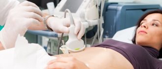
Indications for ultrasound
An ultrasound examination of the thyroid gland is prescribed to the patient by an endocrinologist based on blood tests and general health. The main indications for ultrasound are:
- Harmful working conditions. This means not only hard physical labor, but also constant work on the computer, frequent changes in climate zones;
- Stress, anxiety, insomnia;
- Frequent or constant use of hormonal drugs;
- Presence of endocrine diseases or diabetes mellitus;
- Unfavorable heredity, if close relatives were found to have pathological changes in the organ;
- Age over 40 years, since after this milestone the risk of developing neoplasms almost doubles;
- For prevention purposes. A preventive examination will not be superfluous even if there are no indications for it, since ultrasound will identify all abnormalities in the early stages, if any.
Which doctor should I contact?
In order for the treatment to be successful, it is necessary to initially contact a qualified specialist who will conduct a consultation and help with deciphering the diagnosis. I am such a highly qualified specialist in the field of endocrine health, Georgy Nikitich Romanov. I am a candidate of medical sciences, I have been practicing medicine for almost a quarter of a century, both in public and private hospitals. institutions, I have extensive experience in treating patients with thyroid nodules.
In addition to face-to-face appointments at the clinic, I conduct paid online consultations, which anyone can attend and ask questions of concern. To contact me and make an appointment, you can write to me via email on one of the social networks or messenger: Viber, Telegram, Instagram, WhatsApp, Skype, VKontakte.
Briefly about the disease
A nodule is an area that is different in density or color on ultrasound (ultrasound) from the rest of the thyroid tissue and has clear boundaries.
For a long time, the presence of nodes does not affect the functions of the gland. However, an increase in the size of the nodes leads to compression of tissues, deformation of organs appears, which can be accompanied by discomfort and unpleasant sensations.
Research shows that these formations are the most common lesion of the thyroid gland, they occur in 30-50% of the world's inhabitants, and in older people their presence in humans is almost the norm. Nodes are more common in women over 60 years of age: the likelihood of their occurrence is 6-8 times higher than in men.
There are 2 main types of nodes: benign and malignant. In our region, benign nodes are found in 95% of cases, and the remaining 5% are represented by malignant tumors. According to the morphological picture, benign nodes are colloid nodes, follicular adenomas, cysts and inflammatory processes of the thyroid gland. They can actively produce hormones or remain non-toxic for many years. The picture of the disease largely depends on the patient’s lifestyle, his state of health and other factors.
The most popular questions from patients with answers from Romanov Georgy Nikitich
Galina: I have had nodules on my thyroid gland for 7 years now. Some say to have surgery, others say to wait. I do not know what to do.
Romanov G.N: Indeed, this is a controversial issue, even among doctors. The previous generation of doctors saw a period when ALL nodes in the thyroid gland had to be removed. They were very afraid of degeneration into cancer. Times change, science does not stand still. Today, the practice of node puncture has been actively introduced. If the node is benign, then the tactic is watchful (just observation). If there is the slightest suspicion of an oncological process, prepare for surgery. In addition to oncology, today there are two more indications for the removal of nodes - a cosmetic defect and compression of surrounding tissues and organs (trachea and esophagus). In addition, your consent is also required...
Galina: Is it true that puncture provokes the growth of nodes in the thyroid gland and should not be done often?
Romanov G.N: Such data does not exist in the world. It has never been proven anywhere that puncture of nodes stimulates their growth, much less degeneration. The doctor will offer you repeated punctures only in two cases: when a new node has appeared or the old node has changed during a repeated ultrasound examination (it has grown quickly, changed its contours or its characteristics).
Elena: Hello, doctor! I have been under observation for thyroid nodules for 20 years. In 2003, I had a consultation with you. There were always 2 nodes in the right lobe, which did not tend to increase, they grew a little only with me. They punctured it 2 times - everything was fine. Hormones are normal - the golden mean, the last ultrasound showed that one node (13*11*13mm) became fuzzy, heterogeneous, with uneven edges. I donated blood for hormones - again the golden mean, the thyroid gland itself is not enlarged. I made an appointment for a puncture, wait a week. I'm very worried. I have a question: can a 20-year-old quiet nodule suddenly degenerate into a malignant one? And why do such rebirths occur??
Romanov G.N: This happens extremely rarely. If it does, it will be a follicular tumor, which very rarely behaves aggressively. I think everything is going to be alright!
Tatyana23: Age 40 years old, ultrasound of the thyroid gland size 9.7 mm on the right, almost 5 mm on the left, tests for hormones tsn-1.2 ft4-16.2 anti-tpo 15.8 puncture of the gland (they said it was good), the doctor did not give any advice: see for yourself. But he said in a month and a half to do the puncture again, is this necessary?? Please advise me when to perform a repeat puncture and whether surgery is necessary. Do I need to take any medications or vitamins???
Romanov G.N: You don’t need to take anything! Ultrasound monitoring of the thyroid gland after 6 months and that’s it!
Oksana (Odessa): Hello! I have been diagnosed with a nodule in the thyroid gland, 3 cm. There are ultrasound tests, a test for thyroid hormones, and a biopsy test.
1. Ultrasound results: the thyroid gland is located in a typical manner, the lobes are asymmetrical, with clear contours. Dimensions according to Brunn (age norm 12 cc): right lobe - 22x26x50 mm (volume - 13.7 cc); left lobe - 15x17x44 mm (volume - 5.4 cc), total volume 19.1 cc. The isthmus is 4mm thick. The structure of the glandular tissue is homogeneous, the tissue is isoechoic. Occupying the lower and middle segments of the right lobe, an oval-shaped node of moderately increased echogenicity with clear contours measuring 31x21 mm is determined, containing an oval-shaped anechoic focus measuring 6.5x4 mm. Enlarged lymph nodes are not detected.
2. Study of the content of thyroid hormones and antibodies to thyroid peroxidase in human serum using the RIA method:
Determination of free thyroxine (FT4) - 19.0 nmol/l
Determination of total triiodothyronine (TTZ) - 2.1 nmol/l
Determination of thyroid-stimulating hormone (TSH) - 3.2 mIU/l
Determination of antibodies to thyroid peroxidase - not detected.
3.FNA of the thyroid gland under ultrasound control
Diagnosis: stage 2 nodular goiter
Material: Thyroid node punctate.
Cytogram of a follicular tumor (hypercellular colloid goiter? adenoma?)
Cytodiagnosis is recommended.
Please tell me your opinion, maybe there are some signs that you don’t understand?
Romanov G.N: Remove the right lobe with the node and forget... Unfortunately, there are no other options here.
Yulia: Hello. After laser destruction of the node, on the 4th day my throat began to hurt inside at the level of the node itself. This is fine? I am very worried.
Romanov G.N: This is a burn and it should hurt. Until the wound inside heals...
Oksana: Good evening, ultrasound results: in the lower third on the right there is an isoechoic node with clear contours, heterogeneous structure, size 5x6 mm, with signs of blood flow. This is with normal hormones and normal tissue. Are there indications for puncture?
Romanov G.N: It is necessary to consider other risk factors for cancer to solve (Chernobyl, irradiation of the head and neck, relatives, etc.).
Nadezhda: Hello! Please decipher the results of the test, what does it mean - no signs of malignancy were found? I have a node of almost 2 cm, in the right lobe.
Romanov G.N: This means that everything is fine.
Marina: Hello, doctor. In 2013, I was diagnosed with a thyroid nodule. The gland itself is enlarged. The right lobe was 23x22x50, the left lobe was 18x16x42, the total volume was 19.5. And the knot was 16.1 by 16.8 by 22 mm. Being a coward (age 42 at the moment), I did not do a biopsy. And this year I had a repeat ultrasound. The size of the right lobe is length 56, width 29, thickness 31. The left lobe is length 50, width 18, thickness 16. Total volume 31.6. The node in the right lobe is 36 by 23.5 by 29, with intra and perinodular blood flow. And in the left lobe a small one appeared, 4.7 mm. TSH is now 0.5. I went to the surgeon, and he scolded me, said that I should have operated on three years ago. That I had low TSH (although I thought it was within normal limits). Now I’ll only go for a biopsy, and the surgeon said that regardless Based on its results, I have no other options other than surgery. Please tell me your opinion. Is it really as bad as the surgeon said? What normal recommendations can you give?
Romanov G.N: The growth is really significant. Your history also matters (Chernobyl, heredity?). A biopsy will dot all the i's... It should definitely be performed in your case.
Natalya: Good afternoon! My ultrasound of the thyroid gland: Total volume 7 cm cubic. The contours are smooth, the structure is homogeneous. Knot: 6mm*7mm*5mm, volume 0.13 cm cubic. the shape is round, the contours are smooth, the echogenicity is hypoechoic, the structure is heterogeneous. There are no regional lymph nodes. Please tell me whether it is worth doing a biopsy now or if we can observe.
Romanov G.N: After 6 months, ultrasound control and then a decision on TAB.
Olga Basharenkova: Good afternoon, Georgy Nikitich! I am 36 years old. I examined the thyroid gland a long time ago, the result was a total volume of 18.3, hormone tests were all normal, but in the right lobe there was a hypoechoic 3*2*2 node with mixed blood flow. They advised me to do an ultrasound every year and take hormone tests. In 2021, I did an ultrasound - in the segment of the right lobe - a hypoechoic heterogeneous node 4.5 * 2.4 * 3.5 mm with mixed blood flow, in the IV segment - a cyst 1.4 * 2.7 mm. In the s/segment of the left lobe there is an isoechoic node with a hypoechoic “halo” 6.7 * 12.2 * 8.4 mm with perinodular blood flow. TPO tests - 7.98; TSH-1.370,FT3-4.37, FT4-14.1. I'm worried about the new node, and I'm going to the doctor next week. Can you recommend something on how to reduce it? Thanks a lot.
Romanov G.N: You were born in 1980, which means you are at risk for Chernobyl. All nodes larger than 1 cm will have to be punctured.
Anna: Good day! The background is this: I did a general blood test in the fall - TSH was increased to 6, while the normal level was 4, all other hormones were normal, dizziness started a month ago, my cycle was off (or rather, it hasn’t been there for a couple of months). The endocrinologist diagnosed hypothyroidism and prescribed L-thyroxine 25 mg, an x-ray of the sella turcica and an ultrasound of the thyroid gland. I had an x-ray, the results will be out next week, the ultrasound found a nodule 8x11mm, hypoechoic, smooth fuzzy contours, perinodular blood flow, everything else is normal, he advises doing a biopsy, I doubt it, I’m thinking of visiting an endocrinologist again. I am 28 years old, my mother also has nodes (normal), the men in the family do not have such problems, there are no hereditary diseases. What do you advise? Thanks in advance)
Romanov G.N: Restore TSH with thyroxine and observe.
Svetlana: Good afternoon! Answer please. An ultrasound scan found two nodes in the left lobe. One is 15mm in size with low density, the other is 10mm calcified. They did a biopsy of which node I don’t know. Conclusion - colloid goiter. Is it necessary to do a biopsy of another node? Can we say that the second node is also good? Thank you.
Romanov G.N: They are different in structure, so it is necessary to puncture two nodes.
Olga K: Hello! About 5 years ago, primary hypothyroidism was discovered due to A&T. Eutirox dosage 50 was prescribed. Latest tests: Antibodies to TPO = 5.68, Thyroid-stimulating hormone = 5.46, thyroxine 14.3. Ultrasound: 12/17/14 thyroid lobe right lobe 7.5, left 6.0. echostructure is heterogeneous. mixed hyperechogenicity. In the right lobe there is hyperechogenicity, structure 4.4*3.8 mm. The shape is round, the contours are clear, halo. The structure is homogeneous.
Ultrasound 7.09.12: thyroid gland - right lobe 6.0, left lobe 4.2. The echostructure is heterogeneous. There is no dilatation of blood vessels. Hyperechoic heaviness - no. Echogenicity - isoechoic, uneven. There are no anomalies. The nodular formation in the right lobe is 12.7*8.1 mm. The shape is round. The contours are unclear. Echogenicity - hypoechoic. The structure is homogeneous. What are the forecasts? (they did not suggest performing a puncture; repeat ultrasound in 3 months)
Romanov G.N: Forget about ultrasound - start normalizing TSH.
Elena: Hello, Georgy! I am 57 years old. For the purpose of prevention, an ultrasound scan of the thyroid gland was performed. The right lobe is 4.6 cm cubic. Left - 6.6 cm cube, gland volume 11.2 cm cube. In the middle third of the left lobe there is an oval hypoechoic formation with clear contours, a solid-liquid structure, measuring 2.8-2.1 cm. With CDK, pronounced peripheral and intranodal blood flow TIRADS 3 is determined, mobility during swallowing is preserved. In the lower third of the left lobe, there is an oval hypoechoic formation with clear contours of a solid-liquid structure, measuring 0.9-0.5 cm; with color circulation, moderate peripheral blood flow is TIRADS 3. The mobility of the lobes during swallowing is preserved. Lymph nodes without signs of pathological changes. Hormones are all within normal limits. Is it worth doing node puncture? Or can I be observed?
Romanov G.N: Very similar to a simple cystic nodular goiter. You can still watch.
Interpretation of ultrasound results
The diagnostician describes the results obtained in the study protocol. Typically, the time to prepare a written report does not exceed 15 minutes.
Determination of exact geometric dimensions
The location of the gland will normally be typical or low, the shape will be classic, the contour will have a clear outline. It consists of two lobes with an isthmus that connects them. Sometimes a pyramidal lobe may be present.
Minor (less than one centimeter) tissue growths may be visible. If during the period of intrauterine growth the gland develops with pathology, the tissue may not be divided into two sides, but may move to one side. This is called aplasia of one lobe.
In the case of a completely undeveloped gland, they speak of complete aplasia. The length of the lobe should be from 4 to 6 cm, width from 1.3 to 1.8 cm, thickness from 1.5 to 1.8 cm; the shares are normally the same; the lintel has a thickness of 4 to 8 cm.
The volume of a healthy gland depends on the patient’s weight and is:
- weight 50 kg, gland volume 15.5 cm³
- weight 50-60 kg, volume 18.7 cm³
- weight 60-70 kg, volume 22 cm³
- weight 70-80 kg, volume 25 cm³
- weight 80-90 kg, volume 28.4 cm³
- 100 kg or more, volume 32 cm³
Non-compliance with standard sizes will indicate possible pathologies.
Definition of structure
The echogenicity of a healthy thyroid gland, without any peculiarities, the structure of the jelly tissue is fine-mesh, homogeneous, echogenic granularity is 1 mm or less. Fibrous and connective tissues are not detected. In inflammatory processes, the structure is heterogeneous.
Focal education. There should be no new growths. If the formation is present, it must be classified. If the size is up to 10 mm, then it is a focal formation, more than 10 mm, then it is a node.
Blood flow analysis. The parameters of blood flow, its nature and density, and parameters of the lymph nodes are determined. Normal nodes have clear boundaries, the width is approximately 2 times less than the length.
Contours. Clear contours are the norm. Fuzzy outlines indicate an inflammatory process or tumor.
Focal formations. Assessed for the presence of nodes, cysts, and calcinitis.
Echogenicity is the grayscale image of the tissue being examined and its tone on a monitor.
Parameters of visible lymph nodes (if present), their structure, size, structure. By their presence, the onset of tumor formation can be diagnosed.
The structure of the salivary gland and the level of response to ultrasound.
The size and structure of the soft tissues of the neck and larynx. Those areas that are located near the thyroid gland are examined.
Women over 35 years of age are recommended to have their thyroid gland examined at least once a year. This category of people is at risk for thyroid diseases.
The human neck is quite complex; it contains many nerve trunks and large vessels, the esophagus, trachea, many lymph nodes, and other glands. Therefore, the specialist performing the ultrasound procedure must be highly qualified.
The doctor conducting the research records the data obtained in a special protocol, which describes the geometric parameters of the lobes and isthmus, calculates the volume of the gland, and writes a conclusion.
The document evaluates the position and contours, tissue structures, analyzes the parathyroid glands and lymph nodes, and the images taken are also attached to the protocol. In case of normal indicators, a corresponding entry is made in the document. This takes no more than 10 minutes.