Pirogova Irina Yurievna
Deputy Chief Physician for Organizational and Methodological Work, Head of the Center for Gastroenterology and Hepatology, Gastroenterologist
Now, in the era of interferon-free therapy for hepatitis C, the problem of treating this disease is considered solved. Modern life, full of comfort, has led to the fact that more and more people are faced with such a disease as fatty liver hepatosis. With fatty hepatosis, the liver tissue becomes overgrown with excess fat, which leads to disruption of the functioning of this important organ, which acts as a filter in the human body. This disease often precedes diabetes mellitus, atherosclerosis, hypertension, gout, liver cirrhosis, and can cause pancreatitis. Fatty hepatosis is the main component of the metabolic syndrome, that is, a complex of disorders that occur with obesity.
Experts consider fatty liver disease a new “gentle killer.” Why?
Firstly, because fatty hepatosis is a condition prone to progression and the formation of liver cirrhosis and its complications.
Secondly, fatty hepatosis is the tip of the iceberg of existing metabolic metabolic disorders, which is a springboard for the development of diabetes mellitus, arterial hypertension, sleep apnea syndrome and other life-threatening conditions.
Fatty hepatosis (steatosis) of the liver is a rather insidious disease, which is very difficult to identify. The disease is practically asymptomatic; only by external signs can one suspect something is wrong. Laboratory tests, as a rule, do not give any result at the early stage of hepatosis. Since the symptoms of this disease do not cause much discomfort to a person, fatty hepatosis goes unnoticed for a long time.
General information
Speaking about diffuse changes in the liver , you should first of all understand what this concept means. This condition is not a disease, but a symptom of diseases that manifest themselves as a result of decreased immunity , poor nutrition, alcohol abuse, taking toxic medications, etc. Due to negative effects, connections between molecules are disrupted.
The functions of hepatocytes also change, as a result of which the fatty and connective layer increases. In fact, this is one of the manifestations of liver pathologies, and the correct diagnosis can only be established after a comprehensive examination and consultation with a hepatologist.
This article will discuss how to diagnose this pathology and what needs to be done to improve the condition of the liver.
Causes of diffuse changes in the organ
Diffuse changes in the liver appear due to the following diseases and conditions:
- Diabetes and obesity. During ultrasound, you can see an increase in organ density and echogenicity.
- Hepatitis in chronic form.
- Cirrhosis. During an echographic examination, a large number of damaged areas of parenchyma are revealed, the echogenicity increases several times.
- Any neoplasms (benign or malignant) lead to changes in the structure of tissues in one of the lobes of the liver.
- Viral infections of various etiologies. After recovery, the organ is regenerated.
- Infection with parasites is one of the causes of diffuse changes.
Hepatomegaly is quite severe - a significant increase in the size of the liver. The causes of this pathology are:
- The habit of eating junk food - fast food and processed foods, mayonnaise and hot sauces. With such nutrition, the iron always works in emergency mode.
- Alcohol abuse. The gland's enzymes break down ethyl alcohol into aldehydes, which are extremely harmful to liver cells. Regular alcohol consumption destroys the liver: healthy tissue degenerates into fatty tissue. Untimely treatment of alcoholic hepatosis leads to liver cirrhosis.
- Incorrect or uncontrolled use of medications, including antibiotics.
Some patients prefer to make their own diagnoses and prescribe therapy. Such people do not think about the side effects of self-medication. Any medication must be taken knowing the dosage, taking into account drug compatibility factors (if you need to take several medications at the same time).
The liver is a large filtration station in the body. All toxic substances enter the liver. Here they accumulate and are gradually neutralized. When the amount of harmful substances constantly exceeds permissible standards, the functioning of the gland is disrupted. She can no longer fully cope with her task. Poorly purified blood enters other organs, poisoning the entire body, and various pathologies arise in the gland itself.
There are cases when an organ suffers despite a relatively healthy lifestyle. This indicates that the person is stressed most of the time or lives near businesses, large highways, etc.
Pathogenesis
Parenchyma is liver tissue that consists of hepatocytes. This tissue is involved in the process of purifying the blood, which, in turn, supplies all vital organs with necessary substances and ensures the normal process of bile .
If the liver is healthy, then the cell structure is homogeneous. Fats that enter the body with foods are broken down by enzymes , after which they enter the liver. In turn, there they are converted into cholesterol , triglycerides , etc. When the amount of triglycerides in tissues increases, a change in the liver parenchyma occurs.
Diffuse changes in the liver and pancreas are quite often observed. Since the organs of the digestive system are connected by ducts, often when one organ fails, another suffers. hepatomegaly may develop , that is, a significant enlargement of the liver .
Such changes in the parenchyma can be observed during primary and secondary metabolic disorders, acute or chronic infectious processes.
Degrees of liver fibrosis in adults
Experts distinguish 5 degrees of scarring of liver tissue.
No fibrosis.
In this case, there is no evidence of liver cell death (necrosis) or scarring, despite liver inflammation (hepatitis).
Portal (mild) fibrosis.
In this case, there are areas of necrosis and scarring affecting the small and medium branches of the portal vein, which carries blood from the small intestine. The structure and function of the liver remain normal.
Periportal (moderate) fibrosis.
This is an option with an increase in foci of necrosis, scarring and impaired liver function.
Bridging (severe) fibrosis.
At this stage, scarring has disrupted normal blood flow to the liver and function is further impaired.
Cirrhosis.
This is permanent scarring and irreversible loss of liver function.
Classification
Depending on the severity of the pathological process, the following types of changes are distinguished:
- Pronounced - as a rule, develop as a consequence of oncological processes, viral hepatitis , fatty degeneration , alcoholic illness. In this case, treatment involves surgical intervention.
- Moderate - are the result of nutritional disorders, chemical poisoning, metabolic and endocrine disorders. Moderate diffuse changes in the liver parenchyma are treated by taking medications, following a gentle diet, and blood purification.
- Minor – symptoms may not appear for a long time or may be mild. As a rule, they are observed at the very beginning of the pathological process.
The pathological process can be localized in any part of the liver:
- fabrics;
- parenchyma;
- duct wall.
Taking into account the nature of diffuse changes in the liver, the following are distinguished:
- dystrophic;
- heterogeneous;
- reactive.
The following types of parenchyma structure are also distinguished:
- According to the steatotic type , the formation of fat cells occurs chaotically, in separate areas. If cells accumulate in one place, cysts can form.
- According to the type of hepatosis , fat cells spread throughout the tissue, disrupting the normal functioning of the liver. Diffuse changes in the liver, such as hepatosis, appear both as a result of diseases and due to poor nutrition and alcohol abuse.
Taking into account the reasons for such manifestations, the following types of changes in the liver are distinguished:
- sclerotic;
- swelling;
- dystrophic;
- hypertrophic.
4.Treatment
The therapeutic strategy is developed based on the established causes and rates of dystrophy. In most cases, treatment begins with normalizing the diet and prescribing hepatoprotective agents, usually of plant origin (Essentiale, Hepabene, etc.). Early detection of a pathological process and timely treatment of it leads to good results and therapeutic success. In more complex cases, regular cleansing enemas, “intestinal” antibiotics, vitamins, glucocorticosteroids, potassium supplements, blood transfusions, hemodialysis for renal failure, and antipsychotics are necessary for mental disorders. The rapidly increasing functional failure of the liver leads to general intoxication, and the only way to save the patient’s life is often an immediate liver transplant - which, for obvious reasons, is extremely problematic, and most often impossible.
At the same time, fatty infiltration of the liver, i.e. the accumulation of lipid compounds in large volumes inside hepatocytes, sometimes with breakthrough of the latter and the formation of fatty cysts, allows the reverse development of the pathological process and can be successfully treated. The condition is strict adherence to the diet and adherence to all medical prescriptions.
Causes
The development of diffuse changes in the liver can develop due to the following diseases and conditions:
- Diabetes mellitus and obesity .
- Chronic hepatitis.
- Cirrhosis of the liver.
- The appearance of various neoplasms.
- Viral infectious diseases. Moreover, after recovery, the normal structure of the organ is restored.
- Parasitic infestation.
- Too much fat in the body.
- Autoimmune processes in the liver.
- Long-term use of certain medications, in particular antibiotics .
- Alcohol abuse.
- Unbalanced diet.
- Hereditary tendency.
- Constant stressful situations.
- Unfavorable environmental conditions.
In addition to diffuse changes, the patient may also develop hepatomegaly - enlargement of the liver.
Symptoms of liver fibrosis in adults
Scarring from liver fibrosis can affect the liver's ability to function effectively.
However, fibrosis itself does not cause any symptoms. You may have it and not know it. If fibrosis progresses to cirrhosis, the main symptoms may include:
- constant bruises on the body and bleeding;
- fatigue, confusion and weakness;
- itching;
- poor appetite, nausea and weight loss;
- swelling of the abdomen (ascites) and legs (pasty);
- yellowing of the skin and eyes (jaundice).
Liver cirrhosis is a serious disease that can lead to life-threatening complications. Seek medical help immediately if you experience the above symptoms.
Symptoms
The main signs of diffuse changes in the liver are as follows:
- Excessive sweating and increased fatigue. If sweating is associated specifically with damage to the liver and pancreas, the sweat has an unpleasant, pungent odor. The person complains of drowsiness, weakness, and extreme fatigue even with light exertion.
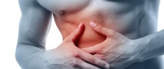
- Aching pain and discomfort in the right hypochondrium. If the pain is very severe, this may indicate inflammatory processes or malignant formations.
- Disruption of the digestion process. Signs of parenchymal damage are frequent nausea and heartburn , loose and frequent stools, changes in the color of stool and urine.
- Yellow coating and cracks on the tongue.
- Yellowness of the eyes and skin.
- Increased body temperature. As a rule, the temperature rises insignificantly and lasts for several weeks.
- Deterioration of the condition of the skin. A rash, papillomas, itching, and peeling may appear.
- Hematomas, bleeding.
- Headaches, irritability, frequent mood swings.
Symptoms of fatty hepatosis:
- Overweight - body mass index over 30
- Nausea in the morning.
- Skin itching.
- Digestive tract dysfunction – bloating, unstable stool
- Fatigue, irritability.
- Pain or heaviness in the right hypochondrium.
Alcohol abuse, fatty, smoked, spicy foods, smoking, weight gain are the most common causes of fatty liver. It is lifestyle, and not heredity, that plays the leading role in the formation of fatty hepatosis.
Tests and diagnostics
In order for the course of treatment to be as effective as possible, it is necessary to carry out all the prescribed studies that will help confirm the diagnosis and determine the characteristics of the course of the pathological process.
First of all, patients undergo an ultrasound of the liver. It is advisable to conduct such a study periodically and as a preventive measure for healthy people. Echography allows you to determine liver pathologies and their features.
Also during the diagnostic process the following studies can be carried out:
- Radionuclide scanning - carried out if liver damage is suspected after injury, to determine metastases .
- Computed tomography - allows you to determine the presence of tumors, foci of parenchymal bleeding, etc.
- biopsy - using this procedure, tissue is taken for histological examination.
- Biochemical blood test with liver tests.
- Test for antibodies to the hepatitis virus.
Non-alcoholic fatty liver disease (steatohepatosis)
Non-alcoholic fatty liver disease (NAFLD) is a disease or spectrum of diseases resulting from excess accumulation of fats in the liver.
NAFLD includes steatosis, non-alcoholic steatohepatitis (NASH), liver fibrosis and cirrhosis. An important criterion that distinguishes NAFLD from alcoholic liver disease is the absence of alcohol consumption by patients in hepatotoxic doses, i.e. more than 40 grams of pure ethanol per day for men and more than 20 grams for women. Most cases of NAFLD are associated with metabolic syndrome.
From the point of view of endocrinology, the main cause of fatty hepatosis is the insusceptibility of liver cells to the action of the hormone insulin (insulin resistance). One of the functions of insulin is to transport glucose into cells from the blood and tissue fluid. With insulin resistance, liver cells do not receive the glucose they need and die, being replaced by unpretentious, but useless, fat cells. The exception is cases of secondary, or “specific” NAFLD, the development of which is associated with exposure to specific toxins, medications, or complicated diseases of other organs and systems. Insulin resistance can be genetically programmed, appear as a result of metabolic disorders, be caused by erroneous immune aggression to insulin and other factors.
Associated factors influencing the replacement of liver tissue with fat: intoxication (alcohol, occupational, etc.), increased fat content in food, sedentary lifestyle.
Obesity is the most significant factor associated with steatohepatosis. The combination of obesity and non-alcoholic fatty liver disease is of scientific interest in understanding the pathogenesis of metabolic syndrome, since impaired liver function aggravates insulin resistance, closing a “vicious circle”. Both obesity in general and the topographical features of adipose tissue deposition in the body play a key role in the pathogenesis of non-alcoholic fatty liver disease, not only due to excessive formation of adipose tissue, but also the secretion of adipocytokines and inflammatory mediators (leptin, adiponectin, TNF-α, etc. ), which may contribute to the development of insulin resistance.
As recent scientific studies have shown, steatohepatosis is positioned as an independent risk factor for cardiovascular diseases. Undoubtedly, the liver plays a critical role in the development of atherogenic dyslipidemia, while at the same time being a target organ.
The role of intestinal microflora has also been proven, which indirectly, due to the entry of lipopolysaccharides of Gram-negative bacteria into the portal bloodstream, activates the immune response through receptors and leads to the development of inflammation and activation of fibrous matrix production.
As recent scientific studies have shown, steatohepatosis is positioned as an independent risk factor for cardiovascular diseases. Undoubtedly, the liver plays a critical role in the development of atherogenic dyslipidemia, while at the same time being a target organ.
Non-alcoholic fatty liver disease develops in four stages:
1. Fatty liver, in which there is an accumulation of a large number of lipid droplets in hepatocytes and the intercellular space. It is worth saying that in many patients this phenomenon does not lead to serious damage to the liver, but in the presence of negative factors, the disease can move to the next stage of development.
2. Non-alcoholic steatohepatitis, in which the accumulation of fat is accompanied by the appearance of an inflammatory process.
3. Fibrosis is the result of a long-term inflammatory process. Functional liver cells are gradually replaced by connective tissue elements. Scars form, affecting the functioning of the organ.
4. Cirrhosis is the final stage of development of fibrosis, in which most of the normal liver tissue is replaced by scars. The structure and function of the organ are disrupted, which often leads to liver failure.
Diagnostics:
An objective examination usually reveals signs of obesity. With steatosis and NASH, a moderate enlargement of the liver is detected, its edge is rounded, and the consistency is doughy. With severe fibrosis, the liver becomes dense; at the stage of cirrhosis, “liver signs”, splenomegaly, and ascites can be detected.
· A biochemical blood test may reveal an increase in the activity of serum transaminases (ALT and AST), GGT, alkaline phosphatase and bilirubin levels.
GGT activity is increased in most patients, usually no more than twice; in some cases this may be the only deviation in the biochemical analysis. An increase in alkaline phosphatase activity is observed in a third of patients and is also usually not
exceeds the norm by more than twice. In approximately 20% of cases, a moderate increase in the content of total bilirubin due to the direct fraction (1.5-2 times) is detected. In patients with liver cirrhosis, along with the changes described above, when the synthetic function of the liver is impaired, a decrease in albumin levels and an increase in prothrombin time, an increase in the level of total bilirubin and INR are detected.
Lipid profile. Diagnostically significant deviations characteristic of NAFLD within MS are an increase in triglycerides (≥ 1.7 mmol/l) and a decrease in HDL-C levels (<0.9 mmol/l in men and <1.0 mmol/l in women ).
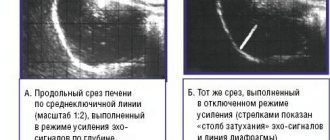
· Ultrasound examination (ultrasound) of the liver. Ultrasound signs of NAFLD can be considered: • diffuse hyperechogenicity of the liver parenchyma and heterogeneity of its structure • blurred and/or emphasized vascular pattern • distal attenuation of the echo signal • higher echogenicity of the liver in comparison with the renal cortex. Ultrasound has advantages in diagnosing NAFLD at the stage of liver cirrhosis, especially in patients without clinical symptoms of liver damage. In the diagnosis of NAFLD, it is possible to use computed tomography and magnetic resonance imaging (CT and MRI)
· Liver biopsy is the modern “gold standard” for diagnosing steatosis, inflammation and fibrosis stage in NAFLD. This method makes it possible to confirm with a high degree of certainty the presence of NAFLD, make a differential diagnosis between steatosis and NASH, assess the stage of fibrosis and, based on histological data, predict
further course of the disease, as well as exclude other causes of liver damage.
· Non-invasive methods for diagnosing NAFLD: The FibroMax test includes 5 non-correlated biochemical parameters: α-2-macroglobulin, haptoglobin, apolipo-protein A1, γ-glutamyl transpeptidase (GGT) and total bilirubin, which allow assessing the severity of fibrosis. With its help, you can differentiate fibrosis (Fl-F3) from liver cirrhosis (F4) FibroMeter test: includes 5 indicators of biochemical and clinical blood tests - alpha-2-macroglobulin, GGT, urea, prothrombin index (%), platelets, which allow you to assess the severity fibrosis.
FibroMeter allows you to differentiate moderate fibrosis (Fl-F2) from severe fibrosis (F3) and from liver cirrhosis (F4)
Treatment:
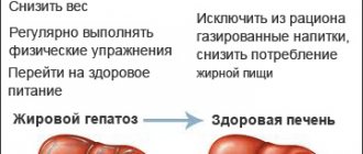
Currently, no strict algorithms for the management of patients with NAFLD have been developed. It has been shown that gradual, moderate weight loss accompanied by a hypocaloric diet, reduced fat intake and increased physical activity are the most important components of the treatment of those who are overweight or obese. There are effective and efficient drug treatments for NAFLD.
Timely consultation with specialists at our clinic and timely treatment prevents the development of cirrhosis and liver fibrosis.
Treatment with folk remedies
For diffuse changes in the liver parenchyma in mild to moderate form, some traditional methods that help improve the condition may be an additional treatment method. But before using any of these remedies, you should consult your doctor and get his approval.
- Herbal decoction . To prepare it, you need to take milk thistle and strawberry leaves, dandelion roots, rose hips and corn silk in equal proportions, grind everything in a blender. 2 tbsp. l. Pour 500 ml of boiling water over the mixture and leave for several hours. Strain and add boiled water so that you end up with 500 ml of decoction. Take 100 ml before meals three times a day.
- Oat jelly . Pour crushed oat grains into a three-liter jar so that the grain occupies a third of the jar, and completely fill it with water. For sourdough, you can use a crust of rye bread or half a glass of kefir. The starter must ferment for 4 days. Next, it is filtered and infused for another 12 hours. After this, the liquid is drained and the grounds are kept in the refrigerator. To prepare jelly you need to take 8 tbsp. l. grounds, pour 2 glasses of water over them and cook over low heat for 5 minutes. Use in the morning on an empty stomach.
- Infusion of sage and calendula . Mix 3 tsp. calendula and 2 tsp. sage, pour 250 ml of boiling water. After 30 minutes, strain and drink in two doses a day.
- Herbal infusions . They can be prepared from rose hips, milk thistle, string, chamomile, and lemon balm. To prepare the infusion, 2 tsp. of the selected product or mixture thereof, add 250 ml of water.
- Pumpkin , flaxseed , olive oil . The selected oil should be drunk on an empty stomach, 1 tbsp. l. for 1-2 months.
New treatments
Radiofrequency ablation
Modern surgery allows this method to be used for metastatic liver disease. A source (electrode) of high-frequency radiation is brought through the skin and muscles to the tumor node. The accuracy of the electrode location is controlled by electroanatomical mapping. Radiation directly affects the metastasis and destroys tumor cells. At the same time, the impact on surrounding tissues and the risk of bleeding are minimal. The procedure can also be performed under local anesthesia.
Chemoembolization of tumor
Used when ablation is contraindicated. Having provided access to the vessels feeding the tumor, a chemotherapy drug is injected into them through a needle. This substance “solders” the walls of the arteries, blood stops flowing through the vessels into the metastasis, and without nutrition the tumor node dies. Aseptic necrosis and death occurs.
Targeted therapy with biologics
Specific drugs obtained as a result of high-tech synthesis (Sorafenib, in particular) selectively act specifically on cancer cells. Substances toxic to tumor cells accumulate in them and prevent further growth of cancerous foci. The method is used in complex therapy, as an addition to other treatment methods, in the presence of contraindications to radical treatment.
Diet
Diet 5th table
- Efficacy: therapeutic effect after 14 days
- Duration: from 3 months or more
- Cost of products: 1200 - 1350 rubles per week
During the treatment and rehabilitation process, it is extremely important to adhere to the principles of dietary nutrition. After all, a proper diet helps restore the normal state of liver tissue and improve well-being. If the pathological changes are weakly degenerated, then dietary nutrition will help eliminate the problem without additional medication.
If the patient is undergoing treatment for such a pathology, the doctor recommends following the diet Table No. 5 .
It is recommended to include the following products and dishes in the menu:
- Day-old bread, crackers.
- Lean meat.
- Lenten soups, borscht.
- Puree of fruits and vegetables, boiled vegetables.
- Low-fat fermented baked milk, kefir, cottage cheese.
- Fresh fruit and vegetable juices.
- One boiled egg a day.
- Weak tea, rosehip decoction, dried fruit compote.
You should eat in small portions, 5-6 times a day. All dishes need to be steamed, boiled, stewed, baked.
The following should be completely excluded from the diet:
- Lard, fatty meat, fatty broths.
- Fatty fish.
- Legumes.
- Smoked meats.
- Mushrooms.
- Canned food.
- Coffee.
- Soda, alcohol.
- Sour berries and fruits.
- Confectionery.
Prognosis and prevention
If the cause of pathological changes in the liver is an unhealthy lifestyle or alcohol abuse, then if treatment is started in a timely manner, recovery occurs in almost all patients. If you follow the doctor's recommendations, liver function is restored almost completely.
Liver cirrhosis is considered life-threatening for patients. With this disease, the prognosis for recovery is only 50%. The remaining patients die within 5 years. The reason for this outcome is irreversible pathological processes in the gland. To prevent this, you need to consult a doctor in a timely manner, lead a healthy lifestyle, undergo regular medical examinations, and, if necessary, get vaccinated against hepatitis.
List of sources
- Diseases of the liver and biliary tract: A guide for doctors / Ed. V.T. Ivashkina. M.: M-Vesti, 2002. 416 p.
- Ivashkin V.T., Yushchuk N.D. Diagnosis and treatment of diffuse liver diseases. Method, manual for doctors. M.: 2003. - 49 p.
- Mizandari M., Mtvardze A., Urushadze O. et al. Complex radiodiagnosis of diffuse liver pathology // Med. visualization. - 2002. - No. 1. - P. 60-68.
- Panfilov S.A., Panfilova E.V. Diagnosis of liver and biliary tract diseases with a course of pathological anatomy. - M.: Binom. Knowledge Laboratory, 2003. 211 p.
- Stepanyan I.A., Kobinets Yu.V., Izranov V.A., Ovchinnikov O.I. Diffuse liver changes: assessment of diagnostic efficiency using standardized arfi-elastometry. Radiation diagnostics and therapy. 2018;(1):30-35.
Treatment
Treatment of steatosis consists of eliminating the causes of the disease, followed by normalization of metabolic processes. In this case, the doctor in any case assesses the nutritional status and prescribes a diet. The basis of therapeutic nutrition is limiting the consumption of animal fats and fast carbohydrates. In parallel with this, patients are recommended to perform feasible physical activity, due to which the utilization of fatty acids is accelerated.
Drug treatment often plays a supporting role. Patients are prescribed:
- hepatoprotectors that improve liver function;
- ursodeoxycholic acid preparations to prevent the formation of stones.
These drugs include Phosphogliv*. It can eliminate the causes of damage and promote the restoration of liver cells.