Non-alcoholic liver steatosis, diagnosis, treatment approaches
Non-alcoholic liver steatosis (non-alcoholic fatty liver disease (NAFLD), fatty liver, fatty liver, fatty infiltration) is a primary liver disease or syndrome formed by excessive accumulation of fats (mainly triglycerides) in the liver. If we consider this nosology from a quantitative point of view, then “fat” should be at least 5–10% of the liver weight, or more than 5% of hepatocytes should contain lipids (histologically) [1].
If you do not intervene during the course of the disease, then in 12–14% NAFLD transforms into steatohepatitis, in 5–10% of cases into fibrosis, in 0–5% fibrosis transforms into cirrhosis of the liver; in 13% of cases, steatohepatitis immediately transforms into liver cirrhosis [2].
These data make it possible to understand why this problem is of general interest today; if the etiology and pathogenesis are clear, then it will be clear how to most effectively treat this common pathology. It is already clear that in some patients this may be a disease, and in others it may be a symptom or syndrome.
Recognized risk factors for developing NAFLD are:
- obesity;
- diabetes mellitus type 2;
- fasting (sharp weight loss > 1.5 kg/week);
- parenteral nutrition;
- presence of ileocecal anastomosis;
- bacterial overgrowth in the intestines;
- many drugs (corticosteroids, antiarrhythmic drugs, antitumor drugs, non-steroidal anti-inflammatory drugs, synthetic estrogens, some antibiotics and many others) [3–5].
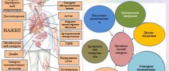
The listed risk factors for NAFLD show that a significant part of them are components of metabolic syndrome (MS), which is a complex of interrelated factors (hyperinsulinemia with insulin resistance - type 2 diabetes mellitus (type 2 diabetes), visceral obesity, atherogenic dyslipidemia, arterial hypertension, microalbuminuria, hypercoagulability, hyperuricemia, gout, NAFLD). MetS forms the basis of the pathogenesis of many cardiovascular diseases and indicates their close connection with NAFLD. Thus, the range of diseases that form NAFLD is significantly expanding and includes not only steatohepatitis, fibrosis, cirrhosis of the liver, but also arterial hypertension, coronary heart disease, myocardial infarction and heart failure. At least, if the direct connections of these conditions require further study of the evidence base, their mutual influence is undoubtedly [6].
Epidemiologically, there are primary (metabolic) and secondary NAFLD. The primary form includes most conditions that develop with various metabolic disorders (they are listed above). The secondary form of NAFLD includes conditions that are formed by: nutritional disorders (overeating, starvation, parenteral nutrition, trophological deficiency - kwashiorkor); medicinal effects and relationships that are realized at the level of hepatic metabolism; hepatotropic poisons; intestinal bacterial overgrowth syndrome; diseases of the small intestine accompanied by indigestion syndrome; resection of the small intestine, small bowel fistula, functional pancreatic insufficiency; liver diseases, including genetically determined ones, acute fatty disease of pregnant women, etc. [7–9].
If the doctor (researcher) has morphological material (liver biopsy), then three degrees of steatosis are morphologically distinguished:
- 1st degree - fatty infiltration <33% of hepatocytes in the field of view;
- 2nd degree - fatty infiltration of 33–66% of hepatocytes in the field of view;
- Grade 3—fatty infiltration of >66% of hepatocytes in the field of view.
Having given the morphological classification, we must state that these data are conditional, since the process is never uniformly diffuse, and at each specific moment we are considering a limited fragment of tissue, and we are confident that in another biopsy we will get the same Most likely, no, and, finally, the 3rd degree of fatty liver infiltration should be accompanied by functional liver failure (at least in some components: synthetic function, detoxification function, biliary capacity, etc.), which is practically not characteristic of NAFLD.
The above material highlights factors and metabolic states that may be involved in the development of NAFLD, and proposes the “two-hit” theory as a modern model of pathogenesis:
the first is the development of fatty degeneration; the second is steatohepatitis.
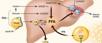
With obesity, especially visceral obesity, the supply of free fatty acids (FFA) to the liver increases, and liver steatosis develops (the first blow). Under conditions of insulin resistance, lipolysis in adipose tissue increases, and excess FFA enters the liver. As a result, the amount of fatty acids in the hepatocyte increases sharply, and fatty degeneration of hepatocytes is formed. Oxidative stress develops simultaneously or sequentially—a “second blow” with the formation of an inflammatory response and the development of steatohepatitis. This is largely due to the fact that the functional capacity of mitochondria is depleted, microsomal lipid oxidation in the cytochrome system is turned on, which leads to the formation of reactive oxygen species and an increase in the production of pro-inflammatory cytokinins with the formation of inflammation in the liver, death of hepatocytes caused by the cytotoxic effects of TNF-alpha1 - one of the main inducers of apoptosis [10, 11]. Subsequent stages of the development of liver pathology and their intensity (fibrosis, cirrhosis) depend on the remaining factors in the formation of steatosis and the lack of effective pharmacotherapy.
Diagnosis of NAFLD and its progression conditions (liver steatosis, steatohepatitis, fibrosis, cirrhosis)
Fatty liver degeneration is a formally morphological concept, and it would seem that diagnosis should be reduced to a liver biopsy. However, such a decision has not been made by international gastroenterological associations and the issue is being discussed. This is due to the fact that fatty degeneration is a dynamic concept (it can be activated or undergo reverse development, and can be both relatively diffuse and focal in nature). The biopsy is always represented by a limited area, and the interpretation of the data is always quite conditional. If we recognize biopsy as a mandatory diagnostic criterion, then it should be performed quite often; the biopsy itself is fraught with complications, and the research method should not be more dangerous than the disease itself. The absence of a decision on a biopsy is not a negative factor, especially since today liver steatosis is a clinical and morphological concept with the presence of many factors involved in pathogenesis.
From the data presented above, it is clear that diagnosis can begin at different stages of the disease: steatosis → steatohepatitis → fibrosis → cirrhosis, and the diagnostic algorithm should include methods that determine not only fatty degeneration, but also its stage.
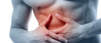
Thus, at the stage of liver steatosis, the main symptom is hepatomegaly (discovered by chance or during a clinical examination). The biochemical profile (aspartate aminotransferase (AST), alanine aminotransferase (ALT), alkaline phosphatase (ALP), gammaglutamyl transpeptidase (GGT), cholesterol, bilirubin) determines the presence or absence of steatohepatitis. If the level of transaminases increases, it is necessary to conduct virological studies (which will either confirm or reject viral forms of hepatitis), as well as diagnosis of other forms of hepatitis: autoimmune, biliary, primary sclerosing cholangitis. Ultrasound examination not only reveals an increase in the size of the liver and spleen, but also signs of portal hypertension (by the diameter of the splenic vein and the size of the spleen). Less commonly used (and perhaps even known) is the assessment of fatty infiltration of the liver, which consists of measuring the “attenuation column”, the dynamics of which at different periods of time can be used to judge the degree of fatty degeneration (Fig.) (ultrasound technique is described) [12].
Earlier models of ultrasound devices assessed densitometric indicators (the dynamics of which could be used to judge the dynamics and degree of steatosis). Currently, densitometric indicators are obtained using computed tomography of the liver. When considering the pathogenesis of NAFLD, a general examination and anthropometric indicators are assessed (determining body weight and waist circumference - WC). Since MS occupies a significant place in the formation of steatosis, in diagnosis it is necessary to evaluate: abdominal obesity - WC > 102 cm in men, > 88 cm in women; triglycerides > 150 mg/dl; high-density lipoprotein (HDL): <40 mg/dL in men and <50 ml/dL in women; blood pressure (BP) > 130/85 mm Hg. st; body mass index (BMI) > 25 kg/m2; fasting blood glucose > 110 mg/dL; glycemia 2 hours after a glucose load 110–126 mg/dL; Type 2 diabetes, insulin resistance.
The data presented above is recommended by WHO and the American Association of Clinical Endocrinologists. An important diagnostic aspect is also the establishment of fibrosis and its degree. Despite the fact that fibrosis is also a morphological concept, it is determined by various calculated indicators. From our point of view, the Bonacini discriminant scoring scale, which determines the fibrosis index (IF), is a convenient method corresponding to the stages of fibrosis. We conducted a comparative study of the calculated IF index with the results of biopsies. These indicators are presented in table. 1 and 2.
Practical meaning of IF:
1) IF, assessed using a discriminant scoring scale, significantly correlates with the stage of liver fibrosis according to puncture biopsy; 2) studying IF makes it possible to assess the stage of fibrosis with a high degree of probability and use it for dynamic monitoring of the intensity of fibrosis formation in patients with chronic hepatitis, NAFLD and other diffuse hepatic diseases, including for assessing the effectiveness of therapy [13].
And finally, if a puncture biopsy of the liver is performed, it is prescribed, as a rule, in the case of differential diagnosis of tumor formations, including the focal form of steatosis. At the same time, in the liver tissue of these patients the following is detected:
- fatty liver degeneration (large-droplet, small-droplet, mixed);
- centrilobular (less often portal and periportal) inflammatory infiltration with neutrophils, lymphocytes, histiocytes;
- fibrosis (perihepatocellular, perisinusoidal and perivenular) of varying severity.
The diagnosis of NAFLD (liver steatosis) is formulated based on the combination of the following symptoms and conditions:
- obesity;
- MS;
- malabsorption syndrome (as a consequence of ileojejunal anastomosis, biliary-pancreatic stoma, extended resection of the small intestine);
- long-term (more than two weeks parenteral nutrition).
Diagnosis also involves excluding the main hepatic nosological forms:
- alcoholic liver damage;
- viral infection (B, C, D, TTV);
- Wilson–Konovalov disease (blood ciruloplasmin levels are examined);
- congenital alpha1-antitrypsin deficiency disease);
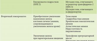
- hemachromatosis;
- autoimmune hepatitis;
- drug-induced hepatitis (drug history and withdrawal of a possible drug that forms intermediate-density lipoproteins (IDL)).
Thus, the diagnosis is formed from the definition of hepatomegaly, the identification of pathogenetic factors contributing to steatosis, and the exclusion of other diffuse forms of liver damage.
Treatment principles
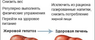
Since the main factor in the development of non-alcoholic liver steatosis is excess body weight (BW), reducing BW is a fundamental condition for the treatment of patients with NAFLD, which is achieved by lifestyle changes, including dietary measures and physical activity, including in cases where the need is absent in reducing BM [14]. The diet should be hypocaloric - 25 mg/kg per day with a limitation of animal fats (30-90 g/day) and a reduction in carbohydrates (especially those that are quickly digestible) - 150 mg/day. Fats should be predominantly polyunsaturated, which are found in fish and nuts; It is important to consume at least 15 grams of fiber from fruits and vegetables, as well as foods rich in vitamin A.
In addition to the diet, you need at least 30 minutes of daily aerobic physical activity (swimming, walking, gym). Physical activity itself reduces insulin resistance and improves quality of life [15].
The second important component of therapy is the impact on metabolic syndrome and insulin resistance in particular. Of the drugs aimed at its correction, metformin has been the most studied [16, 17]. It has been shown that treatment with metformin leads to an improvement in laboratory and morphological indicators of inflammatory activity in the liver. Insulin sensitizers are used in type 2 diabetes, but a meta-analysis did not show any benefit from their effect on insulin resistance [18].
The third component of therapy is to avoid the use of hepatotoxic drugs and drugs that cause liver damage (the main morphological substrate of this damage is liver steatosis and steatohepatitis). In this regard, it is important to take a drug history and avoid drug(s) that damage the liver.
Since bacterial overgrowth syndrome (SIBO) plays an important role in the formation of liver steatosis, it must be diagnosed and corrected (drugs with antibacterial action - preferably non-absorbable; probiotics; motility regulators, liver protectors), and the choice of therapy depends on the initial pathology , which forms SIBO.
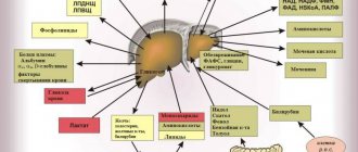
Today, the issue of using liver protectors is not resolved entirely correctly. There are works that show their low efficiency, there are works that show their high efficiency. It appears that their use does not take into account the stage of NAFLD. If there are signs of steatohepatitis, fibrosis, or cirrhosis of the liver, then their use seems justified. I would like to present analytical data on the basis of which and depending on the number of factors involved in the pathogenesis of NAFLD, a hepatoprotector can be selected (Table 3).
From the presented table it is clear (the most commonly used protectors are introduced, if desired, it can be expanded by introducing other protectors) that ursodeoxycholic acid preparations (Ursosan) act on the maximum number of pathogenetic links of liver damage.
We would like to present the results of Ursosan treatment of patients with NAFLD. 30 patients were studied (15 of them had obesity as the basis, 15 had MS; there were 20 women, 10 men; age from 30 to 65 years (average age 45 ± 6.0 years).
The selection criteria were: increase in AST level - 2–4 times; ALT - 2–3 times; BMI > 31.1 kg/m2 in men and BMI > 32.3 kg/m2 in women. Patients received Ursosan at a dose of 13–15 mg/kg body weight per day; 15 patients for 2 months, 15 patients continued taking the drug for up to 6 months. The results of treatment are presented in table. 4–6.
The exclusion criteria were: viral nature of the disease; concomitant pathology in the stage of decompensation; taking drugs that can potentially form (maintain) fatty liver degeneration.
Group 2 continued to receive Ursosan at the same dose for 6 months (with normal biochemical parameters). At the same time, appetite stabilized, and body weight gradually (1 kg/month) decreased. According to ultrasound data, the structure and size of the liver did not change significantly, the dynamics along the “attenuation column” continued (Table 6).
Thus, according to our data, the use of liver protectors in patients with NAFLD in the stage of steatohepatitis is effective, which is reflected in the normalization of biochemical parameters and a decrease in fatty infiltration of the liver (according to ultrasound, a decrease in the “attenuation column” of the signal), which in general is an important justification for their use.
Literature
- Morrison YA et al. Metformin for weight loss in pediatric patients taking psychotropic drugs // Am. Y Psychiatry. 2002. vol. 159, p. 655–657.
- Quote by: Shchekina M.I. Non-alcoholic fatty liver disease // Cous. Med. T. 11, No. 8, p. 37–39.
- Isakov V. A. Statins and the liver: friends or enemies // Clinical gastroenterology and hepatology. Russian edition. T. 1, No. 5, 372–374.
- Diche AM NaSH: bench to bedside — lesons from aminal models. Presentation at the Session Falk Symposium 157, 2006.
- Lindor KD Jn on behalf of the UDCA/NASH Study group. Ursodeoxycholic acid for treatment of nonalcocholic steatohepatitis: results of a randomized, placebo-controlled // Trial gastroenterology. 2003, 124 (Suppl): A-708.
- Drapkina O. M., Korneeva O. N. Non-alcoholic fatty liver disease and cardiovascular risk: the influence of female gender // Pharmateka. 2010, no. 15, p. 28–33.
- Shchekina M.I. Non-alcoholic fatty liver disease // Cons. Med. T. 11, No. 8, 37–39.
- Bueverov A. O., Bogomolov P. O. Non-alcoholic fatty liver disease: rationale for pathogenetic therapy // Clinical perspectives of gastroenterology and hepatology. 2000, no. 1, 3–8.
- Savelyev V.S. Lipid distress - a syndrome in surgery. Materials of the 8th open session of the Russian Academy of Medical Sciences. M. S. 56–57.
- Carieiro de Mura M. Non-alcoholic steatohepatitis // Clinical perspectives of gastroenterology, hepatology. 2001, No. 3, p. 12–15.
- Augulo P. Non-alcoholic fatty liver disease // New Engl. Y Med. 2002, vol. 346, p. 1221–1231.
- Sokolov L.K., Minushkin O.N. et al. Clinical and instrumental diagnosis of diseases of the organs of the hepatopancreato-duodenal zone. M., 1987, p. 30–39.
- Minushkin O. N. et al. Possibilities of clinical and laboratory assessment of liver fibrosis. In the book: Selected issues of clinical medicine. T. III. M., 2005, p. 96–102.
- Berrram SR, Venter Y, Stewart RY Weight loss in olese women exercise v. dietary education // S. Afr. Med. Y. 1990, 78, 15–18.
- Hickman Yg et al. Modest weight loss and physical activity in overweight patient with chronic liver disease result in suctaned improvements in alanine aminotransferase, fasting insulin, and quality of life // Gut. 2004, 53, 413–419.
- Bugianesi E. et al. A randomized controlled trial of merformin versus vitamin E or prescriptive diet in nonalcoholic fatty liver disease // Am. G. gastroenterol. 2005, vol. 100, No. 5 b, t. 1082–1090.
- Uygun A. et al. Metformin in the treatment of patients with non-alcoholic steatogepatitis // Phormacol Ther. 2004, vol. 19, no. 5, p. 537–544.
- Augelico F. et al. Drugs improving insulin resistance for non-alcogolic fatty liver disease and/or non-alcogolic steatogepatitis // Cochrane Database Syst Rev. 2007. CD005166.
O. N. Minushkin, Doctor of Medical Sciences, Professor
FSBI UMTS Administrative Department of the President of the Russian Federation, Moscow
Contact information about the author for correspondence
Treatment of NAFLD
Medications can help with liver disease, but it is also important to follow your doctor's recommendations for lifestyle changes.
- Play sports. It sounds scary, but you can start by walking briskly for 30 to 40 minutes every day. Then gradually add aerobic (dancing, running), and later strength exercises to strengthen the muscles. Reducing weight by at least three to five percent can already reduce the severity of steatosis3,5. Just make sure that your body weight does not decrease too rapidly; It is enough to lose weight by 0.5–1 kilogram per week3.
- Adjust your diet. Avoid sweet, fatty, fried foods and baked goods. It is best to adhere to the principles of the Mediterranean diet. Add to your diet foods with a high content of plant fiber, fresh vegetables and fruits (taking into account their calorie content), focus on vegetable fats and proteins (nuts, legumes, grains), fish and seafood 3.5.
- Stop smoking, alcohol, and sweet carbonated drinks3.5. Drug therapy includes taking hepatoprotectors based on essential phospholipids (EPL). They are embedded in the membranes of liver cells, restore them and protect them from damaging influences8. In addition, EPLs are involved in the processes of normalizing metabolism and help slow down the progression of the disease8.
Weight loss, proper nutrition, and medications can all help with steatosis. But remember that the most important thing when dealing with a disease is its timely detection. Pay close attention to your health and do not forget to get examined regularly.
Reviews and results
According to reviews from most patients, the diet allows you to quickly get rid of bitterness in the mouth, heaviness or discomfort in the right hypochondrium. As a result of therapeutic nutrition, the condition of patients improves significantly, and reviews confirm its necessity. Patients experience reduced heaviness in the right hypochondrium and discomfort in the intestines. Patients with chronic liver diseases, who are forced to constantly adhere to therapeutic nutrition, note that the diet is easily tolerated for a long time, but consider the need for individual preparation to be an inconvenience.
- “... Steatosis was discovered by chance on an ultrasound, although I began to notice heaviness in my right side if I lay on it or was in an uncomfortable position. I had an ultrasound twice with different specialists, took tests, and visited different gastroenterologists. No one can say about the reasons for this pathology in me. The most annoying thing is that I’m not into alcohol and gluttony, I don’t like fatty foods, my weight is normal and I don’t have diabetes either. They prescribed a diet, Ursosan and Essentiale. I've been drinking for three months now. From my experience, I can say that changes in the liver go away very slowly and sluggishly, so you will have to take medications for at least six months and be on a diet constantly. It’s not that difficult to ensure that your diet is free of spicy, fatty, smoked and fried foods, so it’s doable. Soups only with vegetable broths and boiled meat and fish. In this regard, you have to cook for yourself separately, and this is a little annoying. On the positive side, during these three months I have never felt heaviness or bloating under my rib”;
- “... This condition in me is most likely associated with jaundice suffered in childhood. My weight is now more than normal (all my relatives are overweight), plus hormonal disorders associated with menopause. I drink alcohol like everyone else - on holidays. I realized that the diet should be constant. Almost all products are possible, but you will have to do without fried and smoked ones, and cakes can be replaced with biscuits and healthy dried fruits. You will have to eat right for the rest of your life, but as soon as you relax, everything will come back”;
- “... We discovered it by chance, since there were no complaints. The doctor warned that the diet is permanent and that hepatoprotectors need to be taken periodically. I was prescribed Phosphogliv and Milk Thistle. I drink oat infusion for months intermittently, and then I switch to rosehip decoction. I scold myself for not being able to lose weight, even though I’ve been eating right for 4 months. The diet is healthy and the doctor says that if you stick to it and don’t drink alcohol, you can restore your liver to normal without medications. I cook everything in a double boiler. I gave up mayonnaise, ready-made sauces and ketchup. I eat meat or fish with vegetables, and I can also eat potatoes with vegetables separately. I steam the cabbage in a steamer with a small amount of corn oil. Almost no salt. I make compote from dried fruits. Turns out it's hard to break old eating habits."
Treatment tactics for patients with NAFLD and metabolic syndrome due to hepatitis C
If concomitant liver damage is detected in patients with HCV as a result of metabolic syndrome (non-alcoholic fatty liver disease - steatosis), it is necessary to conduct additional examination for indicators of metabolic and hormonal disorders characteristic of this disease.
To assess the degree of liver damage, it is recommended to use a blood test - Fibromax, which makes it possible to separately assess the degree of liver damage by the virus and separately by metabolic syndrome.
Treatment tactics depend on the degree of liver damage as a whole, and separately for each damaging factor. Treatment with antiviral drugs can be started immediately, and further treatment of metabolic syndrome after receiving SVR.
If the degree of liver damage from the virus is significantly less than from metabolic syndrome, it is possible to begin antiviral therapy after treatment of metabolic syndrome.
In cases of concomitant liver diseases, it is necessary to set the goal of treatment not only to obtain SVR, but also to preserve and restore the liver damaged by other pathological factors.
The most important component of successful treatment of NAFLD and fatty liver disease is proper nutrition.
up
Fatty hepatosis of pregnant women
Separately, there is hepatic steatosis, which occurs without a significant cause in women bearing a child. Acute fatty hepatosis of pregnancy is diagnosed in one out of 13,000 women. 20–25% of cases end in death, according to foreign authors, and up to 60%, according to domestic sources.
The disease progresses quickly, over 2–3 weeks. Occurs mainly in late pregnancy - 32–36 weeks. The exact cause of this pathology has not yet been established.
Symptoms that may indicate acute fatty hepatosis in pregnant women: excessive vomiting, severe nervous disorders, general weakness, drowsiness.
The only treatment method is emergency delivery. Within 5 weeks after birth, the disease regresses.
Causes of fatty hepatosis
There are three main reasons for the development of hepatic steatosis:
- Obesity. If the body mass index exceeds 30, the likelihood of dystrophy is about 40%.
- Drinking alcoholic beverages. Moreover, hepatosis can occur even in people who drink a little, but every day. There is a direct relationship between the duration of alcohol consumption and the severity of liver dystrophy and, as a consequence, the risk of developing cirrhosis.
- Long-term use of medications with hepatotoxic effects.
Less commonly, hepatosis develops against the background of diseases that occur with metabolic disorders; this category includes hyperlipidemia, hypercholesterolemia, diabetes mellitus, thyrotoxicosis, myxedema, Itsenko-Cushing syndrome, malignant neoplasms, chronic diseases of the gastrointestinal tract, in which the absorption process is disrupted, etc. .
Liver dystrophy can be caused not only by obesity, but also by poor nutrition - overeating, abuse of fatty foods and foods containing hydrogenated fats and simple carbohydrates.
The disease is sometimes diagnosed in people who have a deficiency of enzymes involved in lipid metabolism (hereditary disease).
Most often, more than one factor leads to the development of steatosis. In most cases, there is a multifactorial factor, for example, the consumption of alcohol by a person who is eating poorly or taking toxic medications.
Menu (Power Mode)
Compliance with this diet must be constant or long-term until results are achieved. The diet is balanced, so it can be used constantly. The menu can be compiled according to your preferences, observing the basic principles of cooking and excluding prohibited foods. It is recommended to consume vegetables daily, the fiber of which helps eliminate excess cholesterol, and fermented milk drinks, which improve intestinal microflora.
| Breakfast |
|
| Lunch |
|
| Dinner |
|
| Afternoon snack |
|
| Dinner |
|
| For the night |
|
| Breakfast |
|
| Lunch |
|
| Dinner |
|
| Afternoon snack |
|
| Dinner |
|
| For the night |
|
| Breakfast |
|
| Lunch |
|
| Dinner |
|
| Afternoon snack |
|
| Dinner |
|
| For the night |
|