An analysis for immunoglobulins G to the spike (S) protein of SARS CoV-2 allows you to determine the level of immune protection to the virus. It is advisable to perform such a study after vaccination.
In connection with the spread of coronavirus infection COVID-19, special attention is paid not only to treatment, but also to timely detection. Most medical institutions began testing using the PCR (polymerase chain reaction) method. It is aimed at identifying viral RNA in the epithelium of the upper respiratory tract. That is, if a person has already had a coronavirus infection, the PCR result will be negative.
Further, the diagnosis of COVID-19 began to be carried out using enzyme-linked immunosorbent assay (ELISA). Unlike PCR, ELISA allows you to detect specific antibodies in the blood. This implies one important advantage of the method. With its help, you can identify not only active forms of the disease, but also determine whether a person has had coronavirus or not.
What are antibodies to coronavirus?
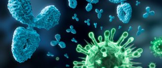
Antibodies (also known as immunoglobulins) are special proteins that are produced and/or produced by plasma cells.
What do immunoglobulins do?
Immunoglobulins are formed in response to foreign bacteria or viruses entering the body. They interact with the antigen (specific site of the pest) and neutralize it.
Thus, our immunity guards our health.
What classes of immunoglobulins are there?
There are 5 classes of immunoglobulins
, some of which contain subclasses.
IgA - secreted on the surface of the epithelium and present in saliva, tears, and on the surface of mucous membranes.
IgM - detected upon initial exposure to antigen.
Indicates an acute infectious process in humans.
IgG is the main class of immunoglobulins that protects against viruses, bacteria, and toxins.
IgD is found on the surface of developing B lymphocytes. The function is not installed.
IgE - secreted during an immediate allergic reaction.
What are immunoglobulins and how are they related to COVID-19
Immunoglobulins (Ig) are specific proteins that are released by cells of the immune system in response to the presence of pathogens. The main functions of immunoglobulins are:
- Binding to pathogen antigens.
- Neutralization of the pathogen.
- Involvement of other “components” of the immune system to suppress foreign agents.
There are several classes of immunoglobulins, but if we talk about coronavirus infection (and other diseases), the most interesting are two of them - IgG and IgM.
Class M immunoglobulins are the first to be produced in response to the penetration of a particular pathogen into the body. Therefore, the determination of these antibodies is important in diagnosing the acute stage of the disease when the pathogen is present in the body.
Immunoglobulins G provide further protection against re-entry of the pathogen. Their maximum concentration is observed approximately three weeks after infection with COVID-19.
Thus, IgM indicates the presence of an acute infection, and IgG helps confirm the fact that a person has already been ill.
Anyone can be tested for COVID-19 at ProfMedLab. You can call our specialist to conduct a test at home, and also enter into an agreement with the clinic to conduct testing for employees on behalf of a legal entity. ProfMedLab has already performed more than 30,000 tests for coronavirus infection.
What to choose – qualitative or semi-quantitative analysis?
Why is qualitative and quantitative analysis carried out?
Qualitative analysis allows us to answer 2 questions:
- Were you sick or not?
- How long have you been sick?
A semi-quantitative test can answer these questions and also determine the amount of immunoglobulins in the body.
Why determine the amount of immunoglobulins?
Determining the amount of antibodies allows you to determine whether long-term immunity has been formed. It is what protects the body from re-infection with coronavirus.
Decoding the results of the test for antibodies to coronavirus
How to decipher a qualitative coronavirus antibody test
| Yes/No | Ig G (Eat) | Ig G (No) |
| Ig M (Yes) | 1 option | Option 2 |
| 5-10 weeks have passed since infection. IgM is still present in the blood, but IgG is already being formed | Acute phase of the disease. 1-3 weeks have passed since infection | |
| Ig M (No) | Option 3 | Option 4 |
| Several months have passed since the illness. You may have had the disease asymptomatically | You have not encountered any viruses or no more than 7 days have passed since infection |
Antibodies to coronavirus - table with interpretation of results
In the 2nd and 4th version
(if there are suspicious symptoms), it is recommended to take a PCR test for coronavirus to identify the pest.
The complete absence of immunoglobulins may also mean that:
1) The preparation conditions were not met and the result was distorted
2) The symptoms of influenza or ARVI were similar to the symptoms of coronavirus,
3) The patient suffered a mild form of the disease and did not develop antibodies.
4) The patient suffered a severe form of the disease, and the antibodies quickly disappeared.
Cost of the study
The ProfMedLab clinic offers several types of tests for antibodies to COVID-19:
- Quantitative determination of class G immunoglobulins. The analysis allows you to understand whether a person has had a coronavirus infection or not. The material for the study is venous blood. Deadlines - 24 hours.
- Quantitative determination of immunoglobulins class G and M. We recommend combining it with PCR in order to obtain the most objective information. The material for the study is venous blood. Time frame: 2–3 days.
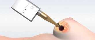
- Rapid test SARS-CoV-2 Antibody Test. You can do it yourself at home or in our clinic, and then consult a doctor. The material for the study is capillary blood (from a finger). The result will be ready in 20 minutes.
The research preparation period is from 1 day from the moment of material collection. Prices for these and other types of analyzes are presented in the table:
| Name of service | Type of analysis | Research price, rub. |
| Determination of RNA coronavirus SARS-CoV-2, quality delivery time - 2 days | PCR, smear | 1800 |
| IgG antibodies to coronavirus infection SARS-CoV-2 | ELISA, blood | from 1200 |
| IgM antibodies to coronavirus infection SARS-CoV-2 | ELISA, blood | from 1200 |
| Antibodies IgM+ IGg to coronavirus infection SARS-CoV-2 (complex) | ELISA, blood | from 2200 |
| CORONAVIRUS SARS CoV-2, RNA, delivery time 2-3 days | PCR, smear | 2200 |
| Determination of SARS-CoV-2 coronavirus RNA, quality. Russian language. urgent | PCR, smear | from 2200 |
| Determination of SARS-CoV-2 coronavirus RNA, quality. English. urgent | PCR, smear | 3000 |
| Rapid test for COVID-19, IgG, IgM (panel) | ELISA, blood | 2900 |
| PCR analysis for coronavirus infection SARS-Co-V-2 + home visit of the Central Administrative District | PCR, smear | 3500 |
| PCR analysis for coronavirus infection SARS-Co-V-2 + home visit within the Moscow Ring Road | PCR, smear | 5500 |
| PCR analysis for coronavirus infection SARS-Co-V-2 + travel up to 35 km from the Moscow Ring Road | PCR, smear | 7000 |
| Determination of IgG antibodies to coronavirus infection SARS-Co-V-2 - departure for up to 10 people | ELISA, blood | 3900 |
| Determination of IgG antibodies to coronavirus infection SARS-Co-V-2 - departure of 10-30 people | ELISA, blood | 3500 |
| Determination of IgG antibodies to coronavirus infection SARS-Co-V-2 - departure of 30-75 people | ELISA, blood | 3200 |
| Determination of IgG antibodies to coronavirus infection SARS-Co-V-2 - departure of 75-150 people | ELISA, blood | 3000 |
| IgG antibodies to coronavirus SARS CoV 2, spike (S) protein (after vaccination or previous COVID-19) | blood | 1300 |
Ig G - interpretation of the test for antibodies to coronavirus
To determine class G immunoglobulins in the blood, a qualitative and quantitative method for determining antibodies is used. Below is a table with a breakdown of the enzyme immunoassay for coronavirus (igG indicator for coronavirus).
Coronavirus antibody index - transcript of captions
| Result | Index | Meaning |
| Negative | Less than 0.8 | The patient either did not encounter the disease or underwent the procedure in the acute phase. |
| Border | 0,8-1,1 | You need to do a repeat test after 14 days. Perhaps the test was done at the onset of the disease or during the recovery process. |
| Positive | More than 1.1 | This level of antibodies to coronavirus in adults is normal. The person had coronavirus infection several months ago, and humoral immunity had time to develop. |
Class G antibodies to coronavirus - interpretation of results
An antibody titer to coronavirus of 1,800 (positive result) is normal. In this case, immunoglobulins of class A and M should be absent in the blood. Their presence indicates the stage of recovery.
Linked immunosorbent assay
Enzyme-linked immunosorbent assay (ELISA) is one of the types of immunochemical analysis. It is based on a highly specific immunological reaction of an antigen (AG) with a corresponding antibody (AT) with the formation of an immune complex. In this case, one of the components is conjugated to the enzyme. As a result of the reaction of the enzyme with a chromogenic substrate, a colored product is formed, the amount of which can be determined spectrophotometrically.
Any version of ELISA contains 3 mandatory stages :
- The stage of recognition of the test compound by a specific antibody;
- The stage of forming the connection of the conjugate with the immune complex or with free binding sites;
- The stage of converting the enzyme label into a recorded signal.
The reaction involves:
- Solid phase;
- AG and AT;
- Conjugate (antigen or antibody labeled with an enzyme);
- Enzyme marker;
- Substrate;
- Stop reagent (sulfuric acid is most often used).
Solid phase
Various materials can be used as a solid phase for ELISA: polystyrene, polyvinyl chloride, polypropylene and others. The solid phase can be the walls of a test tube, 96-well and other plates, balls, beads, as well as nitrocellulose and other membranes that actively absorb proteins.
Antigens and antibodies
AG and AT used in ELISA must be highly purified and highly active. Ags must have high antigenicity, optimal density and number of antigenic determinants. The sensitivity of ELISA depends on the concentration, activity and specificity of the antibodies used. The antibodies used can be poly- or monoclonal, of different classes (IgG or IgM) and subclass (IgG1, IgG2). The sensitivity and specificity of the method increases when using monoclonal antibodies. In this case, it becomes possible to detect low concentrations of Ag (AT) in samples.
Enzyme markers: the most widely used are horseradish peroxidase (HRP), alkaline phosphatase (ALP) and β-D-galactosidase.
Substrates
The choice of substrate is primarily determined by the enzyme used as a label, since the enzyme-substrate reaction is highly specific.
- For HRP, 3,3',5,5'-tetramethylbenzidine (TMB chromogen) is used as a substrate. A color reaction occurs, the intensity of which depends on the amount of bound analyte;
- For ALP the substrate is 4-nitrophenyl phosphate;
- β-D-galactosidase catalyzes the hydrolysis of lactose to form glucose and galactose.
ELISA classification
The classification of ELISA methods is based on several approaches:
1 — by type of reagents present at the first stage of ELISA:
- In competitive ELISA, at the first stage, the system contains both the analyzed compound and its analogue, labeled with an enzyme and competing for specific binding sites with it;
- Non-competitive methods are characterized by the presence at the first stage of only the analyzed compound and binding sites specific to it.
2 — according to the principle of determining the test substance:
- Direct determination of the concentration of a substance (AG or AT). Antibodies to the test substance combined with a specific label are used. In this case, the label will be located in the formed specific AG-AT complex. The concentration of the analyte will be directly proportional to the recorded signal;
- Indirect determination of the concentration of a substance is based on the difference in the total number of binding sites and the remaining free binding sites. In this case, the concentration of the analyte will increase, and the recorded signal will decrease; therefore, in this case there is an inverse dependence on the magnitude of the recorded signal.
3 — by type of results:
- Quantitative;
- Semi-quantitative;
- Qualitative.
Competitive ELISA
- Specific monoclonal antibodies are immobilized on the solid phase;
- An antigen labeled with an enzyme and the test sample are added to the wells of the panels in a known concentration. In parallel, positive and negative controls are placed in adjacent wells. To construct the calibration, a standard unlabeled antigen in various dilutions is used. Incubation and washing are carried out;
- Add the substrate, incubate, stop the reaction when optimal staining develops in the positive control wells;
- The results are recorded on an ELISA reader. The concentration of the analyte is inversely proportional to the optical density.
Competitive ELISA for the determination of antibodies: the desired antibodies and enzyme-labeled antibodies compete with each other for antigens adsorbed on the solid phase.
Non-competitive “sandwich” - ELISA option
The main advantage of the method is its high sensitivity, which exceeds the capabilities of other ELISA schemes.
- Monoclonal antibodies or affinity-purified polyclonal antibodies are immobilized on the solid phase;
- The test sample is added to the wells of the panels, and a positive control sample and a negative control sample in various dilutions are placed in parallel. Incubate and wash;
- Enzyme-labeled monoclonal or polyclonal antibodies—conjugate—are added to the wells. After incubation, washing is carried out to remove unbound antibodies;
- The substrate is added and incubated. The reaction is stopped when optimal staining is achieved in the positive control wells;
- The results are recorded on an ELISA reader. The concentration of the analyte is directly proportional to the optical density.
Qualitative analysis is often used in screening studies and diagnosis of infectious diseases. The result of the study is determined by comparing its optical density with the calculated value of the critical optical density (OPcrit., “Cut-off”).
The formula for calculating “Cut-off” is indicated in the instructions for the test system. The “Cut-off” calculation can involve both the averaged values of the optical densities of positive and negative controls, as well as the optical density of a special control sample—cut level control. If the optical density of the sample is higher than the Cut-off, the sample is considered positive for specific antibodies.
For the semi-quantitative version of the technique, the ratio between the average optical density of the sample and the Cut-off optical density is calculated.
Samples are considered as:
- positive if the ratio is more than 1.1;
- doubtful if the ratio is 0.9–1.1;
- negative if the ratio is less than 0.9.
Doubtful test results cannot be unambiguously interpreted, and to clarify the result, it is necessary to repeat the examination after 1–2 weeks.
The ELISA method determines:
- Hormones;
- Tumor markers;
- Antibodies (IgG, IgM) to pathogens - ToRCH infections, tuberculosis, syphilis, hepatitis C virus (HCV antibody), mycoplasma, chlamydia, giardia, Helicobacter pylori, etc.;
- Antigens of viruses - hepatitis D, E, A (HD, HEV, HAV), hepatitis C (HCVcore), hepatitis B (HBs, HBe HBcore);
- Autoimmune diseases.
Sources:
- Lectures on laboratory diagnostics, Siberian State Medical University, 2015;
- https://www.bosterbio.com/protocol-and-troubleshooting/elisa-principle.
BAU/ml - new interpretation of the test for antibodies to coronavirus
The World Health Organization has approved a new international standard for the determination of immunoglobulins for coronavirus, with the unit of measurement BAU (translated as “binding antibody coefficient”).
Interpretation of quantitative analysis for antibodies to coronavirus
Laboratory diagnostics
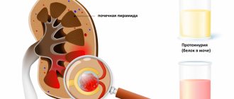
the level of immunity protection against coronavirus is carried out by studying immunoglobulins to the S-protein of the virus. To facilitate the interpretation of the study results, it was proposed to use a universal measurement system.
Conversion table for antibody test results from different manufacturers in BAU/ml
| Manufacturer | Conversion factor to BAU/ml |
| Abbott ARCHITECT | 0,142 |
| Roche | 1 |
Preparing for the test:
No special preparation is required (donating blood on an empty stomach is not necessary, you can smoke). A coronavirus test can be taken during the day, no earlier than 3 hours after eating. A person who takes tests to determine the presence of antibodies to the Covid-19 coronavirus should not have clinical manifestations of an infectious disease: fever, cough, runny nose, muscle pain. If symptoms of Covid-19 are detected, we ask you to inform the administrator before visiting the clinic and taking the test. Analysis is not taken if the patient is in self-isolation due to illness.