Electrocardiogram (ECG): interpretation of results
By: Administrator |
Tags: decoding ECG results, decoding ECG, ECG, ECG normal, Electrocardiogram, electrocardiogram decoding results | Comments: | June 24, 2021 Electrocardiography is a simple and informative study that determines heart rate indicators. A cardiograph records heart activity and records its parameters on paper. To evaluate them and draw conclusions about the patient’s condition, it is necessary to decipher the cardiogram. ECG interpretation is performed manually by comparing the characteristics of the graph with special tables or using computer programs to interpret the ECG results.
When and who needs to undergo heart diagnostics?
The doctor prescribes this diagnostic test in the following cases:
- high blood pressure;
- chest pain, shortness of breath;
- dizziness or fainting;
- heart murmurs;
- heart rhythm is disturbed;
- rheumatism;
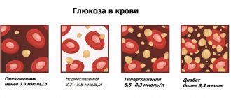
- diabetes.
An electrocardiogram is also prescribed in case of overdose of certain medications. An ECG is part of the examination during medical examination, prof. examination, pregnancy, preparation for operations.
This is not an exhaustive list of cases when an ECG is necessary. In addition, it is performed without a doctor’s referral.
Principle of operation
Electrocardiography is a graphical visualization of the functionality of the heart muscle. To perform the procedure, special equipment is used - an electrocardiograph. This equipment records the currents that occur when the heart muscle relaxes and contracts. The obtained data is displayed on paper tape in the form of a special graph called a cardiogram.
Since the ECG records and displays electrical currents arising in the heart, let us consider the nature of the occurrence of the electrical impulse. Very complex biochemical processes continuously occur in the muscle cells of the heart. Their result is the formation of a large number of microcurrents, which are combined into a single pulse.
Electrocardiography allows you to display the dynamics of the electrical impulse that acts on the heart muscle and ensures its contraction. To obtain a more accurate and informative result of the study, indicators are taken simultaneously from different parts of the heart muscle. The device's sensors are attached to certain areas of the chest, wrists, and ankles. The data is transmitted on the screen of the ECG machine in the form of 12 complex graphs and simultaneously displayed on paper.
Content:
- Principle of operation
- Which specialist should you contact?
- Indications for ECG
- Features and types of ECG
- Contraindications and restrictions
- Preparing for the study
The duration of the study is 5-10 minutes. The information obtained during this period is quite sufficient to assess the function of the cardiovascular system.
Pathologies during ECG
The ECG is normal if all indicators are within a certain range. Otherwise, they speak of a deviation from the norm, but the presence of pathology must be determined by a doctor. Heart cardiogram decoding makes it possible to identify the following pathologies:
- Sinus arrhythmia suggests a physiological disorder, but is normal in children and adolescents.
- Atrial fibrillation can occur periodically or constantly, and is accompanied by a feeling in the patient of heart fluttering, anxiety, and panic.
- Sinus bradycardia, when the heart rate is about 50 beats per minute, is observed in healthy people during sleep and in athletes.
- Sinus tachycardia manifests itself in excess of the standard heart rate (90 beats per minute). In healthy people, it is temporarily observed during physical and emotional stress, drinking strong coffee, alcohol, and energy drinks. The pathology is indicated by a rapid heartbeat at rest.
- Extrasystole is characterized by a chaotic heartbeat, too frequent or too rare. Patients feel tremors behind the breastbone, tingling, empty stomach or sudden fear.
- Paroxysmal tachycardia manifests itself with periodic rapid heartbeat, while the pulse can reach 200-250 beats per minute. The duration of the attack can be from several minutes to several days.
- WPW syndrome is accompanied by a lack of air, a feeling of cardiac arrest for a moment, and a strong heartbeat.
Types of ECG of the heart
There are several types of ECG of the heart, thanks to which you can evaluate the work of the heart muscle in various conditions and conditions.
Classical electrocardiography
It takes 5-10 minutes and allows you to study the condition of the heart at the time of the procedure. It is used to determine the size of the chambers, heart rate, conductivity of the heart muscle, and evaluate the blood supply to the myocardium at rest. However, many cardiac pathologies appear only during sleep or physical activity, so it is extremely difficult to identify them during a short visit to a cardiologist.
Holter monitoring (Holter ECG)
A complex, in-depth study that provides continuous recording of cardiac muscle parameters for 24-72 hours or up to 10 days (if necessary). Recording is carried out with a special device that is carried with you on a belt or belt. Electrodes are attached to the surface of the skin in certain places. After which the person leads a normal life (works, walks). At the same time, he notes in a notebook when he experiences pain in the heart area, takes medications, and begins active physical activity. The data obtained is reviewed and corrected by the doctor, after which he draws conclusions and prescribes treatment.
Interesting! There are portable devices that are implanted under the skin near the heart to record information throughout the year.
Independent interpretation of ECG: algorithm of actions
Cardiogram analysis algorithms summarize practical experience and data taken from specialized literature. It is especially important to show how ECG interpretation is performed for students, interns, and paramedics who are starting their careers.
Sequence of actions when independently analyzing ECG results:
- Evaluate the rhythm and its regularity.
- The intensity of contractions of the heart muscle.
- The electrical axis of the heart or the frontal projection of the excitation vector of the ventricles and the direction of the electric wave along the ventricles during contraction are determined. The electrical axis of the heart is normal from 30° to 70°, direction down to right.
- The parameters of the P wave are determined.
- The QRS complex is analyzed.
- Determine the parameters of the ST segment.
- The characteristics of the T wave are analyzed.
- The characteristics of the remaining intervals and segments are analyzed.
Contraindications and restrictions
Electrocardiography is considered an absolutely safe diagnostic method that allows you to obtain maximum information about the condition and function of the heart muscle. Among the disadvantages of the procedure, one can highlight the low information content in certain cases. For example, coronary heart disease is diagnosed exclusively with an exercise ECG. With extensive injuries and deformations of the chest, the accuracy of diagnosis decreases.
There are contraindications only for ECG with stress. Physical and medicinal stress is contraindicated in patients with suspected aortic aneurysm dissection, severe heart failure, pathological heart rhythm disturbances, or the development of infection in the body. If a person has recently suffered a myocardial infarction, he may only be prescribed a standard electrocardiogram.
Best materials of the month
- Coronaviruses: SARS-CoV-2 (COVID-19)
- Antibiotics for the prevention and treatment of COVID-19: how effective are they?
- The most common "office" diseases
- Does vodka kill coronavirus?
- How to stay alive on our roads?
How to make an appointment with a cardiologist
- It is advisable to undergo an ECG with interpretation and get advice from a qualified doctor by visiting a diagnostic and treatment center. Regular customers note the following benefits of service:
- Modern diagnostic equipment, which makes it possible to obtain an accurate diagnosis and speed up the examination.
- Patients are advised and accompanied by specialists with scientific degrees, doctors of the highest category with many years of practice.
- Carrying out an ECG in the clinic or at home, issuing the result immediately after the study.
- Completion of the study on the day of treatment or any convenient time.
To make an appointment with a cardiologist, just use the feedback button on the Loritom clinic website.
Which specialist should you contact?
In order to undergo an ECG procedure, you can contact your local physician and receive the appropriate referral. The examination itself is usually performed by a nurse who has completed special qualification courses. Electrocardiography is a simple diagnostic method, so the requirements for specialists performing it are minimal. Here, the cardiogram is deciphered by a certified doctor, who must have a specialization as a cardiologist. Knowledge in this area will allow him to correctly assess the functionality of the heart muscle and issue an informative conclusion.
What affects the accuracy of indicators
Sometimes the results of a cardiogram may be erroneous and differ from previous studies. Errors in results are often associated with many factors. These include:
- incorrectly attached electrodes. If the sensors are poorly attached or become dislodged during an ECG, the test results can be seriously affected. That is why the patient is recommended to lie still during the entire period of taking the electrocardiogram;
- extraneous background. The accuracy of the results is often influenced by extraneous devices in the room, especially when the ECG is performed at home using mobile equipment;
- smoking, drinking alcohol. These factors affect blood circulation, thereby changing the cardiogram parameters;
- meal. Another reason that affects blood circulation and, accordingly, the correctness of indicators;
- emotional experiences. If the patient is worried during the study, this may affect the heart rate and other indicators;
- Times of Day. When conducting a study at different times of the day, the indicators may also differ.
The specialist must take into account the above-described nuances when interpreting the ECG; if possible, they should be excluded.
Decryption features
To record a cardiogram, special sensors are attached to a person’s body, which transmit electrical impulses to an electrocardiograph. In medical practice, these impulses and the paths they take are called leads. Basically, 6 main leads are used during the study. They are designated by the letters V from 1 to 6.
The following rules for deciphering a cardiogram can be distinguished:
- In lead I, II or III, you need to determine the location of the highest region of the R wave, and then measure the gap between the next two waves. This number should be divided by two. This will help determine the regularity of your heart rate. If the gap between the R waves is the same, this indicates normal contraction of the heart.
- After this, you need to measure each tooth and interval. Their standards are described in the article above.
Most modern devices automatically measure your heart rate. When using older models, this has to be done manually. It is important to consider that the ECG recording speed is usually 25 – 50 mm/s.
Heart rate is calculated using a special formula. At an ECG recording speed of 25 mm per second, it is necessary to multiply the R - R interval distance by 0.04. In this case, the interval is indicated in millimeters.
At a speed of 50 mm per second, the R - R interval must be multiplied by 0.02.
For ECG analysis, 6 of 12 leads are usually used, since the next 6 duplicate the previous ones.
Normal values in children and adults
In medical practice, there is the concept of an electrocardiogram norm, which is typical for each age group. Due to the anatomical characteristics of the body in newborns, children and adults, the study indicators are slightly different. Let's take a closer look at them.
ECG norms for adults can be seen in the figure.
Normal ECG in adult patients
A child's body is different from an adult's. Due to the fact that the organs and systems of the newborn are not fully formed, electrocardiography data may differ.
In children, the mass of the right ventricle of the heart prevails over the left ventricle. Newborns often have a high R wave in lead III and a deep S wave in lead I.
The ratio of the P wave to the R wave in adults is normally 1:8; in children, the P wave is tall, often more pointed, in relation to R is 1:3.
Due to the fact that the height of the R wave is directly related to the volume of the ventricles of the heart, its height is lower than in adults.