Pain syndrome
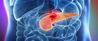
The appearance of pain is typical for large cysts, as they compress the surrounding tissues, including the nerve plexuses. Small cysts do not exert such pressure, therefore, as a rule, there are no complaints of pain. This symptom is especially characteristic for the period of formation of false cysts during acute and exacerbation of chronic pancreatitis, and it is caused to a greater extent by destructive processes. Over time, the intensity of the pain decreases, it is characterized as “dull” or rather “discomfort”. A characteristic symptom is the “lucid interval” (temporary improvement and absence of pain after acute pancreatitis or injury). The most severe pain is caused by cysts located on the posterior surface of the gland and compressing the solar plexus area. Impact on this nerve plexus creates in patients a very intense, long-lasting burning pain that radiates to the back. Movement, compression by clothing, a belt, or a belt increase its intensity. The condition is somewhat alleviated when taking the knee-elbow position (“on all fours”). A pronounced increase in pain - “dagger” pain can indicate the occurrence of complications (for example, rupture of a cyst), gradual progression of pain along with an increase in body temperature and the appearance of intoxication - about its suppuration.
Dyspepsia
Other manifestations of pancreatic cysts may include dyspeptic disorders: nausea, vomiting (which may result in an attack of pain), and stool instability. As a result of the decrease in the amount of pancreatic juice entering the intestines, the digestion of food and the absorption of nutrients are disrupted. As a result, the patient loses weight, loses weight, and becomes weak.
Intestinal obstruction
Sometimes large pancreatic cysts cause compression of neighboring organs, impairing their patency. When a cyst is located in the head of the gland, obstructive jaundice may occur (yellowishness of the skin, sclera, and itchy skin appears); if the portal vein is compressed, swelling in the legs or ascites develops. Very rarely, large pancreatic cysts compress the intestinal lumen (duodenum), which interferes with the passage of food. In these cases, incomplete high intestinal obstruction may form.
Diagnosis of pancreatic cyst
When examining the abdomen, its asymmetry is possible: the appearance of a bulge or protrusion in the area where the formation is located.
Laboratory tests show a slight increase in leukocytes in the blood, an increase in ESR, and sometimes the level of enzymes in the blood increases: amylase, phosphatase and bilirubin, which is associated with exacerbation of pancreatitis and damage to working pancreatic tissue.
More specific and meaningful information can be obtained by using instrumental diagnostic methods.
- Ultrasound examination
allows not only to detect a formation, see its location, measure its size, evaluate its contents, but also to identify indirect signs of complications: for example, in the case of suppuration of a cyst, ultrasound can see the unevenness of the echo signal against the background of the cavity; in case of malignancy (malignancy), heterogeneity of the contours . - Magnetic resonance imaging and computed tomography
provide more detailed information about the size, localization of the cyst, and its connection with the bed of the ducts. Contrast tomography helps to recognize cystic tumors. - Endoscopic retrograde cholangiopancreatography
(ERCP) has a unique place in the diagnosis of pancreatic cysts. This endoscopic examination provides detailed information about the connection of the cyst with the ducts of the gland, which determines treatment tactics. However, this examination requires the injection of a contrast agent directly into the pancreatic duct, which causes ductal hypertension and is accompanied by a high risk of infection. Therefore, at present, ERCP is performed according to strict indications: for example, if an obstructive nature of the cyst is suspected, in order to remove the stone from the pancreatic duct or install a stent in the obstruction area. - Endosonography
(a modern diagnostic method that combines the advantages of endoscopy and ultrasound) allows not only to clearly visualize the location, size of the cyst and the nature of its contents, but also to take a targeted biopsy if the presence of a cystic tumor is suspected.
Pathogenesis
The development of pancreatic cysts occurs under the influence of provoking factors, resulting in discomplexation of pancreatocytes in the pancreas with the development of small diffuse necrosis of acinar (secreting) cells. As a consequence, there is a violation of secretion into the lumen of the ducts and parapedesis of secretory granules in the interstitium. Destroyed cells promote interstitial/intracellular activation of trypsin, which activates pancreatic hydrolytic/proteolytic enzymes, which in turn contribute to autoenzyme aggression and the development of foci of necrosis, which is manifested by regional rheological disorders and severe interstitial edema.
Resorption of pancreatic enzymes from the interstitium contributes to autolytic damage to blood vessels with the subsequent development of thrombohemorrhagic local syndrome.
The formation of fluid accumulations is based on the processes of lysis of foci of infiltration/necrosis. The formation of fluid accumulations is ensured mainly due to the exudative reaction of inflamed tissues and, to a lesser extent, due to pancreatic juice arriving due to a violation of the outflow of juice along the main pancreatic duct. As a result of a pronounced violation of capillary permeability, almost within 1–2 hours after the destruction of the acini, many mast cells/leukocytes appear in the tissues, actively secreting biologically active substances, which leads to the formation of a demarcation shaft around the focus of necrosis, consisting of leukocytes, fibrin, histeocytes, lymphoplasma cells components and nuclear detritus.
Activation of fibroblasts promotes the intensive formation of elements of connective tissue structures/collagen, which serve as the basis for the formation of a barrier, which subsequently becomes the base of the postnecrotic cyst capsule.
Treatment of pancreatic cyst - surgery
The clear answer is surgery only! Modern technologies make it possible in many cases to avoid major surgery and limit ourselves to minimally invasive endoscopic or endovideosurgical intervention.
The following methods of treating cysts are possible:
- Removal of the cyst itself or a cystic part of the pancreas.
- Internal drainage of the cyst.
- External drainage of the cyst.
With the first method, as a rule, the cyst is removed along with a section of the pancreas. The volume of intervention depends on the size of the formation, on the location where the cyst is located, on the condition of the tissues adjacent to it. Distal resection, distal or pancreatoduodenal resection, are quite complex and require appropriate technical and medical support. Currently, these operations are included in the list of types of high-tech medical care performed under quotas from the Ministry of Health of the Russian Federation or under compulsory medical insurance policies. These operations can be performed in our Clinic both traditionally and laparoscopically.
Drainage operations are considered the most physiological and less traumatic, which are aimed at creating an outflow from the cyst into the stomach, duodenum or small intestine using internal drainage. An anastomosis is created, which ensures the delivery of gland juice to food, which relieves the pain reaction and rarely leads to relapses. Currently, it is also possible to perform these operations using endoscopic techniques, which are performed endovideosurgically or endoscopically with ultrasound guidance.
External drainage of cysts in planned surgery has recently been rarely used. The choice of this type of intervention is most often forced in emergency situations. Indications for this method:
- if the process of cyst formation has not completed;
- serious condition of the patient;
- suppuration of the cyst.
Such interventions are called palliative, they do not solve the problem, but can lead to relapse, to fistulas. They are used as one of the stages of patient treatment. Drainage operations can be carried out only after confirmation of a non-tumor cause of the formation.
Conservative treatment of the underlying disease is mandatory. For pancreatitis, it is imperative to follow a diet, the purpose of which is to reduce the secretion of pancreatic juice as much as possible.
Enzyme replacement drugs and analgesics are used; secretion suppressants. It is imperative to monitor the level of glycemia and, if necessary, correct it.
Stages
The process of formation of a post-acrotic cyst of the pancreas goes through 4 stages. At the first stage of the appearance of a cyst, a cavity is formed in the omental bursa, filled with exudate due to acute pancreatitis. This stage lasts 1.5-2 months. The second stage is the beginning of capsule formation. A loose capsule appears around the unformed pseudocyst. Necrotic tissue with polynuclear infiltration remains on the inner surface. The duration of the second stage is 2-3 months from the moment of occurrence.
At the third stage, the formation of the fibrous capsule of the pseudocyst, firmly fused with the surrounding tissues, is completed. The inflammatory process is intense. It is productive. Due to phagocytosis, the liberation of the cyst from necrotic tissue and decay products is completed. The duration of this stage varies from 6 to 12 months.
The fourth stage is the isolation of the cyst. Only a year later, the processes of destruction of the adhesions between the wall of the pseudocyst and the surrounding tissues begin. This is facilitated by the constant peristaltic movement of organs that are fused with a fixed cyst, and the long-term effect of proteolytic enzymes on scar adhesions. The cyst becomes mobile and is easily separated from the surrounding tissue.
Forecast
The prognosis for pancreatic cysts is quite favorable. It depends both on the cause of the disease and on the timeliness of diagnosis and surgical treatment.
In our clinic you can undergo a full examination. The clinic’s specialists have all the necessary equipment for diagnosing and treating cystic formations of the pancreas. The full range of necessary surgical (both traditional and minimally invasive) interventions is performed. Most of them are included in the list of types of high-tech medical care (HTMC), performed under quotas of the Ministry of Health of the Russian Federation or compulsory medical insurance policies.
Causes
The development of pancreatic pseudocyst is based on polyetiological factors, the main of which are:
- Acute/chronic destructive pancreatitis .
- Traumatic injuries of the pancreas.
- Blockage of the bile ducts (congenital or caused by the presence of stones (cholelithiasis), tumors, strictures, scars, persistent narrowing of the walls (stenosis).
- Parasitic infestations ( echinococcosis , opisthorchiasis , cysticercosis ).
Risk factors include:
- Abuse of alcoholic beverages.
- Obesity with lipid metabolism disorders, accompanied by increased cholesterol .
- Cholelithiasis (stones in the bile duct/gallbladder).
- Diabetes.
- Surgeries on the pancreas or adjacent organs.
Consequences and complications
Why is a cyst in the pancreas dangerous? The danger lies in the high risk of complications. The most common complications of unresolved false cysts are severe pain and extrahepatic cholestasis / duodenal obstruction caused by external pressure, rupture into the abdominal cavity, pseudoaneurysm , bleeding and formation of a pancreatic abscess .
What will the transcript of an abdominal ultrasound show?
T.N. Marambey July 24, 2021 Ultrasound examination
First, let's look at what this ultrasound shows.
Behind the anterior wall of the abdomen there is a large space - the abdominal cavity. There are quite a few organs located in it, which an ultrasound of the abdominal cavity will show. This:
- stomach
- intestines
- pancreas
- liver
- bile ducts: intra- and extrahepatic
- spleen
- gallbladder
- kidneys
- adrenal glands
- abdominal part of the aorta and its branches
- lymph nodes
- lymphatic trunks and vessels
- division of the autonomic nervous system
- nerve plexuses.
The abdominal cavity is lined with two layers of a thin membrane - the peritoneum. It is this inflammation that is called peritonitis and is a life-threatening condition. The organs are covered in different ways by the peritoneum: some are wrapped in it, some do not even touch it, but are located within the boundaries outlined by it. Conventionally, the cavity is divided into the abdominal cavity itself and the retroperitoneal space. The latter includes the bottom of the list of organs, starting with the kidneys.
All these organs - both the abdominal cavity and the space behind the peritoneum - are examined during an ultrasound examination of the abdominal cavity. This study can detect the presence of structural damage, inflammation, pathological formations, enlargement or reduction of the organ, and disruption of its blood supply. Ultrasound does not see how a sick or healthy organ copes with its functional responsibilities.
What does ultrasound give? The study helps to find the cause of the disease in such cases:
- pain or discomfort in the abdomen
- bitterness in the mouth
- feeling of a full stomach
- intolerance to fatty foods
- increased formation of gases
- frequent bouts of hiccups
- feeling of heaviness in the right or left hypochondrium
- jaundice
- high blood pressure
- lower back pain
- fever not due to a cold
- weight loss not related to diets
- belly enlargement
- as control over the effectiveness of treatment of pathologies of the digestive system
- and also as a routine examination, including for existing anomalies of organ development, cholelithiasis.
Pathology determined by ultrasound
What does abdominal ultrasound diagnose? Using this study, the following diseases can be identified:
1. From the gallbladder side:
- acute and chronic cholecystitis
- empyema of the bladder
- gallstone pathology
- during a choleretic breakfast, the motor function of the bladder can be assessed
- developmental anomalies (kinks, septa).
2. From the liver:
- cirrhosis
- hepatitis
- abscesses
- tumors, including metastases
- hepatosis
- "stagnation" in the liver due to cardiopulmonary diseases
- fatty liver change.
3. From the kidneys and urinary system:
- kidney tumors
- "wrinkled bud"
- pyelonephritis
- narrowing of the ureters
- stones and “sand” in the kidneys.
4. From the side of the spleen, ultrasound of the abdominal cavity reveals:
- cysts
- tumors
- abscesses
- heart attacks
- organ enlargement in infectious and parasitic diseases
5. From the pancreas:
- cysts
- tumors
- abscesses
- stones in the ducts
- signs of acute and chronic pancreatitis.
6. Ultrasound reveals free fluid in the abdominal cavity
7. From the abdominal part of the aorta or its branches, an aneurysm and its dissection, vasoconstriction may be visible
8. From the side of the retroperitoneal lymph nodes, their enlargement and homogeneity of structure are visible
How to understand the research results
To do this, consider the ultrasound form (protocol). It indicates points that relate to each organ separately.
Liver
Interpretation of abdominal ultrasound in relation to this organ includes:
| Parameter | What is written on the form | Normal ultrasound findings in adults |
| Dimensions of the entire organ | Normal, decreased, increased (underline as appropriate) | Norm |
| right | The numbers are indicated in cm for each item | Up to 12.5 |
| left | Up to 7 | |
| caudate | 30-35 | |
| Oblique-vertical dimension (OVR) of the right lobe | Numbers in mm | Up to 150 mm |
| Outlines | It is emphasized whether they are even or not | Smooth |
| Capsule | It is emphasized whether it is differentiated or not, thickened or not | Differentiated, not thickened |
| Left lobe thickness | Number in mm | 50-60 |
| Right lobe thickness | 120-125 | |
| Echostructure of parenchyma | Emphasized, normal, increased or decreased | Norm |
| Focal formations | Yes or no | Must not be |
| Portal vein | Size in mm | Up to 14 mm |
| Vascular pattern | Depleted, normal or enhanced | Ordinary |
| Inferior vena cava | Size in mm | Anechoic, diameter 20 mm |
| Hepatic veins of the first order | Size in mm | Up to 1 mm |
Decoding the results
- Fatty hepatosis is indicated by an increase in the echo density of the organ in the form of small foci. The edge of the liver is rounded. In the final stages, due to compaction of the organ, it is impossible to see the portal vessels.
- With cirrhosis of the liver, its enlargement and dilation of the portal and splenic veins are visible. The lower edge of the organ will also be rounded, the contours will be uneven. The increase in echo density in this case will be large-focal. Free fluid in the abdominal cavity (ascites) is also determined.
- If an increase in size, rounding of the edges, as well as expansion of the vena cava and the absence of narrowing during inspiration are described, this indicates congestion in the liver due to cardiac or pulmonary disease.
- If lesions are described in which there is a violation of the normal echostructure, this may indicate malignant or benign tumors, cysts or abscesses.
In the video, a specialist talks about errors that occur during ultrasound examination of the abdominal organs.
Gallbladder
Ultrasound norm based on the results of examination of this organ:
- Shape: various – pear-shaped, cylindrical.
- Dimensions: width 3-5 cm, length 6-10 cm.
- Volume: 30-70 cubic meters cm.
- Walls: up to 4 mm thick.
- Formations in the lumen: normally there are none.
- Acoustic shadow from formations: this applies to stones and bladder tumors. Based on the presence of this shadow, the types of stones are deciphered (they come in different compositions).
- Whether they move or not: the stones are usually movable, but can be soldered to the wall or be large in size. Based on this and some other signs, one can judge whether the formation is a tumor.
Signs of gallbladder pathology
- In acute cholecystitis, thickening of the organ wall is noted, and the dimensions can be normal, reduced or enlarged. The wall may also be described as a "double contour" and the presence of fluid around the bladder indicates that local peritonitis has already developed and urgent surgery is needed.
- Thickening of the wall will also occur with chronic cholecystitis. The contour in this case is clear and dense.
- In conclusion, various deformations of the organ can be described. This is not a disease, but a structural feature.
- If echo-negative objects are described that leave an acoustic shadow, while the wall of the bladder is thickened and the contour is uneven, we are talking about calculous cholecystitis. In this case, the expansion of the bile ducts indicates that the stone is blocking the exit of bile.
Interpretation of ultrasound of the bile ducts
Normally, on ultrasound, the bile ducts have the following characteristics:
- common bile duct: diameter 6-8 mm
- intrahepatic ducts: should not be dilated
Norms of the pancreas on ultrasound
- There should be no additional education.
- head: up to 35 mm
- body: up to 25 mm
- tail: about 30mm
- contour: smooth
- echostructure: homogeneous
- echogenicity: neither reduced nor increased
- Wirsung duct: 1.5-2 mm
- education: normally there are none.
A decrease in the echo density of the gland indicates acute pancreatitis, an increase in it indicates chronic pancreatitis or cancer. The expansion of the Wirsung duct also indicates chronic inflammation. The “favor” of cancer is indicated by a segmental increase in size and unevenness of the contour of the gland, depression on the surface of the liver, as well as displacement or compression of the inferior vena cava or aorta.
Interpretation of ultrasound of the spleen
- Dimensions: length – up to 11 cm, thickness – up to 5 cm, longitudinal section – up to 40 sq. cm
- splenic index: no more than 20 cm2
- structure: normally – homogeneous
- splenic vein at the hilum.
- You can see an increase in the size of the organ. This is associated with both certain blood diseases and liver diseases (such as cirrhosis) or infectious diseases.
- Densified (less often, less dense) tissue indicates a splenic infarction, that is, that as a result of thrombosis or injury, the death of some part of the organ occurred.
- Ultrasound also allows you to see a rupture of the spleen, which usually occurs either with a severe injury or with a minor bruise, but in the case of an enlarged organ.
Ultrasound of hollow organs (stomach, small, large and rectal intestines)
It only indicates whether there is a symptom of an “affected organ” (there should not be one) and whether there is fluid deposition in the intestinal lumen (this should not happen either).
If an ultrasound scan of the kidneys was also performed, then a description of this organ is also included in the conclusion of the study. The results of an ultrasound examination of the kidneys are normal:
- width: 5-6 cm
- length – about 11 cm
- organ thickness: 4-5 cm
- kidney parenchyma - no more than 23 mm thick
- the pelvis should not be dilated
- There should be no structures in the lumen of the pelvis and ureters.
Lymphatic structures with ultrasound imaging
Ultrasound of the retroperitoneal lymph nodes normally suggests the following conclusion: “Lymph nodes are not visualized.” That is, if they are of normal size, ultrasound “does not see” them. An increase in these immune organs indicates either an infectious disease present in the abdominal cavity or a malignant formation. In the latter case, they can increase due to the fact that cancer cells of the hematopoietic system “live” in them, as well as with metastases of any nearby organ tumor.
Sonologist's conclusions
At the conclusion of the ultrasound, the sonologist (ultrasound doctor) indicates the presence of pathology: he describes what the echo signs look like. If in the referral the doctor indicates that it is necessary to conduct an examination for some disease, but the ultrasound did not visualize it (for example, calculous cholecystitis), then there may be the phrase “Echo signs of the disease were not identified.” The final diagnosis is made only by the doctor who refers you for examination.
Who needs to undergo Doppler ultrasound of the abdominal vessels
This examination, which is also called Doppler ultrasound (Doppler ultrasound) of the abdominal vessels, is often performed together with ultrasound. The patient's sensations are not differentiated and are not more harmful than ultrasound. It allows you to evaluate the anatomy and characteristics of blood circulation in vessels such as:
- abdominal aorta
- common hepatic artery
- iliac arteries
- celiac trunk
- splenic artery
- superior mesenteric artery
- portal vein of the liver and its branches
- inferior vena cava.
Ultrasound of the vessels of the abdominal cavity makes it possible to timely identify early abnormalities in the vessels, identify and evaluate the degree of increase in pressure in the portal vein (with cirrhosis, “congestive” liver), and evaluate the result of implantation of a vena cava filter.
Ultrasound of the abdominal aorta and its branches helps in the diagnosis of:
- fainting states
- frequent headaches
- epileptic seizures
- high blood pressure
- repeated strokes (sometimes blood clots can “fly off” from this large vessel)
- leg pain
- potency disorders
- aortic aneurysm
- atherosclerotic lesions
- vasoconstriction
- anomalies in the development of large vessels.
Duplex scanning
Vascular examination during ultrasound using modern equipment almost always includes duplex angioscanning. This is the “gold standard” in assessing blood circulation in the venous vessels. It allows you to identify pathological blood flow, obstructions to blood flow, assess their location, extent and severity.
With this type of study, the sonologist receives a color two-dimensional image of the abdominal vessels, where red indicates the movement of blood towards the sensor, and blue, on the contrary, away from the sensor. Based on the intensity of the red and blue colors, the doctor draws conclusions about the speed of blood flow in any part of the vascular system.
Additional Study Details
Reviews about ultrasound are mostly positive: the study is painless, harmless, and very informative. The negative point is that before the procedure you need to carefully prepare so that gases in the intestines (“flatulence phenomena”) do not interfere with the correct diagnosis.
How much does it cost to conduct this research? A complete examination of all organs (including the kidneys and urinary system) with duplex angioscanning is estimated by clinics at an average of 2000-2500 rubles. An examination of individual organs with an assessment of the blood flow in them costs about 800-1000 rubles.
Thus, interpretation of abdominal ultrasound should be carried out by a specialist, taking into account not only the “norm” numbers, but also based on clinical manifestations. The above values will help you understand a little about the pathology identified in you, but the final assessment should be given by a specialist therapist or gastroenterologist.
We also looked here: ultrasound of the abdominal cavity, interpretation is normal, they don’t see fluid on ultrasound, form for ultrasound of gastrointestinal organs, splenic vein is normal in adults
← All articles
Cysts in women and men: symptoms and treatment
In the initial stages of development, cystic elements do not have symptoms. When they grow and suppurate, they manifest themselves differently, depending on the etiology, location, composition of the contents and size. For example, when the ovary is damaged, women complain of a feeling of heaviness and compression of the lower abdomen, pain during menstruation, cycle disruption, etc. With kidney pathology, the lower back hurts and aches, it is difficult to empty the bladder, and jumps in blood pressure are noted.
Diagnostics
Diagnostic methods depend on the location of cystic formations. Most often this is an ultrasound, but MRI, CT, and radiography can also be prescribed. Laboratory tests are also carried out - blood and urine.
Surgery
The intervention tactics are determined by the doctor based on the location, size of the cystic formation and the patient’s complaints. The classic technique is scalpel excision of the tumor along with adjacent tissues. Today, such an operation is performed mainly laparoscopically using an endoscope. Laparoscopy of a cyst is less traumatic, the patient’s recovery lasts one day. You can also choose the method of aspiration (suction) of the contents with further cauterization of the capsule with a laser, liquid nitrogen, radio wave or a special drug for sclerosis of the walls. The removed lesion must be sent for histological examination.
Is surgery necessary?
When making such a diagnosis, many patients wonder whether the cyst needs to be removed. Some tumors, if they are small and do not bother the person, can remain under observation. Others, such as cervical cysts, can have consequences: bacteria accumulate in them, which sooner or later will lead to inflammation or suppuration. The need for removal should be discussed with your doctor.
Symptoms
The symptoms of a pancreatic cyst are extremely ambiguous, since it is largely determined by the size, type, shape, and localization of the cystic neoplasm. Cysts of small diameter on the pancreas (up to 3 cm) in most cases are asymptomatic, since they do not compress the adjacent nerve plexuses/vessels and are diagnosed accidentally. Most often, a pancreatic cyst manifests itself with weakness, a feeling of discomfort in the abdominal cavity, loss of appetite, moderate/occasionally acute pain, less often nausea and vomiting, and fever.
The enzymatic deficiency syndrome is characterized by progressive loss of body weight, the appearance of symptoms of steatorrhea , dehydration, and anemia , which is caused by a disruption in the breakdown of proteins/fats with the development of protein-energy deficiency. During an objective examination, asymmetry of the abdomen may be noted; upon palpation, tension in the upper/middle part of the abdomen may be noted.
As the size of the cyst increases, compression syndrome of adjacent organs is characteristic: when the cyst is localized in the head of the gland, the development of obstructive jaundice, which is manifested by skin itching and icterus of the sclera/skin; with pressure on the portal veins - swelling in the lower extremities; If the cyst is large and interferes with the flow of urine, urinary retention is typical. When the nerve plexuses are compressed, a burning, burning pain occurs. When the cyst suppurates, intoxication syndrome appears.
Benefits of endoscopic drainage
Within 6 weeks after the onset of an attack of pancreatitis, cysts tend to disappear on their own. Many patients limit themselves to periodic monitoring by a doctor, CT scans and following a low-fat diet. Indications for surgical treatment arise only in certain cases:
- If the cyst increases in size.
- If the patient has symptoms.
- If complications develop.
The purpose of the operation is to remove the contents of the pseudocyst. This can be done in different ways:
- External drainage - the pseudocyst is pierced through the anterior abdominal wall with a special needle directly through the skin, a catheter is placed into it, and its second end is brought out. This method is usually used in the absence of endoscopic drainage techniques, as well as for diagnostic purposes or as a temporary measure. In 54% of cases, external drainage is ineffective, and relapses occur in 63% of cases. High risk of infection. However, if an infectious process is detected in the pseudocyst, this type of drainage is suitable as an emergency measure.
- Surgical drainage . During surgery (usually laparoscopic - without incisions, through punctures), the cavity of the pseudocyst is connected to the stomach or small intestine. This method is effective in 85–90% of cases, but the risk of complications is high – 24%.
The modern method of treating pancreatic pseudocysts is endoscopic drainage. It is performed without incisions or punctures on the body.
One option is endoscopic transpapillary drainage . The idea is that a plastic drainage is installed through the pancreatic duct into the lumen of the cyst. This ensures the outflow of the contents of the pseudocyst. This procedure is effective in more than 80% of cases, relapse occurs only in 10–14%. However, its implementation is possible only if there is communication between the pancreatic duct and the cyst cavity.
In some cases it is necessary to resort to transmural drainage . This method is still used in some clinics. During endoscopy, the doctor detects a bulge on the wall of the stomach or duodenum - an approximate projection of a pseudocyst. A puncture is made in this place, an artificial connection is created and a stent is installed. If the endoscopist has the proper experience, the effectiveness of this method reaches 90%. However, the risks associated with this method are unreasonably high. This is due to bleeding, which is often an indication for emergency surgery. The risk of relapse is from 6 to 18%.
Endoscopic drainage under endosonography guidance is a modern method for draining pancreatic pseudocysts. It is considered the world standard for treatment of this category of patients. The development of any complications associated with the operation is minimal, since drainage is performed under X-ray, endoscopic and endosonographic control. The combination of these techniques provides patients with minimal risks and maximum information content of the operation. Also, the use of modern instruments and innovative systems ensures optimal drainage and the course of the postoperative period.
Euroonco has everything you need to perform endoscopic drainage of a pancreatic pseudocyst as successfully as possible:
- Expert class endoscopy equipment from leading manufacturers.
- Possibility of performing the intervention in a specialized operating room, where there is all the necessary X-ray, endoscopic and ultrasound equipment
- Extensive experience: our doctors have installed more than 1,500 stents in various organs.
- The endoscopy department is headed by Mikhail Sergeevich Burdyukov, MD, an expert endoscopist with 16 years of experience.
- Innovative drainage systems Hot AXIOS stents from the American company Boston Scientific Corporation. At the moment, this is the best solution for drainage of pancreatic pseudocysts. The Hot AXIOS stent has a dumbbell shape. Due to this, it stays firmly in the right place. The wide opening minimizes the risk of obstruction.
Euroonco is a place where you can get professional advice and medical care at the level of the world's leading medical centers. Contact us to learn more about endoscopic treatment of pancreatic pseudocysts and to schedule a preliminary consultation with a doctor:
Book a consultation 24 hours a day
+7+7+78
Diet
Diet 5th table
- Efficacy: therapeutic effect after 14 days
- Duration: from 3 months or more
- Cost of products: 1200 - 1350 rubles per week
It is the most important component of the treatment process both during conservative therapy and after surgical intervention. The main one is Table No. 5 (pancreatic) for the entire period of treatment and No. 5, which is prescribed if necessary for up to 1 year. Most patients with this pathology (forum “pancreatic diseases”) believe that it is dietary nutrition that helps minimize the risk of complications.


