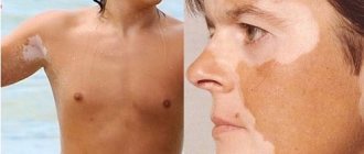
Microsporia is a fungal disease of the skin. Very often transmitted from infected pets. Another name for the disease is ringworm.
Microsporia is the most well-known disease among young children, caused by fungi of the genus Microsporum. Due to the high contagiousness and multiple routes of transmission of infection, the pathological process can spread to adults. The characteristics of the course of the disease directly depend on the type of fungus that provoked the onset of the disease. Correct and timely treatment eliminates the risk of an inflammatory process of the skin with irreversible consequences, for example, the appearance of deep ulcers, scars, and widespread infection on the skin.
Microsporia as a disease
Microsporia in humans is the subject of research by dermatologists and infectious disease specialists. According to its etiological characteristics, microsporia belongs to an infectious disease from a large group of dermatophytosis, when skin lesions can occur on smooth and hairy parts of the body.
The localization of the infectious process can be either focal or generalized, which completely depends on the degree of development of the disease and the type of fungus from the genus Microsporum . Another name for the disease is ringworm, but the term cannot be equally applied to microsporia of all varieties.
According to clinical data, children are susceptible to infection due to frequent neglect of personal hygiene and frequent contact with street animals (dogs, cats and kittens). The child interacts more often with other people’s animals when playing on the playground. In younger children, infection is possible through contact, due to the need to learn about the world around them “through the mouth.” The main pathogens are:
- Microsporum canis (canine microsporum);
- Microsporum ferrugineum (rusty microsporum).
Canine microsporium is a zoophilic fungus. The vegetative body of the fungus is thin and resembles a reed in appearance. The fungus affects domestic animals and humans. Rusty microsporia most often affects the human body, and under certain factors, the infection can spread to animals. The vegetative body of the fungus is wide and its shape is flat.
When grown in artificial environments, colonies of the fungus have a rich brown color, reminiscent of rust on metal. Microsporum ferrugineum is a particularly contagious form of the fungus of the genus Microsporum. For infection, it is enough for a small part of the fungus to come into contact with human skin. The fungus is quite resistant to the external environment, where it can remain viable for about 3–4 months.
The huge family of microorganisms of the genus Microsporum is not represented only by fungi such as Microsporum canis and Microsporum ferrugineum. Particular attention is due to the key practical significance in medical practice.
When and how can a child become infected?
Microsporia is a highly contagious (highly contagious) disease caused by dermatophyte fungi (literally, growing on the skin) of the genus Microsporum.
There are several types of this fungus that cause the development of three forms of the disease.
Anthropophilic and zoophilic forms of microsporia are widespread in Russia.
The disease occurs throughout the year. However, zoophilia is characterized by seasonality: the largest number of cases are recorded in late summer and autumn.
Classification of fungal infection
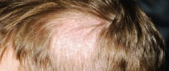
Microsporia in humans, initial stage photo
The classification of microsporia is important for clinicians to identify an infectious disease from skin lesions of another origin. Microsporia is classified according to the following main areas.
According to the location of the lesion:
- fungal infection of smooth skin;
- microsporia of the scalp;
- appearance on the nail plates.
Nail damage is a rare clinical situation and can only be caused by an advanced form of fungal infection of the human body. Fungal colonies can grow, affecting the nail plate. The rarity of the appearance of an infectious focus on the surface of the nails is explained by the lack of a nutrient medium for the fungus in this anatomical zone.
By pathogen type:
- Zoonotic microsporia. It is considered a type of zoophilic microorganisms carried by animals.
- Anthropotic form. The main carriers are people.
- Geophilic. This type of Microsporum fungus lives in soil substrates.
Classification according to the type of infectious agent does not have significant clinical significance in therapeutic practice, since the symptoms, diagnosis and treatment tactics are the same in all cases. This classification is used by epidemiologists to compile statistical values, as well as to determine the sources of infection in a certain area.
By type of clinical course:
- superficial;
- purulent (otherwise, infiltrative-suppurative).
The superficial form is characteristic of the anthropotic form of the fungus. The lesion covers the upper epidermal layers, causing peeling, redness of the skin, hair thinning, breakage, and loss in the affected areas. The infiltrative form of microsporia is one of the most severe clinical cases. Animals and much less often people are susceptible to infection, and only animals are the main carriers. In areas of skin lesions, large focal fragments with purulent exudate are recorded, and the patient’s general well-being suffers.
Symptoms of microsporia, photo
The duration of the incubation period depends on the type of fungus that provoked microsporia (see photo).
Thus, when infected with zoophilic and geophilic species of Microsporum fungi, the incubation period lasts 5–14 days. And when infected with anthropophilic forms, the incubation period of microsporia lasts much longer - from 4 to 6 weeks.
But since microsporia is most often provoked by a fungus of the Microsporumcanis species, which belongs to the zoophilic species, in most cases the incubation period of the infection is 1 - 2 weeks.
Development and transmission routes
The high degree of infectiousness is due to the presence of the fungus in the body of animals, in soil substrates, and in some plants. The pathogenic fungus Microsporum is a type of anaerobic microorganisms that require oxygen to maintain their vital functions. Fungi receive nutrition only on structures with a high content of proteins and microelements, so lesions are recorded only on the surface of the epidermis, as well as in hair follicles.
The vegetative body of the fungus is made up of mycelium - a multinucleated cell obtained as a result of the welded concentration of many fungal cells. The mycelium forms fungi, and when favorable conditions arise, spores. Spores are known to be a way for a fungal colony to reproduce.
Once on the surface of the epidermis, the fungus immediately begins its life activity in the vellus hairs of smooth skin, in the hair follicles. The main source of infection in the human body is contact with sick animals (guinea pigs, livestock, cats, dogs and their offspring).
Carriers can also be representatives of wild nature (monkeys, foxes, arctic foxes, lions, tigers). Infection can occur not only through direct contact, but also through the use of animal care equipment (bowls, food, toilet, wool). There are several main routes of transmission:
- contact (through direct or indirect contact with animal carriers or humans);
- through soil or plants (infection is possible through contact with soils and plants with which the hosts of the fungus have had any interaction).
Sometimes infection can occur from a sick person to a healthy one. Often, when in contact with sick animals, a person is not aware that they have a fungal disease. The disease in animals is often asymptomatic and occurs in a latent form. Epidemiologists note spikes in infection in the fall, as well as in the summer months, when cats give birth. Often it is kittens that become hidden carriers of a pathogenic fungus. To become infected, it is enough to pick up a kitten, stroke its fur, and put it to bed.
What is the difference between microsporia and trichophytosis?
| Peculiarities | Microsporia | Trichophytosis |
| Skin condition | Covered with plaque | Smooth |
| Hair condition | Broken at 2-3 mm | Broken at the base, only black spots visible |
| Transfer method | From person to person | From a person, an animal, an object affected by spores |
| Danger | In older children it goes away on its own | Requires special therapy; ignoring symptoms can lead to complications |
| Therapy methods | Antifungal drugs for local and systemic use (Batrafen, Mikoderil, Exoderil, Mikosan, Lotseril, Lamisil and others) | |
Related Articles
Trichophytosis, ringworm: symptoms, treatment, prevention
Symptoms and treatments for scalp fungus
Dermatophytosis: types, characteristic symptoms, treatment methods
Terbinox for mycoses: release forms, instructions for use, precautions
Fungus on the face: photos of symptoms, characteristic features, treatment methods
Sun fungus on the skin: causes, symptoms and treatment
Symptoms and clinical picture
The first signs of a fungal infection depend on the location of the infectious focus. Symptoms on smooth skin differ from the manifestations of fungus on hair structures. The clinical picture in children may be similar to the adult course and may differ from it.
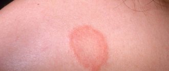
Symptoms of smooth skin lesions
Microsporia of smooth skin is characterized by the formation of a spot with clearly defined boundaries. As the disease progresses, such a spot increases in diameter. The edge of the spot resembles a defined roller, rising above the surface of the skin, having a bubbly structure with crusts and nodules. The center of the spot is a focal inflammatory process with a pink tint and pronounced peeling. The number of ring-shaped lesions when affecting smooth skin is from 1 to 3, and the diameter varies from 0.5 to 3.5 cm. More often, lesions are recorded on the neck, face, shoulders or forearms. Significant symptoms include moderate itching and flaking.
In children during the neonatal period or in young women, peeling may be completely absent, but the inflammatory process will be more significant. With a complicated allergic history, the Microsporum fungus can be mistaken for atopic dermatitis or other chronic skin diseases, so it is not always detected and identified in a timely manner. If you continue hormonal treatment of the underlying disease, fungal colonies begin to grow rapidly. When the pathological process spreads to the nail plate, the outbreak is usually localized along the edge of the nail from the cuticle. The form of the disease is characterized by the appearance of a whitish spot, gradual thinning and destruction of the nail structure.
Signs of fungus on the scalp
Microsporia of the scalp is often recorded in children aged 4 to 13 years. The frequency of occurrence in children is explained by the absence of special organic acids in the hair, which prevent the spread of fungal colonies and are found in sufficient quantities in the hair of adults. If a small patient is a carrier of a fungus of the genus Microsporum with its localization in the scalp, then spontaneous recovery is possible when he reaches puberty. An interesting fact is the rarity of the occurrence of lesions on hair structures in red-haired children. Characteristic signs are the following manifestations:
- the formation of 1 or 2 large lesions on the crown, temples or crown;
- the diameter of the lesions varies from 2.5 to 6 cm;
- the shape of the pathological spot can be round or oval;
- the presence of screenings along the edge of the lesion with a diameter of about 1 cm;
- peeling;
- hair fragility;
- slight itching.
As the disease progresses, the hair in the affected area breaks off and resembles a “hedgehog” after a haircut. Hence the name “ringworm”. The pathological ring exhibits slight swelling and redness.
Symptoms of the suppurative form
The purulent form is characterized by the formation of a focus with visible inflammation, the formation of soft nodules of a bluish tint. Pustules form on the surface of the affected skin area. With slight pressure, purulent exudate is released. The development of a suppurative form is provoked by inadequate treatment of the superficial form of microsporia, the presence of a burdened clinical history and chronic diseases, and advanced forms of microsporia.
Making a diagnosis of microsporia only based on primary signs and the appearance of the pathological formation may be unreliable. If there is any doubt, doctors resort to different methods of differential diagnosis.
Means and methods of therapy
Microsporia cannot be left untreated - an infected dog poses a danger to people and other animals . Therapy should be aimed at reducing the spread of pathogens in the environment. It is necessary to localize the lesion and eliminate it as quickly as possible. Treatment includes local treatments and systemic medications; symptomatic and compensatory therapy may also be required, as well as the fight against secondary pathologies.
Remains of hair are removed from the affected areas of the skin and around them - this is necessary for better contact of medicinal substances with the skin. Various ointments, lotions and sprays are used for treatment:
- miconazole;
- clotrimazole;
- ketaconozole;
- salicylic ointment;
- xeroform;
- kubatol (tar-based spray);
- Wilkinson's ointment;
- 2% chlorhexidine solution.
It is useful to perform a complete treatment of the dog’s coat once. For this purpose, special shampoos containing drugs against microsporia are used. It is recommended to wash the dog at the beginning of treatment. In small breed dogs, hair is completely removed from the body and the entire surface of the skin is treated. For bathing, enilconazole solutions are used in a dose of 20 ml/l of water, 2% chlorhexidine - 25 ml/l.
If there is no pronounced inflammatory reaction, then the use of ointments with corticosteroids is not recommended. These drugs reduce the effectiveness of antifungal agents. They also often cause atrophy of hair follicles and thinning of the skin, which provokes complications of the pathology.
Griseofulvin is used as a systemic means of control, penetrating into the subcutaneous layers and suppressing microsporia on the entire surface of the body, which avoids the hidden spread and persistence of infection. The drug has severe side effects - vomiting, diarrhea, especially in puppies. Griseofulvin should not be used in pregnant bitches. Ketocanozole and itrocanozole can be used as alternatives.
A vaccine against fungal infections can be used as an adjuvant. Despite the name, this drug does not have a pronounced preventive effect, but it suppresses the activity of microspores. The vaccine has a detrimental effect on the fungus and also accelerates the restoration of skin and hair.
Treatment of microsporia is a very long procedure. Regardless of the drug chosen, the course is 2-3 months. Therapy is continued for another two weeks after the disappearance of clinical signs and negative microscopic examinations.
Diagnostic measures
Some symptoms of the disease, especially if there is a history of other skin diseases, may be similar to each other. Diagnostic measures make it possible to accurately determine the development of the disease, exclude similar pathologies, and begin timely treatment. The main diagnostic methods include:
- laboratory tests (urinalysis, blood);
- biochemical blood test to determine liver function;
- scraping of fungi from the surface of the lesion to identify the type of pathogen;
- illumination with a fluorescent lamp.
The main task of differential diagnosis is to establish an accurate diagnosis in the shortest possible time, as well as to exclude the exacerbation of some chronic skin diseases.
Treatment tactics
If the lesion affects only the surface of smooth skin and is not localized on the scalp, then systematic treatment with drugs for topical use (balms, ointments, liniments) is sufficient. Microsporia of smooth skin can be treated with the following drugs:
- Lamisil, Terbizil;
- Sulfuric ointment;
- salicylic-sulfuric;
- tar ointment;
- bifosin and others.
Ointments are recommended to be used simultaneously with antiseptic solutions to achieve a better therapeutic effect. The causative agents of microsporia are particularly resistant to antimycotics, so treatment requires duration and the use of large doses of drugs. If the disease is localized on the hair structures, then tablets are added to the use of local remedies. The main ones are:
- Griseofulvin (at the rate of 22 mg per kg of body weight 3 times a day with a spoon of any oil);
- Terbinafine (about 250 or 500 mg 1 time per day);
- Itraconazole (100 to 200 mg per day 1 time).
For microsporia, the treatment of which is agreed upon with the doctor individually, it is important to comply with the isolation regime. The patient must be in an isolated box, and all things must be disinfected. Microsporia in children has the same principles in treatment, and the main difference is the revision of the dosage and medications with the least amount of side effects. In case of aggravated course of the disease, in the presence of a complicated medical history and chronic diseases, the course of treatment is selected by specialists collectively according to the profile of concomitant pathologies.
Folk remedies for treating microsporia
The following traditional medicines are used:
- apply napkins moistened with fresh onion juice to the affected areas;
- lubricate the affected areas with tincture of common lilac flowers: pour two tablespoons of dried flowers with 100 milliliters of 70% alcohol, leave and strain;
- wash the affected areas with a decoction of celandine herb: pour one tablespoon of dry herb with a glass of water and boil over low heat for 10-12 minutes, cool, strain; alternate with other means;
- lubricate affected areas with propolis oil: chop 15-20 grams of propolis with a knife, pour in 50 grams of vegetable oil and heat in a water bath or in the oven until the oil boils, stirring occasionally; let the oil boil two or three times; the wax will settle to the bottom of the dish, and the propolis will dissolve in the oil; when the prepared oil has cooled, carefully drain it from the sediment;
- lubricate the affected areas with an ointment prepared using the following mixture: burdock roots - two parts, hop cones - two parts, calendula officinalis flowers - one part; Preparation of the medicine: grind 10-15 grams of the dry mixture in a mortar into powder and mix with 40 grams of Vaseline.
Prevention and prognosis
Prevention of fungal skin infections comes down to maintaining hand and skin hygiene, and avoiding contact with other people's animals. Your own pets should be checked for various infections at least 2 times a year. This applies to families with small children. When a disease is detected, the patient must be isolated for adequate treatment and to prevent spread to other family members and surroundings. Household items and other personal belongings of the patient are subjected to antiseptic treatment and disinfection. With prompt consultation with a doctor and proper therapy, the prognosis for patients is favorable.
The disease does not pose any threat to the lives of patients. Proper treatment, organized on time, helps prevent irreversible damage to the structure of the skin (scars, scars). An untreated fungus on the scalp can cause baldness in the affected area and cause inferiority of the scalp. In addition to aesthetic defects, the disease can cause deep emotional distress, including depression and an inferiority complex. Timely therapeutic treatment can eliminate the risk of developing such negative complications.
Who is at risk?
Children of preschool and school age most often
Whereas in adults the disease is rare. This is due to the fact that after puberty, the sebaceous glands of the skin produce more fatty acids, which have a detrimental effect on the fungus.
In addition, the disease is more common in patients with predisposing factors :
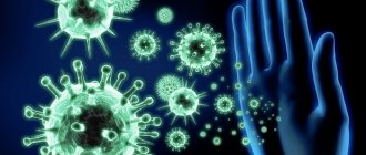
• Immune system dysfunction
• Endocrine diseases: diabetes mellitus, decreased thyroid function
• Lack of vitamins
• Poor housing, communal and living conditions
• High humidity and hot weather
• Presence of abrasions, cracks or scratches on the skin
• Tendency to excessive sweating
This is due to the fact that in these conditions, local immunity on the skin decreases, which facilitates the penetration of the fungus.
On a note
In children with skin allergic manifestations, microsporia can “masquerade” as the underlying disease - for example, with atopic dermatitis. What's happening? The fungus invades the site of existing rashes, significantly complicating diagnosis and treatment.
In addition, local use of hormonal ointments reduces the protective ability of the skin, which facilitates easy penetration and spread of the fungus.