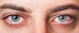
Exogenous allergic alveolitis is a diffuse inflammation of the lung tissue, leading to the development of pneumosclerosis, respiratory failure and cor pulmonale.
- Types of allergic alveolitis
- Pathogenesis
- Symptoms
- Diagnostics Differential diagnostics
- Laboratory diagnostics
Exogenous allergic alveolitis, or hypersensitive interstitial pneumonitis, develops with repeated aerogenic entry into the sensitized body of antigens (allergens) of microbial, animal, plant origin or various low-molecular chemical compounds.
First, sensitization of the body occurs in the form of Tn2 activation and the appearance of IgG antibodies to the causally significant antigen. The reaction can be caused by substances that can enter the alveoli. When a causally significant antigen enters a sensitized organism, the reaction occurs quickly (within 4-8 hours) similar to the Arthus phenomenon with the formation of immune complexes. The processes of deposition of immune complexes and the formation of chemoattractants, anaphylatoxins (C5a, C3) as a result of complement activation lead to acute damage to blood vessels and alveoli with the involvement of neutrophils and macrophages.
On the 2nd day after contact with the antigen, DTH develops with the involvement of CD4+ (Tn1), activated macros and CD8+ cytotoxic T lymphocytes. Subsequently, epithelioid-macrophage granulomas are formed, fibroblasts are activated, and interstitial fibrosis develops.
Some types of exogenous allergic alveolitis and sources of causally significant allergens
| Type of exogenous allergic alveolitis | Source of allergens and allergens |
| Alveolitis caused by medications | Medicines: penicillins and other antibiotics, sulfonamides, nitrofurans, gold salts |
| Alveolitis caused by low molecular weight compounds | Dithioisocyanates, heavy metal salts |
| Barn disease | Wheat, flour (allergens of grain weevil, fungi) |
| Coffee miller's disease | Coffee beans |
| Disease of those washing in the sauna | Damp wood (fungal allergens) |
| Lung disease associated with the use of humidifiers and air conditioners | Water and air containing allergens of fungi and bacteria, including allergens of actinomycetes |
| Cheesemakers' disease | Moldy cheese (penicillium fungi allergens) |
| Weavers cough | Cotton (mold allergens) |
| Tanner's lung | Maple bark (mold allergens) |
| Bird lover's lung | Feathers and droppings of pigeons, budgies (bird and fungal allergens) |
| Lung of a worker processing malt | Rotted barley, malt dust (allergens of Aspergillus fungi - A. fumigatusl A. clavatus) |
| Farmer's lung | Rotted hay (allergens of thermophilic actinomycetes and fungi) |
1.General information
The nosological formulation “exogenous allergic alveolitis” may seem frighteningly complex; to avoid this, we will analyze it “under a microscope” in two different aspects – anatomical and terminological.
We will consider the nasopharynx and oropharynx to be the air intake part of the human respiratory system; the entrance to the visceral, internal part (lower respiratory tract) - the trachea, which descends from the larynx and at the level of 4-5 vertebrae is divided into two main bronchi.
Each of the main air channels further forms many branches, which together resemble a tree turned upside down (without foliage or needles). The terminal, thinnest “branches” are no longer called bronchi, but, in the diminutive sense, bronchioles. Bronchioles, unlike the bronchi of the previous orders, are devoid of a cartilaginous frame and are associated with the most important, at the same time the smallest gas exchange cells of the lungs - alveoli (Latin for “concave bubble”, “depression”). A person has about three hundred million alveoli in one lung.
Alveolitis, according to the principles of the formation of medical terms, should be understood as inflammation of the alveoli. Allergic alveolitis means caused by the body’s painful sensitivity to any specific substance or factor. Finally, the clarification “exogenous” indicates that this factor is external to the body, it acts or comes from the outside. In fact, this clarification is unnecessary, since any allergy is by definition exogenous, and hypersensitivity to internal factors is called differently: autoimmune reactions or diseases. It should also be added that the inflammation caused by hypersensitivity in this case covers not only the alveoli, but also the bronchioles - to reflect this fact in the formulation of the diagnosis, the disease would have to be called, for example, alveobronchiolitis.
Exogenous allergic alveolitis (sometimes written “hypersensitivity pneumonitis”, not to be confused with pneumonia) is informally called farmers' disease or organic dust disease. Like the much better known hay fever (hay fever), exogenous allergic alveolitis shows a certain seasonality and affects (especially in rainy weather) 4-8% of people engaged in agricultural work. However, this is not the only risk group.
The disease was first described less than a hundred years ago (in 1932); in terms of the general population of earthlings, it occurs with a frequency of approximately 0.05% and remains an object of study. Experts identify a number of clinical forms, usually called figuratively and etiologically aptly: “trombonist’s lung”, “sequoiasis”, “laboratory assistant’s lung”, “sauna worker’s lung”, “bagassosis” (literally translated “sugar-cane disease”), “suberosis” (literally “corkosis”), “thatched roof disease”, “pneumonitis of metalworking fluids” and many others.
Sign up for a consultation
A must read! Help with hospitalization and treatment!
Prices
| Name of service (price list incomplete) | Price |
| Appointment (examination, consultation) with a pulmonologist, primary, therapeutic and diagnostic, outpatient | 1750 rub. |
| Consultation with a candidate of medical sciences | 2500 rub. |
| Professor consultation | 4300 rub. |
| Consultation (interpretation) with analyzes from third parties | 2250 rub. |
| Prescription of treatment regimen (for up to 1 month) | 1800 rub. |
| Study of external respiratory function (RPF) with drug tests | 1800 rub. |
| X-ray of the chest organs (survey) | 1900 rub. |
| X-ray of the chest organs in 2 projections | 2900 rub. |
2. Reasons
As follows from the above, the essence of exogenous allergic alveolitis is a violent reaction of the immune system, selectively hypersensitive to fine dust suspensions in the inhaled air. If a person who is otherwise practically healthy is often or constantly exposed to such conditions, the likelihood of developing hypersensitivity pneumonitis reaches 10-15%.
At first, it was believed that the allergic reaction was caused predominantly by the organic component of airborne dust: microscopic fungal spores, particles of bird droppings, invisible house dust mites and their waste products, wood dust, microparticles in the production of beer, tobacco products, flour and any other products that require grinding dried plant materials. However, as allergology developed, more and more evidence accumulated in favor of a general concept: there is no substance that would not become an allergen for someone. Today, hypersensitivity pneumonitis is diagnosed in workers in the mining and chemical industries, paint and varnish and metalworking enterprises, manipulation rooms (“nurse’s lung”), gas stations, furniture stores, and many others. As with other allergies, sensitization to an organic or inorganic allergen is greatly facilitated by repeated, frequent or constant contact with it: this produces about the same effect on the immune system as wearing sandpaper gloves would have on the skin of the hands.
Visit our Pulmonology page
Diagnostics
At the moment, this disease is poorly studied. Its symptoms are similar to other respiratory diseases, so diagnosis can be difficult.
To make a diagnosis, the SM-Clinic pulmonologist performs the following procedures:
- Interviews the patient for complaints
- Collects data on the history of the disease
- Conducts a general inspection
- Takes a sputum sample
- Takes a blood test to determine inflammation
- Orders an x-ray of the lungs
- If necessary, prescribe a computed tomography scan
- Performs spirometry - a procedure for studying breathing
- Conducts provocative tests (the patient is allowed to inhale an aerosol with a suspected allergen)
- Takes a blood test for gas composition
- Performs bronchoscopy - a procedure to examine the bronchi from the inside.
- During bronchoscopy, tissue is collected for biopsy.
3. Symptoms and diagnosis
There are various types of exogenous allergic alveolitis: from acute or subacute attacks to chronic allergic status with a smoldering and periodically flaring clinic.
The picture of acute hypersensitivity alveolitis resembles the symptoms of ARVI: many patients report muscle and joint pain, general malaise, chills, headache, thoracalgia (chest pain) and a feeling of heaviness, expectorant cough, sometimes with blood in the sputum. When contact with the allergen is avoided, the symptoms gradually decrease, but with each resumption they can become more and more severe.
Chronic allergic alveolitis is fraught with progressive cardiopulmonary failure with all its characteristic symptoms: shortness of breath, persistent cough, difficulty breathing, hypertension, “drum stick and watch glass” syndrome (deformation of the terminal phalanges and nail plates), cyanotic skin, etc. With a long-term (ten or more years) course, almost all patients develop serious complications: three quarters of patients suffer from chronic bronchitis, every fourth suffers from pulmonary emphysema.
Diagnosis requires a detailed study of the anamnesis - first of all, living and working conditions, the possible connection of attacks with specific situations, past allergic reactions, etc. are important. An external examination, auscultation, spirometric tests, pulmonary radiography, CT, and in some cases bronchoscopy are performed. Of the laboratory data, the most important are the immunological indicators of blood and sputum obtained by bronchoalveolar lavage (wash).
The extreme nonspecificity of the clinical picture and its instability when the action of the allergen ceases creates a high probability of differential diagnostic errors. It can be assumed that the diagnosis of “exogenous allergic alveolitis” is established many times less often than this condition actually occurs.
About our clinic Chistye Prudy metro station Medintercom page!
EXOGENOUS ALLERGIC ALVEOLITIS
Exogenous allergic alveolitis (EAA) was first described in 1932. Since then, various variants of the course of this disease have been identified, the development of which is caused by exposure to different antigens. Sources of these antigens can be moldy hay, compost, bird and rodent dander, air conditioners, humidifiers, etc. Functional changes are nonspecific and are similar to those in other interstitial lung diseases. The most sensitive change is a decrease in the diffusion capacity of the lungs. The prognosis of the disease does not depend on the functional state at the time of diagnosis. The basis of treatment is the exclusion of contact with the “culprit” agent. Corticosteroids may be prescribed; If complications occur, symptomatic therapy is carried out.
Extrinsic allergic alveolitis was first described in 1932. Since different variants of the disease which are caused by various antigens have been identified. The sources of the antigens may be moldy hay, compost, avian and rodent dandruff, air-conditioners, humidifiers, etc. The functional changes are nonspecific and similar to those of other interstitial diseases of the lung. The decreased diffusion function of the lung is the most obvious change. The prognosis of the disease does not depend on the functional status at diagnosis. The basis of treatment is to exclude contacts with a “guilty” agent. Corticosteroids may be given. Symptomatic therapy is used if complications occur.
O. E. Avdeeva, S. N. Avdeev, A. G. Chuchalin Research Institute of Pulmonology, Ministry of Health of the Russian Federation, Moscow O. Ye. Avdeeva, SN Avdeev, AG Chuchalin Research Institute of Pulmonology, Ministry of Health of the Russian Federation, Moscow
Introduction
Exogenous allergic alveolitis (EAA), or hypersensitivity pneumonitis, includes a group of related interstitial lung diseases characterized primarily by diffuse inflammatory changes in the lung parenchyma and small airways, developing in response to repeated inhalation of various antigens that are products of bacteria, fungi, animal proteins, some low molecular weight chemical compounds. The disease was first described in 1932 by J. Campbell in five farmers who developed acute respiratory symptoms after handling wet, moldy hay. This form of the disease is called "farmer's lung." Variants of EAA associated with other causes were then described. Thus, the second most important form of EAA - “bird lovers' lung” - was described in 1965 by S. Reed et al. [2] in three patients who were breeding pigeons. EAA can have a different course and prognosis: the disease can be completely reversible, but it can also lead to irreversible damage to the pulmonary architecture, which depends on many factors, including the nature of antigen exposure, the nature of the inhaled dust and the patient’s immune response. The incidence of the disease is up to 42 cases per 100 thousand of the total population. It is very difficult to determine what percentage of patients exposed to the culprit agent will develop EAA. However, most experts agree that approximately 5 to 15% of individuals exposed to high concentrations of the etiologic agent will develop hypersensitivity pneumonitis. The prevalence of EAA among people exposed to low-containing agents has not yet been determined.
Etiology
Most often, the development of EEA is associated with professional factors, hobbies, and can also be the result of environmental exposure. Some etiological agents responsible for the development of EEA are presented in the table. The most important of these agents are thermophilic actinomycetes and avian antigens. In agricultural areas, the leading causative agents are thermophilic actinomycetes - bacteria less than 1 micron in size, with the morphological properties of fungi, they are widely found in soil, compost, water, and air conditioners. The most common species of thermophilic actinomycetes associated with EAA are Micropolyspora faeni, Thermoactinomyces vulgaris, Thermoactinomyces viridis, Thermoactinomyces saccharis, and Thermoactinomyces candidum. These microorganisms multiply at temperatures of 50 - 60 ° C, i.e. in those conditions that are achieved in heating systems or during the decay of organic material. Thermophilic actinomycetes are responsible for the development of “farmer's lung”, bagassosis (lung disease in sugar cane workers), “lung of mushroom growers”, “lung of people who use air conditioners”, etc.
Causal factors of exogenous allergic alveolitis
| Syndrome | Antigen source | Possible antigen |
| Farmer's lung | Moldy hay | Thermophilic Actinomycetes: Mycropolyspora faeni, Thermoactinomyces vulgaris; Aspergillus spp. |
| Bagassoz | Sugar cane | Mycropolyspora faeni, Thermoactinomyces sacchari |
| Lung of mushroom growers | Compost | Thermoactinomyces vulgaris, Mycropolyspora faeni |
| Lung of persons using air conditioners | Air conditioners, humidifiers, heaters | Thermoactinomyces vulgaris, Thermoactinomyces viridis, Аmeba, Fungi |
| Suberosis | Cork tree bark | Penicillum frequentans |
| Malt brewer's lung | Moldy barley | Aspergillus clavatus |
| Cheesemakers' disease | Cheese particles, mold | Penicillum caseii |
| Redwood | Sequoia wood dust | Ggaphium spp., Pupullaria spp., Alternaria spp. |
| Lung producing detergents | Detergents, enzymes | Bacillus subtitus |
| Bird lovers lung | Excrement, bird dander | Poultry whey proteins |
| Lung of laboratory workers | Rodent urine and dander | Proteins in rodent urine |
| Lung sniffing pituitary gland powder | Pituitary gland powder | Pork and bovine proteins |
| Lung of those employed in the plastics industry | Diisocyanates | Toluene diisocianate, diphenylmethane diisocianate |
| "Summer" pneumonitis | Dust from damp residential areas (Japan) | Trichosporon cutaneum |
Avian antigens are represented mainly by serum proteins - gamma globulin, albumin. These proteins are found in the excrement and secretions of the skin glands of pigeons, parrots, turkeys, canaries and other birds. People caring for these birds most often become ill through chronic contact with them. Proteins from pigs and cows can also cause EEA, an example of which is the disease that develops in patients with diabetes insipidus who snort pituitary gland powder - “the lung of individuals who snort pituitary gland powder.”
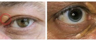
Rice. 1. “Drumstick” syndrome with chronic EEA.
Among fungal antigens in EEA, Aspergillus spp is of greatest importance. Various species of Aspergillus are associated with the development of diseases such as malt cooks lung, cheese makers lung, suberosis (a disease that develops in workers who work with cork bark), as well as farmer's lung, and air conditioner lung. Aspergillus fumigatus can cause the development of alveolitis in urban residents, as it is a frequent inhabitant of damp, unventilated, warm rooms. An example of EAA associated with reactive chemical compounds is the disease in people involved in the production of plastics, polyurethane, resins, and dyes. The most important are diisocyanates and phthalic anhydrite.
Rice. 2. Epithelioid cell granuloma in subacute EAA (hematoxylin-eosin staining; x 400).
The causes of EAA vary significantly between countries and regions. Thus, in the UK, among the forms of EAA, “the lung of budgerigar lovers” predominates, in the USA - “the lung of those who use air conditioners and humidifiers” (15 - 70% of all variants), in Japan - the “summer type” of EEA, etiologically associated with the seasonal growth of fungi of the species Trichosporon cutaneum (75% of all variants). In large industrial centers (Moscow), according to our data, the leading causes are currently avian and fungal (Aspergillus spp.) antigens.
Pathogenesis
A necessary condition for the development of EAA is the inhalation of antigenic material of a certain size in a sufficient dose and over a certain period of time. In order for antigen deposition to occur in the small airways and alveoli, the antigen must be less than 5 microns in size, although the disease may develop with the absorption of soluble antigens from large particles deposited in the proximal parts of the bronchial tree. Most people exposed to antigenic material do not develop EAA, which suggests, in addition to external factors, the participation of endogenous factors in the development of the disease, which have not yet been fully studied (genetic factors, characteristics of the immune response).
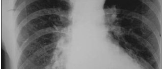
Rice. 3. Plain radiograph for EAA, chronic course. Diffuse infiltration and enrichment of the pulmonary pattern, mainly in the basal regions.
EAA is rightly considered an immunopathological disease, in the development of which the leading role belongs to allergic reactions of types 3 and 4 (according to the Gell, Coombs classification), and non-immune inflammation is also important. Immune complex reactions (type 3) are of primary importance in the early stages of EAA development. The formation of immune complexes (IC) occurs in situ in the interstitium during the interaction of the inhaled antigen and IgG. Local deposition of IR causes acute damage to the interstitium and alveoli, characterized by neutrophilic alveolitis and increased vascular permeability. IR leads to activation of the complement system and alveolar macrophages. Active complement components increase vascular permeability (C3a) and have a chemotactic effect on neutrophils and macrophages (C5a). Activated neutrophils and macrophages produce and release pro-inflammatory and toxic products such as oxygen radicals, hydrolytic enzymes, arachidonic acid products, cytokines (such as interleukin-1-IL-1, tumor necrosis factor a - TNF-a). These mediators lead to further damage and necrosis of cells and matrix components of the interstitium, enhance the body's acute inflammatory response, and cause an influx of lymphocytes and monocytes, which further support delayed-type hypersensitivity reactions. Evidence of the development of immune complex reactions in EAA is: the timing of the inflammatory response after contact with the antigen (4 - 8 hours); detection of high concentrations of precipitating IgG antibodies in the serum and bronchoalveolar fluid (BAL) of patients; detection of immunoglobulin, complement components and antigens in the histological material of lung tissue during acute EAA, i.e. all components of IC; classical skin Arthus reactions in patients with EAA, caused by highly purified preparations of the “culprit” antigens; increase in the number of neutrophil leukocytes in BAL fluid after inhalation provocation tests. T-lymphocyte-mediated immune responses (type 4) include CD4+ T-cell delayed-type hypersensitivity and CD8+ T-cell cytotoxicity. Delayed reactions develop 24 to 48 hours after exposure to the antigen. Cytokines released as a result of immune complex injury, especially TNF-a, induce the expression of adhesion molecules on the cell membranes of leukocytes and endothelial cells, which significantly increases the subsequent migration of lymphocytes and monocytes to the site of inflammation. A distinctive feature of delayed-type reactions is the activation of macrophages by gamma interferon secreted by activated CD4+ lymphocytes. Continued antigenic stimulation supports the development of delayed reactions and leads to the formation of granulomas and activation of fibroblasts by growth factors, and ultimately to excessive collagen synthesis and interstitial fibrosis. Evidence of type 4 reactions is: the presence of memory T-lymphocytes both in the blood and in the lungs of patients with EAA; histological confirmation in subacute and chronic course of EAA in the form of granulomas, lymphomonocytic infiltrates and interstitial fibrosis; animal models with experimental EAA have shown that the presence of CD4+ T lymphocytes is required for disease induction.
Clinical picture
There are three types of disease: acute, subacute and chronic. Acute EAA
usually develops after massive exposure to a known antigen in the home, work environment, or environment.
Symptoms appear within 4 to 12 hours and include fever, chills, weakness, chest tightness, cough, shortness of breath, muscle and joint pain. Patients rarely have sputum, and if present, it is scanty and mucous. Frontal headaches are also a common symptom. When examining the patient, cyanosis is often detected, and when auscultating the lungs, crepitus, more pronounced in the basal sections, is often detected, and wheezing may sometimes be present. These symptoms usually resolve within 24 to 72 hours, but often recur after new contact with the “culprit” antigen. Shortness of breath on exertion, weakness, and general lethargy may persist for several weeks. A typical example of acute EAA is farmer's lung, where symptoms appear within a few hours of exposure to moldy hay. EAA is diagnosed quite rarely; atypical pneumonia of a viral or mycoplasmal nature is often assumed, and the correct diagnosis largely depends on the doctor’s alertness. For farmers, the differential diagnosis of acute EAA is made with pulmonary mycotoxicosis (or toxic organic dust syndrome), which occurs due to massive inhalation of fungal spores. In contrast to patients with acute EAA, almost all patients with mycotoxicosis have a normal radiograph and there are no precipitating antibodies in the serum. The subacute form
develops with less intense chronic exposure to the “culprit” antigens, which often occurs at home.
A typical example is EAA associated with exposure to poultry. The main symptoms are shortness of breath on exertion, fatigue, cough with mucous sputum, and sometimes fever at the onset of the disease. In the lungs, usually in the basal regions, soft crepitus is heard. Differential diagnosis usually includes sarcoidosis and other interstitial lung diseases. If dust inhalation occurs over a long period of time and the dose of inhaled antigen is low, a chronic form of EAA
. Unrecognized or untreated subacute EAA can also become chronic. A characteristic symptom of chronic alveolitis is progressive shortness of breath during physical exertion, sometimes accompanied by anorexia and a pronounced decrease in body weight. Patients subsequently develop interstitial fibrosis, cor pulmonale, respiratory failure, and heart failure. The insidious onset of symptoms and the absence of acute episodes often make it difficult to distinguish EAA from other interstitial lung diseases, such as idiopathic fibrosing alveolitis. Tachypnea and crepitus are also common findings in chronic EAA. Wheezing can be observed with airway obstruction, but is not a characteristic sign of the disease, but in some patients it can lead to erroneous diagnostic conclusions. In the chronic course of EAA, changes in the terminal phalanges of the fingers in the form of “watch glasses” and “drumsticks” are often observed. In a recent study, Sansores (1990) et al. The clubbing symptom was found in 51% of 82 patients with bird lovers lung disease. It should be noted that disease progression was observed in 35% of patients with the “drumstick” symptom and only in 13% of patients without it. Thus, the “drumstick” symptom is a common sign of chronic EAA and can serve as a harbinger of an unfavorable outcome.
X-ray picture
Changes in radiographs of the lungs can vary from a normal picture in the case of acute and subacute clinical forms to a picture of severe pneumosclerosis and “honeycomb lung”. The X-ray picture may be normal even in the presence of hypoxemia, pronounced changes in functional tests and granulomatous changes in the histological material (M. Arshad et al., 1987). In one study analyzing 93 cases of EAA, S. Monkare et al. found that the radiological picture was unchanged in 4% of cases and minimally changed in 25.8%. These minimal changes included a slight decrease in the transparency of the lung fields - a “ground glass” appearance that is easily “visible” during the initial examination. The X-ray picture varies significantly with different variants of the course and stages of the disease. In acute and subacute forms, the most common findings are changes in the form of a decrease in the transparency of the lung fields of the “ground glass” type, widespread nodular-reticular opacities. The size of the nodules usually does not exceed 3 mm and can involve all areas of the lungs. Often the apexes and basal regions of the lungs remain free of nodular lesions (R. Cook et al., 1988). X-ray changes in the acute course of EAA usually resolve within 4–6 weeks in the absence of repeated contact with the “culprit” allergen. As a rule, improvement in the X-ray picture precedes the normalization of functional tests, such as, in particular, the diffusion capacity of the lungs. In chronic alveolitis, well-defined linear shadows, pronounced interstitial changes, nodular opacities, a decrease in the size of the lung fields are often detected, and in advanced stages - a picture of a “honeycomb lung”. Computed tomography (CT) is a more sensitive method for imaging EAA. CT makes it possible to detect nodular opacities, “ground glass” zones, and “cellular changes” that are invisible with conventional radiography. In a study by D. Hansell et al. [3] showed a significant correlation between the severity of the decrease in the transparency of the pulmonary fields according to CT data and functional indicators - the residual volume and its ratio to the total lung capacity.
Laboratory data
During acute attacks of EAA, laboratory blood tests reveal moderate leukocytosis, on average up to 12 - 15 x 103 per 1 ml. Sometimes leukocytosis can reach 20 - 30 • 103 per 1 ml (D. Emanuel et al., 1964). There is often a shift in the leukocyte formula to the left. Eosinophilia is rarely detected and, if present, is often insignificant. Most patients have normal ESR values, but in 31% of cases this figure reaches 20 - 40 mm/h and in 8% more than 40 mm/h (S. Moncare, 1984). Elevated levels of total IgG and IgM are often detected, and sometimes the level of total IgA is also increased (C. Aznar et al., 1988). Some patients also exhibit a moderate increase in rheumatoid factor activity. Quite often, an increase in the level of total LDH is noted, which may reflect the activity of the inflammatory process in the pyrenchyma of the lungs (S. Matusiewicz et al., 1993). Of particular importance in EAA is the detection of specific precipitating antibodies to the “culprit” antigen. The most commonly used methods are double diffusion according to Ouchterlony, micro-Ouchterlony, counter immunoelectrophoresis and enzyme immunoassay methods (ELISA, ELIEDA). Precipitating antibodies are found in most patients, especially in acute cases of the disease. After cessation of contact with the antigen, antibodies are detected in the serum for 1 to 3 years (Y. Cormier et al., 1985). In chronic cases, precipitating antibodies are often not detected. False-positive results are also possible; Thus, among farmers who do not have symptoms of EAA, antibodies are detected in 9 - 22% of cases (Y. Cormier et al., 1989; E. Tercho et al., 1987), and among “bird lovers” - in 51% (C McSharry et al., 1984). In patients with EAA, the level of precipitating antibodies does not correlate with disease activity and may depend on many factors, for example, it is significantly lower in smokers (K. Anderson et al., 1988). Thus, the presence of specific antibodies does not always confirm the diagnosis of EAA, and their absence does not exclude the presence of the disease. However, the detection of precipitating antibodies can help in the diagnosis of EAA when there is an assumption of the presence of EAA based on clinical data, and the nature of the “culprit” agent is unclear.
Functional tests
Functional changes are nonspecific and similar to those in other interstitial lung diseases. The most sensitive functional change is a decrease in lung diffusion capacity (DCL), which is also a good predictor of oxygen transport - a decrease in DCL well reflects the severity of desaturation during exercise. Impaired gas exchange is usually reflected by hypoxemia at rest, aggravated by physical activity, an increased alveoloarterial P(A-a)O2 gradient, and normal or slightly reduced partial tension of CO2 in arterial blood. In the early stages of the disease, as a rule, there is a normal O2 tension in the arterial blood, but a decrease in saturation is already noted during physical activity. Changes in pulmonary function tests in acute EAA usually appear 6 hours after antigen exposure and demonstrate a restrictive type of ventilation impairment. Changes in the function of external respiration can sometimes occur in two phases: immediate changes of an obstructive type, including a decrease in forced expiratory volume in 1 s (FEV1), a decrease in the Tiffno coefficient (FEV1/FVC); these changes persist for about an hour, and then after 4 - 8 hours they are replaced by a restrictive type of ventilation: a decrease in lung volumes - total lung capacity (TLC), vital lung capacity (VC), functional residual capacity (FRC), residual lung volume (RLV) . The Tiffno coefficient is within normal values; there may be a decrease in the maximum average expiratory flow (MSEP 25 - 72), which reflects the presence of obstruction at the level of the small airways. In chronic EAA, the most characteristic change is also a restrictive pattern: a decrease in static lung volumes, a decrease in pulmonary compliance, and lung DFL. Sometimes, with chronic changes, an increase in compliance and a decrease in elastic recoil are described, which is characteristic of airway obstruction in emphysema (R. Seal et al., 1989). Approximately 10–25% of patients show signs of airway hyperresponsiveness. Alveolar damage in interstitial lung diseases reflects decreased clearance of DTPA-labeled technetium (99mTc) from the lungs into the blood. S. Bourke et al. (1990) found that the clearance rate of technetium was altered in 20 non-smoking pigeon fanciers who had normal DCL and TEL values. Further study of this method in a large sample of EAA patients is necessary to confirm the role of the 99mTc-DTPA clearance test in routine clinical practice. So far, no correlation has been shown between changes in respiratory function and the prognosis of EAA. Patients with significant functional changes may recover completely, while patients with minor functional defects at disease onset may subsequently experience progressive disease with the development of fibrosis and small airway obstruction.
Provocative tests
Inhalation tests were first carried out by J. Williams (1963) at the Brompton Clinic; he was able to reproduce the symptoms of acute EAA. Aerosols for the tests were prepared from moldy hay dust, from moldy hay extracts, and from actinomycete extracts isolated from moldy hay. In each case, the disease was "reproduced" in farmers with a history of EAA. Inhalation tests with extracts from "good hay" in patients with "farmer's lung" or with extracts of moldy hay in healthy people did not result in symptoms of the disease. Unlike patients with bronchial asthma, provocative tests in EAA do not cause immediate symptoms or changes in pulmonary function. However, 4–6 hours later, patients with a positive response develop dyspnea, weakness, fever, chills, and crepitus in the lungs. When studying FVD, a significant decrease in vital capacity and DSL is revealed. These changes usually resolve within 10 to 12 hours (J. Fink, 1986). The materials used for the tests are prepared from dust of “suspicious” material or from extracts of mixtures of antigen substances obtained through various chemical processes. In each case, the inhaled agents are a mixture of different materials and often contain nonspecific irritants. There are currently no commercially available standardized, highly purified, specific antigens for challenge tests. Moreover, there are no standardized methods for performing tests or reliable dose-response indicators. Sensitive patients may experience severe exacerbation of the disease after the test. Significant hypoxemia is often observed, which may be why many patients are reluctant to undergo testing. Because of the late onset of symptoms and functional changes, and the need for frequent spirometry and diffusion tests, the provocative test takes quite a long time. Currently, it is customary to evaluate test results by reducing vital capacity, increasing the number of leukocytes in the blood, and increasing body temperature [4]. Fortunately, the diagnosis of EAA rarely requires such procedures, and provocative testing is usually performed only in research settings. However, in some circumstances, when convincing evidence of a causative factor of the disease is required (for economic or social reasons), provocative tests become necessary. One of the options for such tests can be observation of the patient in his natural professional or everyday conditions. Patients with chronic EAA often do not experience significant change in symptoms unless exposed to a massive dose of the culprit antigen, so natural exposure tests can leave patients somewhat skeptical about the cause of their disease.
Histological picture
A common symptom of EAA are non-castifying granulomas, which can be found in 67 - 70% of cases. These granulomas differ from those in sarcoidosis: they are smaller in size, less clearly defined, contain a higher number of lymphocytes, and are accompanied by widespread thickening of the alveolar walls and diffuse lymphocytic infiltrates [5]. Elements of organic material are usually absent, and sometimes small fragments of foreign particles may be detected. The presence of giant cells and Schaumann bodies is a useful finding but is not specific for EAA. Granulomas usually resolve within 6 months without reexposure to the antigen. Another characteristic sign of the disease is alveolitis, the main inflammatory elements of which are lymphocytes, plasma cells, monocytes and macrophages. Foamy alveolar macrophages predominate in the luminal regions, i.e. inside the alveoli, while lymphocytes are in the interstitium. In the early stages of EAA, intraalveolar fibrinous and proteinaceous effusion may be detected. Morphological changes may also occur in the small airways. They include bronchiolitis obliterans, peribronchial inflammatory infiltrates, and lymphatic follicles. Granulomatosis, alveolitis and bronchiolitis make up the so-called triad of morphological signs in EAA, although all elements of the triad are not always found. Vasculitis in EAA is extremely rare and has been described as a fatal outcome of the disease (D. Barrowcliff, 1968). With the development of pulmonary hypertension, hypertrophy of the media of arteries and arterioles is noted. In the chronic course of EAA, fibrotic changes are detected, expressed to varying degrees. Sometimes fibrosis is associated with moderate lymphocytic infiltration and poorly defined granulomas; in this case, the diagnosis of EAA can also be assumed based on morphological examination. However, the histological changes in chronic EAA often do not differ from those in other chronic interstitial lung diseases. So-called nonspecific pulmonary fibrosis may be the final manifestation of universal reactions to a damaging factor in these diseases. At advanced stages, changes in the architectonics of the pulmonary parenchyma are noted, like a “honeycomb lung”.
Bronchoalveolar lavage
Bronchoalveolar lavage (BAL) reflects the cellular composition of the distal airways and alveoli. The most characteristic findings of BAL in EAA are an increase in the number of cellular elements (about 5 times) with a predominance of lymphocytes, which can account for up to 80% of the total number of all BAL cells. Lymphocytes are represented mainly by T cells, most of which are in turn CD8+ lymphocytes (cytological and suppressor T lymphocytes). The CD8+/CD4+ ratio is less than one, while in sarcoidosis it is 4.0 - 5.0. Most often, a similar pattern of BAL is characteristic of the subacute and chronic course of EAA. If lavage is performed within a period of up to 3 days after contact with the “culprit” antigen, then the composition of BAL fluid may look completely different - an increase in the number of neutrophils without concomitant lymphocytosis is detected. Often in BAL fluid with EAA there is also an increased content of mast cells. Their number can exceed the normal level by tens of times. As a rule, mast cells are detected with recent exposure to the antigen (no later than 3 months). It is believed that it is the number of mast cells that most accurately reflects the activity of the disease and the degree of activation of fibrogenesis processes (L. Bjermer et al., 1988). In subacute EAA, plasma cells may be present in the BAL fluid. Of great importance for determining the activity of the disease is the content of non-cellular components of BAL fluid, such as immunoglobulins, albumin, procolagen-3-peptide, fibronectin, vitronectin, mucin antigens (KL-6), surfactant proteins SP-A, SP-D. (Milman N., 1995)
Treatment
The key element and basis of treatment for EAA is avoidance of contact with the “culprit” agent. It must be emphasized that in some patients, remission of the disease may occur despite subsequent contacts with the antigen (S. Bourke et al., 1989). It has been shown in animal models that chronic exposure can lead to desensitisation and the development of immune tolerance [6]. This immune response needs further study. Still, the main attention should be focused on eliminating the “culprit” agent. To achieve adequate control, an industrial hygiene system is required, including the use of masks, filters, ventilation systems, changes in environment and habits. Recognition and early diagnosis of EAA is very important because progression of the disease can be prevented. If contact with the antigen is maintained, a serious and irreversible chronic disease may develop. In acute, severe and progressive forms of the disease, glucocorticosteroids are recommended. Initially high doses are gradually reduced after achieving clinical effect. Since the prognosis of EAA is almost unpredictable during the initial diagnosis of the disease, prednisolone is often prescribed already at the first stage of therapy. In the acute course of EAA, a dose of prednisolone 0.5 mg per 1 kg of patient body weight for 2 - 4 weeks may be sufficient. The empirical regimen for subacute and chronic EAA includes prednisolone at a dose of 1 mg/kg for 1–2 months, followed by a gradual reduction in the dose to a maintenance dose (5–10 mg/day). Prednisolone is discontinued when clinical improvement is achieved or in the absence of clinical and functional response to it. If during the period of reducing the dose of prednisolone the disease worsens, you should return to the previous stage of therapy. There are currently no data on alternative therapy for EAA. When the disease is resistant to corticosteroids, D-penicillamine and colchicine are sometimes prescribed, but the effectiveness of such therapy has not been proven. Patients with proven airway hyperresponsiveness may benefit from the use of inhaled bronchodilators. Encouraging results have been obtained using cyclosporine and lipoxygenase inhibitors in experimental EAA in animal models (W. Kopp et al., 1985). If complications occur, symptomatic therapy is carried out: oxygen for respiratory failure, antibiotics for bacterial bronchitis, diuretics for congestive heart failure, etc.
Recommended reading:
1. Campbell JM. Acute symptoms following work with hay. Br Med J 1932;ii:143-4.
2. Reed CE, Sosman AJ, Barbee RA. Pigeon breeders lung is a newly observed interstitial pulmonary disease. JAMA 1965;193:261-5. 3. Hansell DM, Wells AU, Padley SP, Muller NL. Hypersensitivity pneumonitis: correlation of individual CT patterns with abnormal functionalities. Radiology 1996;199(1):123-8. 4. Hendrick DJ, Marshall R, Faux JA, Krall JM. Positive “alveolar” responses to antigen inhalation provocation test. Their validity and recognition. Thorax 1980;35:145-7. 5. Corrin B. Pathology of interstitial lung disease. Semin Resp Crit Care Med 1994;15:61-76. 6. Selman MR, Chapela Raghu. Hypersensitivity pneumonitis: clinical manifestations, diagnostic and therapeutic strategies. Semin Respir Med 1993;14:353-64.
A complete list of used literature is in the editorial office
4.Treatment
The first step should always be to completely avoid contact with the allergen. In many cases, this is enough (without any treatment) to normalize the condition and functioning of the respiratory system. In more complex cases, when stopping contact with the allergen is impossible or problematic, for example, because it requires a certain amount of time, a change of profession and/or place of residence, or when the symptoms become severe or threatening, drug therapy is prescribed.
Standard antihistamines and desensitizing agents are ineffective in this case; the method of choice is long-term, for a month or more, administration of glucocorticosteroid hormones in gradually decreasing (until complete withdrawal) dosages.
Sign up for a consultation
Treatment
Eliminating the patient’s interaction with the allergen is the first and main stage of treatment for allergic alveolitis. This measure is sufficient for complete recovery without the use of medications. If the symptoms do not go away, the doctor prescribes the following treatment:
- Glucocorticosteroid drugs - to suppress the source of inflammation.
- Antifibrotic drugs - to prevent the proliferation of connective tissue in the lungs.
- Bronchodilators – to treat shortness of breath.
- Cytostatics – to slow down the development of tumors.
People prone to allergies must take preventive measures to prevent the occurrence of allergic alveolitis. Be sure to undergo regular examination by a pulmonologist.
Methods of treating pathology
Allergic alveolitis usually goes away on its own after eliminating contact with the antigen. For recovery to occur, the help of an allergist is required. It identifies the allergen with which the patient should avoid interaction. If this is not possible, as well as in severe cases of the disease, the doctor prescribes drugs from the group of glucocorticosteroids. They eliminate inflammation and reduce the manifestation of an allergic reaction.
If you have complaints about breathing or chest pain, contact our medical office. We have experienced pulmonologists who will conduct a full examination and make the correct diagnosis. We will determine the cause of allergic alveolitis and prescribe its treatment.
Prevention
Prevention of EAA is based on improving technological processes (sealing, mechanization of technologies, moving control panels outside the work premises, air humidification, etc.); upon entry to work - high-quality preliminary preventive medical examinations, in accordance with the order of the Ministry of Health and the MP of the Russian Federation “On approval of lists of harmful and (or) hazardous production factors and work, during the performance of which preliminary and periodic medical examinations (examinations) are carried out, and the Procedure for conducting preliminary and periodic medical examinations (examinations) of workers engaged in heavy work and work with harmful and (or) dangerous working conditions” No. 302 n dated 04/12/2011, periodic allergological examination of workers.
Additional medical contraindications to employment in contact with industrial substances of toxic-allergic action are total degenerative and allergic diseases of the upper respiratory tract; chronic diseases of the bronchopulmonary system; allergic diseases when working with allergenic aerosols; congenital anomalies (malformations) of the respiratory and heart organs.
The rule when determining the ability to work of patients with EAA is the following: the presence of the disease is an absolute contraindication to continuing to work in contact with the causative substance. The patient is recognized as permanently partially incapacitated, permanently disabled in his profession, in need of constant rational employment, in need of medical and social rehabilitation.
Diagnosis
The diagnosis of EAA is established based on the presence of the following criteria in the history of signs of contact during the performance of professional duties with a specific antigen specified in the sanitary and hygienic characteristics of working conditions;
• the presence of episodes of shortness of breath, accompanied by a dry cough, fever and malaise, developing several hours after inhalation of the corresponding antigen, and the disappearance of respiratory symptoms of the disease after cessation of contact with the antigen;
• objective data, auscultatory data, the presence of bilateral crepitus over the lungs;
• consultation data from an allergist, pulmonologist, occupational pathologist;
• data from laboratory research methods: the level of interleukin-8 is increased, which is the most important factor in neutrophil chemotaxis, thereby determining neutrophilia and lymphocytopenia; the level of tumor necrosis factor β is increased, which stimulates the proliferation of fibroblasts and their synthesis of collagen; the erythrocyte sedimentation rate and the level of C-reactive protein increase; rheumatoid factor is determined; eosinophilia is rarely detected; elevated levels of general immunoglobulin G (IgG) and IgM, specific precipitating antibodies to the “culprit” antigen are detected (by double diffusion methods, counter immunoelectrophoresis and enzyme immunoassay methods). data from instrumental research methods.
When studying the function of external respiration, a restrictive type of ventilation disorders is revealed with a decrease in lung volumes; obstructive changes are possible with a decrease in the rate of forced expiration. Hypoxemia is diagnosed both during physical activity and at rest. Positive skin prick tests are determined (the study is carried out by an allergist). The provocative inhalation test is positive. The materials used for tests are prepared from dust of “suspicious” material or from extracts of mixtures of antigen substances obtained through various chemical processes. In sensitive patients, after the test, a severe exacerbation of the disease and significant hypoxemia may develop, so many patients are reluctant to undergo the study.
When X-raying the lungs at the initial stage, extensive darkening of the pulmonary fields is observed, later - multiple small focal shadows or reticular nodular lesions; When contact with organic dust is eliminated, they completely disappear. In acute and subacute forms, there may be a decrease in the transparency of the pulmonary fields of the “ground glass” type, fuzzy spots, diffuse or isolated nodular infiltrates; in the chronic form, a network of diffuse nodular infiltrates appears, as well as disorders such as pleural effusion, compaction or hilar adenopathy.
In the figure we show an x-ray of patient A, 66 years old, a former employee of a nickel ore mining and processing plant (industrial contact with nickel, cadmium, cobalt), whom we observed with a chronic form of EAA. In the clinic of occupational diseases, a diagnosis was made of “chronic cobalt intoxication, exogenous allergic alveolitis resulting in fibrosis, cobalt myocardial dystrophy. Chronical bronchitis. Chronic pulmonary emphysema. Respiratory failure of the second degree. Chronic cor pulmonale. N II A. Occupational disease.”
When performing a high-resolution computed tomography of the lungs, multiple small focal shadows are revealed against the background of a reticular restructuring of the pulmonary pattern. Nodular shadows and “ground glass” zones are revealed. Bullous swelling of the pulmonary parenchyma and transformation of the lung tissue like a honeycomb lung are often detected. Sometimes a symptom of air trapping is observed. Lung scintigraphy with gallium can detect acute alveolitis, however, this research method is nonspecific, its results poorly correlate with the clinical picture of the disease and morphological changes in the lungs, and a negative result does not exclude the presence of the disease.
When analyzing the cytosis of bronchoalveolar lavage after lavage, 5-fold increased levels of various populations of T-lymphocytes (mainly CD8+) can be detected; in “amiodarone lung” - foamy macrophages.
A lung biopsy is indicated if other criteria are insufficient to make a diagnosis. If a biopsy is obtained in the active phase of the disease, it reveals an interstitial alveolar infiltrate consisting of plasma cells, lymphocytes, sometimes eosinophils and neutrophils and granulomas, mild interstitial fibrosis. Giant cells and Schaumann bodies may be present, but are not specific for EAA.
Obliterative bronchiolitis and peribronchial inflammatory infiltrates often develop. Granulomatosis, alveolitis and bronchiolitis constitute the so-called. a triad of morphological characteristics in EAA, while all elements are not always found.
Differential diagnosis
A similar X-ray picture is observed in pulmonary disseminations of a tumor nature (bronchioloalveolar cancer, carcinomatosis; lung lesions in lymphogranulomatosis, leukemia). It is also necessary to carry out a differential diagnosis with idiopathic fibrosing alveolitis, pulmonary granulomatosis: pulmonary sarcoidosis, disseminated pulmonary tuberculosis (Table 3), pneumomycosis; pulmonary manifestations in systemic vasculitis and angiitis (periarteritis nodosa, Wegener's granulomatosis, idiopathic pulmonary hemosiderosis, Goodpasture's syndrome) [6–8].
Thus, verification of sarcoidosis is carried out on the basis of histological examination of lung tissue biopsies, which makes it possible to identify specific changes. It is possible to perform a Kveim test.
Lung carcinomatosis is more severe, accompanied by intoxication and severe respiratory failure. X-rays of the lungs reveal polymorphic focal shadows with unclear contours, and there is no “chopping” of the roots of the lungs. There is rapid progression of the process in the lungs and changes in the peripheral lymph nodes. To clarify the diagnosis, it is necessary to examine the sputum for the presence of atypical cells, and if the peripheral lymph nodes are enlarged, a biopsy is indicated.