Trichophytosis: symptoms of the disease
Trichophytosis of the scalp manifests itself as follows:
- small lesions appear (less than 2 cm in diameter);
- they have a reddish color, clearly defined boundaries; not prone to fusion;
- their surface may be covered with bubbles, which over time turn into crusts;
- slight itching is felt (however, it may be absent).
If there is a chronic form of trichophytosis of the scalp, the symptoms are less pronounced, only mild peeling is observed. The lesion is most often located on the back of the head. A careful examination reveals black dots of broken hair.
With the infiltrative-suppurative form of the disease, a long incubation period is observed (up to 2 months). This type of disease is characterized by the fusion of lesions, the appearance of pustules and crusts, and hair damage. In addition, dilated follicles can be seen on the surface of the lesions, from which pus pours out when pressed.
Causes of development and pathogens
Ringworm is the common name for dermatophytosis. It is caused by dermatophyte fungi; the type of disease is determined by their appearance.
- Microsporia is a fungal infection caused by Microsporum fungi. The risk group is mainly cats, but dogs and other animals can become infected through contact with a sick cat. People are also at risk.
- Trichophytosis is a lesion of the body caused by the activity of Trichophyton. Kittens and puppies under one year of age are more susceptible to the disease. The pathogen can be transmitted to humans.
The main cause of dermatophytosis is contact with a sick animal. The risk group includes pets living in groups, rodent hunters and domestic cats on their own. The fungus is active at any time of the year, but peak incidence occurs in the spring and summer, especially in hot and humid climates.
Microsporia
A typical symptom of this pathology is the formation of round balding patches on the head and body. The fur in the affected area breaks off and thins, crusts and small rashes form in the lesion. As a rule, it affects the face, paws, and tail; it is rarely diagnosed on the body.
There are 4 forms of the disease.
- Superficial microsporia - affects only the fur, manifests itself in small round areas of alopecia, without skin inflammation.
- The follicular (deep) form is accompanied by severe lesions of the epidermis. It manifests itself as foci of baldness, inflammation, and the formation of dense crusts. Accompanied by itching, the animal scratches its body, thereby spreading Microsporum spores to healthy areas of the body.
- The atypical form of infection is manifested by small abrasions and injuries, without the characteristic round foci of alopecia.
- Latent lichen is a subclinical form in which the causative agent of the disease is present on the body, but there are no symptoms.
The hidden form poses a danger to all surrounding animals. A carrier cat does not have characteristic symptoms, but carries the fungal spores, infecting others.
Trichophytosis
Accompanied by fragility and hair loss, the clinical picture is very similar to microsporia. Localization of lesions: neck, head, paw pads. The areas are round, regular in shape, and quickly increase in size. The disease is accompanied by severe itching, so the cat scratches the body, the skin becomes inflamed, crusts and weeping wounds form. The addition of infection causes purulent inflammation.
Kinds:
- Superficial trichophytosis - does not affect the dermis, is diagnosed in adults.
- Deep form - affects young animals, accompanied by inflammation of the skin. Areas of baldness become covered with crusts, under which pus accumulates. It is characterized by a protracted course - from two months.
- Atypical form - occurs without dermatitis and itching, mainly in the summer. Small patches of alopecia appear on the body, there is no discomfort.
The disease is highly contagious; an infected cat quickly transmits spores to healthy individuals. All kittens are at risk, especially in spring and summer.
Prevention
The basis of preventive measures is:
- Strict adherence to the rules of personal hygiene: do not use other people’s personal hygiene items, underwear, clothes, wash your hands thoroughly after contact with animals.
- Early (timely) identification of patients with trichophytosis with their isolation and quarantine of contact persons.
- Identification of animals with trichophytosis, carrying out deratization work.
- Carrying out current/final disinfection at infection sites (in schools, kindergartens, at home, bathhouses, hotels, hairdressers, medical institutions, dormitories, etc.).
How and what to treat lichen in cats
Treatment of dermatophytosis with extensive lesions is long-term and labor-intensive. It includes oral administration of antifungals, local treatments (washing with shampoos, external antifungal agents), as well as environmental treatment. All animals that are in close contact with the patient must be treated. As a rule, in uncomplicated cases and for asymptomatic animals, local treatments (washing with shampoos) are sufficient. The end of treatment is determined by two negative cultures at an interval of one month.
It is very important to carry out both sowings before stopping treatments. One can understand the owners who find it difficult to constantly treat all their pets and their apartment with special products when there are no longer any traces of lichen on the cat. However, its spores are very resistant, and if you stop treatment and surface treatment ahead of time, there will be a relapse quite quickly. Moreover, symptoms may reoccur not in the cat, but in children or elderly relatives.
The Vakderm vaccine, which, unfortunately, is widely used by both owners and some doctors, has no proven effectiveness, and even in some cases causes an increase in clinical signs. Common cases of recovery after the use of “vaccines against lichen” are associated with spontaneous self-healing, which is a common and described phenomenon for this group of diseases.
The use of immunostimulants in the treatment of lichen in randomized trials also did not show any results.
(c) Veterinary center for the treatment and rehabilitation of animals “Zoostatus”. Varshavskoe highway, 125 building 1. tel.
8 (499) 372-27-37
Solutions for the treatment of trichophytosis and microsporia
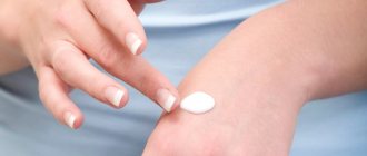
The solutions are used mainly to treat the affected skin on the head. Smooth skin is wiped with medicinal solutions before applying the ointment.
- Yodicirin. The drug contains iodine and glycerin. Accelerates healing, promotes separation of scales. The drug is applied to a napkin and applied to the affected area for 20-30 minutes.
- Vokadin. Contains iodine. Fights fungi and bacteria. Duration of therapy is 2.5 months.
- Nitrofungin. Treat wounds 2-3 times a day until recovery.
Ointments against lichen
Many types of ointments have been developed for the treatment of microsporia and human trichophytosis. They differ in composition, method of action on the parasite and healing rate.
The ointment is thicker than other drugs. It lasts longer on the treated area, penetrates into the deeper layers of the skin, and therefore fights mycosis better.
An alcohol solution of iodine is used in combination with ointment. It has a detrimental effect on fungi and bacteria, disinfects the affected area of the skin:
- Iodine is used in the morning, ointment at night.
- 20% sulfur ointment. The sulfur contained in the preparation dries out the ulcers. Kills the causative agent of lichen and accumulated bacteria.
- Salicylic. Reduces inflammation, kills parasitic mycoses. The surface treated with ointment must be covered with a napkin. It is strictly forbidden to treat wounds on the face.
- Sulfur-tar. Kills the pathogen and disinfects the surface. The product is used to treat the affected area and adjacent tissues. If the focus of inflammation is strong, then apply a bandage.
- Lamisil. Inhibits the reproduction of the parasite and kills it.
- Mikospor. Destroys microsporid cells. The drug is applied to the wound in a thin layer.
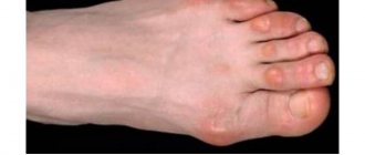
What does ringworm look like in cats?
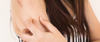
A characteristic symptom is areas of alopecia (baldness), since the fungi infect the hair follicles and their hyphae grow along the entire length of the hair, destroying it. Red, scaly bumps may appear on the skin. In cats, the ears are most often affected. These are the most typical signs of trichophytosis, however, lichen can manifest itself in a variety of ways, on any part of the body, both in the form of a hairless, scaly area, and in the form of a rash, crusts and other lesions.
Almost always, lichen is not accompanied by itching; if the cat actively scratches or licks the affected area, then its origin is most likely not related to dermatophytes, or there is a secondary lesion.
If the pet has recently been in contact with a large number of cats, or a new resident has appeared at home, picked up from the street - either a cat or kitten, or a dog or puppy, the likelihood of lichen becomes higher. An indirect sign can be considered the appearance of similar areas of baldness in other pets, as well as pink spots on the skin of people.
The likelihood of contracting lichen in other animals and humans with good immunity is low.
Pathogenesis
The pathogenesis of dermatophytosis is based on the virulence factors of fungi, due to their ability to secrete enzymes that destroy keratin (keratinolytic activity). The enzymes secreted by dermatomycetes and their metabolic products, as well as the contents of destroyed cells, cause an inflammatory process in the lesion, manifested by redness of the skin, itching and hair breaking. Keratinases have the ability to decompose not only keratin, but also other proteins (collagen and elastin), and it is the severity of keratinase activity that determines the characteristics of the manifestation of diseases when infected with various types of fungi. An important role in the spread of dermatomycetes is played by the presence of a specialized apparatus (perforator organs) for the directed growth of hyphae (the filamentous structure of the fungus). Thanks to this feature, the fungus grows in the direction of the intercellular junctions, which are the points of least resistance. Dermatomycetes, as a rule, do not penetrate deeper than the granular layer of the epidermis, since specific protective factors are activated at a deeper level.
The process of fungal invasion begins with the burrowing of hyphae into the skin and the release of keratinases (enzymes for the breakdown of keratins), which leads to irritation of nerve endings and, accordingly, the occurrence of itching. Dermatomycetes, loosening the cortex, cause acanthosis, parakeratosis, serous inflammation in the skin with the formation of blisters. Further, swelling occurs in the papillary/subpapillary layer, the vessels dilate, which leads to the development of polymorphic perivascular infiltration. The mycelium is usually located between the horny plates. Threads and spores of fungi can also be stuffed with hair follicles, which also undergo inflammatory changes.
List of sources
- Leshchenko V. M. Fungal skin diseases. In the book: Skin and venereal diseases (a guide for doctors). Ed. Yu. K. Skripkina, V. N. Mordovtseva. M., 1999. T. 1. P. 257-311.
- Kubanova A.A., Potekaev N.S., Potekaev N.N. Guide to practical mycology. – Moscow, Financial Publishing House “Business Express”, 200 p.
- Bayazitova A.A., Bayazitova A.A., Kupriyanova-Ashina F.G., Khaldeeva E.V., Ilyinskaya O.N. Primary pathogens - Pathogens of superficial mycoses // International Journal of Applied and Basic Research. – 2015. – No. 11-2. – pp. 241-248.
- Medvedeva T.V. Trichophytosis: modern ideas about the etiology, clinical picture, features of diagnosis and therapy / T.V. Medvedeva, V.B. Antonov, L.M. Leina, T.S. Bogomolova // Klin. dermatology and venereology. - 2007. - No. 4. - P. 70–74.
- Rodionov A.N. Fungal skin diseases (Guide for doctors) St. Petersburg: Peter. 1998. – 288 p.
Principles of treatment
Despite the general clinical picture, different types of ringworm require different approaches to therapy. It is almost impossible to independently distinguish trichophytosis from microsporia. To make a diagnosis, it is necessary to perform three examinations - Wood's lamp examination and analysis of scraping of the affected epidermis and culture for dermatophytes. The first study will confirm the presence of fungi, the second analysis will accurately determine the type of spores. The third analysis will eliminate errors in identifying the pathogen
The treatment is determined by the veterinarian. The treatment regimen depends on the general health of the patient, age and degree of damage to the body and coat.
- Antifungal agents are the basis of therapy. They are used internally and externally. The tablets are given with food. Indications: severe inflammation and profuse hair loss. Superficial forms of lichen are treated with ointments or solutions that are used to treat the lesions. Medicines used: Ketoconazole, Terbinafine, Fungin Forte.
- Iodine-containing products - for treating lesions. They stop the spread of spores to healthy epidermis and reduce infectiousness.
- Vitamin preparations and immunostimulants are prescribed to weakened animals for rapid recovery. They are indicated for kittens and older cats.
- Antibiotics, antiseptics and other means of symptomatic therapy are used as needed. The veterinarian may prescribe antibacterial ointments if wounds become infected and pus forms under the crusts.
Treatment is carried out in several stages. First, you need to give the medicine with food (as prescribed by your doctor), then treat the skin for 2-3 weeks. Treatment with antiseptic and fungicidal agents is carried out at intervals of 2 hours. The treatment is long-term, about 2 months. Treatments must be carried out constantly; the frequency of treatment is described in the instructions for the specific drug.
After a course of therapy, scrapings are taken again. The pet is considered healthy if 2 cultures for dermatophytes are negative.
Incubation period
The fungus enters the human body, adapts, and begins to develop without causing visible changes on the skin for up to 8 weeks.
Superficial lichen (trichophytia) appears 5-7 days after contact with the skin. Infiltrative-suppurative (microsporia) can “dormant” for several months. This is dangerous because the infected person does not yet know about his disease, but is already spreading it.
Diagnostics
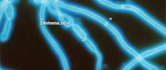
The final diagnosis is made by a dermatologist. The doctor examines the affected areas under a fluorescent lamp. If the pathogen is Microsporum, then the hair glows bright green, and if Trichophyton, then white-blue.
A scraping is taken from the infected area of skin, culture and microscopy are done. After 5 days, a colony grows in a Petri dish by which the pathogen is identified.
Lichen is differentiated from syphilis and alopecia of non-contagious etiology.