Coronary heart disease is considered one of the leading causes of death worldwide. This is a pathology that leads to disruption of the heart and is accompanied by narrowing of the coronary arteries. Coronary angiography is used to diagnose coronary artery disease. The study shows the condition of the heart vessels, blood circulation in them and places of blockage. To carry out such a procedure, there are rules for preparing the patient, contraindications and possible complications.
What it is
The coronary arteries supply blood to the heart, carrying nutrients and oxygen. This is necessary for the normal and uninterrupted functioning of the heart muscle. But sometimes, due to congenital abnormalities, atherosclerotic plaques or spasms, the lumen in the arteries narrows, this is called stenosis. The most dangerous is occlusion, that is, cessation of blood flow. This picture leads to coronary artery disease or myocardial infarction, and in severe cases, death.
Content:
- What it is
- When is coronary angiography prescribed?
- How to prepare
- How it's made
- Contraindications for the procedure
- Is this examination dangerous?
- Where can I get it and how much does it cost?
The condition and functioning of the coronary arteries and vessels can be checked using coronary angiography. This is a type of angiography and fluoroscopy. The study is carried out by filling the arteries with a special substance that does not transmit x-rays - contrast. A catheter delivers contrast to the heart.
When the substance is distributed throughout the vessels, a series of X-rays are taken. In such images, the blood network, arterial patency, and stages of the disease are clearly visible. Based on such a study, the doctor makes a conclusion about treatment methods.
There are the following types of examination:
- CT coronary angiography. The procedure is carried out using computed tomography, contrast is injected into a vein without catheterization of the coronary vessels of the heart. During the examination, the radiologist sees places of narrowing, the thickness of the walls of blood vessels, and the detailed structure of the circulatory system in this area.
- Interventional selective coronary angiography. The catheter is moved through the vein to the coronary artery itself, and contrast is injected. The procedure is carried out under X-ray control, the doctor sees the result in real time on the screen.
- Ultrasound coronary angiography. An unpopular technique, but used for research. It happens in the same way as the previous method, but an ultrasound sensor is attached to the end of the catheter. This is done to assess the condition of the vascular walls.
- The most common method of examination in the CIS is interventional. Computed tomography is increasingly being carried out in private clinics, as this method provides more information. Other examination methods are prescribed in isolated cases.
How is coronary angiography of the heart performed?
- It is carried out strictly in a specially equipped operating room! A type of study - computed tomographic coronary angiography or CT coronary angiography, requires the presence of a computed tomograph.
- On the eve of the study, the patient undergoes a series of tests to ensure that the procedure is safe.
- The doctor will tell you in advance how to prepare for the examination. Read more about preparing for cardiac angiography in the following article.
- Basically, the research is carried out as planned. Urgent coronary angiography of vessels is performed in acute coronary syndrome and myocardial infarction.
- Coronary angiography takes from 15 to 30 minutes, but sometimes the time increases due to various circumstances.
- According to indications, coronary angioplasty and vascular stenting are performed simultaneously with coronary angiography.
When is coronary angiography prescribed?
The purpose of this procedure is to examine in detail the condition of the arteries of the heart, looking for narrowing or blockage. Coronary angiography of blood vessels is usually used by cardiologists and cardiac surgeons. It is needed before complex operations to make the correct diagnosis and choose the right treatment. The patient is referred for coronary angiography in the following cases:
- ineffectiveness of drug therapy in patients with coronary heart disease;
- positive stress test (stress echocardiography, bicycle/treadmill testing);
- before any heart surgery in patients over 40 years of age;
- angina pectoris after a heart attack, stenting or coronary bypass surgery;
- all emergency situations when there is a suspicion of a cardiovascular accident - acute myocardial infarction (in the first 6 hours);
- unstable angina;
- lack of information about other diagnostic methods.
It is also prescribed to evaluate the effectiveness of therapy for the heart and blood vessels. Checking is needed for atypical pain in the chest area without a clear cause. It is often used before coronary artery bypass surgery and other interventions.
FAQ:
Question: I am 56 years old and have coronary heart disease . The cardiologist recommends a coronary angiography . I don't quite understand what this is? Answer: Coronary angiography is a study of the vessels of the heart, which, like a crown, surround the heart, supplying it with oxygen-rich blood. These are called coronary arteries. Narrowing of the coronary arteries leads to a decrease in blood supply to the heart, which leads to oxygen starvation, i.e. to cardiac ischemia. It is very important to know the condition of the arteries of the heart, because... the obstruction to blood flow can be eliminated, radically relieving the person of the symptoms of coronary heart disease . Question: How is coronary angiography performed? Answer: Coronary angiography is an x-ray examination in which a special contrast agent is injected into the vessels of the heart using a flexible thin probe. As the contrast agent passes through the vessels, short-term x-rays are taken with significant magnification using high-resolution equipment. The slightest changes in the coronary bed are visible “at a glance.” The results are recorded in digital format and are available for viewing on any personal computer. Question: Is coronary angiography performed under anesthesia? Answer: No general anesthesia is required. The pulse is determined in the groin area or on the wrist, and a probe or catheter is inserted into the lumen of the artery under local anesthesia. The subject does not feel how the catheter moves through the vessels, because There are no sensory nerve endings inside the arteries. There is no pain, the patient is fully conscious and, together with the operator, monitors the progress of the study on the monitor. The duration of the procedure is no more than 15-20 minutes. Further monitoring of the patient for several hours is necessary. At the CELT clinic, coronary angiography is performed exclusively through the radial artery (at the wrist). Immediately after the examination, the patient can get up and walk, the bandage on his arm does not limit him in any way (see ambulatory coronary angiography ). After 4 hours the patient is discharged home. If the examination is carried out through the groin, then bed rest is necessary and the period of stay in the clinic is extended to a day. Question: If coronary angiography does not reveal narrowing in the vessels of the heart, then there is no coronary heart disease? Answer: Yes, the absence of changes in the coronary arteries practically excludes the diagnosis of coronary heart disease . In rare cases, ischemia of the heart muscle can occur in the presence of “normal” coronary arteries, but the absence of damage to the arteries of the heart is the most reliable predictor of a good prognosis. This is very important information for choosing the right patient management tactics. Question: At what age is coronary angiography , what are the contraindications to coronary angiography? Answer: Coronary angiography is performed at any age, in all cases when there is a need for it, namely if the patient has angina pectoris after myocardial infarction. Coronary angiography, in the first hours of myocardial infarction , makes it possible to determine where the blockage of the vessel is causing the heart attack and immediately eliminate it. Coronary angiography is necessary for all adults with heart defects before surgery, before “major” vascular operations. Some patients at high risk for coronary heart disease , such as diabetes , may not have symptoms of the disease. In this case, coronary angiography is, in fact, the only reliable way to exclude or confirm coronary disease . Question: I need a coronary angiography . I contacted the Federal Center. They gave me a list of tests and put me on the waiting list. They said I needed hospitalization for 2-3 days. Answer: We coronary angiography on the day of admission! All tests and additional studies are carried out within 4-5 hours, including coronary angiography. Question: How can I get a coronary angiography at your center? Answer: You can come any day, appear at the clinic, at the cardiovascular surgery department in the morning at 08:30 on an empty stomach, PRIORLY AGREEING YOUR ADMISSION WITH US BY PHONE 305-34-04 or 788-33-88. Before admission, before performing coronary angiography , you should have on hand the results of the following tests and studies: - General blood test; — General urine analysis; - Biochemical analysis - protein, urea, creatinine, bilirubin and its fractions, lipid profile, potassium and sodium, glucose; — Coagulogram; — Blood type and Rh factor, Rh antibodies; — Blood test for HIV, RV, antibodies to hepatitis B and C; — Ultrasound of the heart, ECG. If you do not need tests and studies, then they can be performed with us on the day of admission. Question: coronary angiography cost you and what are the prices for coronary angiography in Moscow in other clinics? Answer: Coronary angiography including the stay costs 30,000 rubles. Today, if you analyze the prices for coronary angiography in Moscow, this is the minimum cost.
How to prepare
Coronary fluoroscopy is performed after preparing the patient. The procedure can be emergency or planned. Emergency diagnostics are carried out with minimal analyzes and tests; the type and number of such checks is selected by the attending physician depending on the situation. For routine examination the following is prescribed:
- consultation of specialists: neurologist, cardiologist, surgeon;
- blood tests: coagulability, Rh factor and group, biochemical analysis;
- fluorography;
- allergy tests;
- Analysis of urine;
- electrocardiogram.
A full list of examinations is prescribed based on the clinical picture. The procedure is performed on an empty stomach to avoid nausea and the gag reflex. If the contrast agent will be injected into the thigh, you will need to remove hair from that area the day before.
A few days before the scheduled examination, the patient is shown a large amount of water, alcohol is prohibited. You cannot eat on the day of the event.
Before the examination itself, you need to warn the doctor about what medications you took and how you feel. You also need to take a change of clothes and personal hygiene items for your stay in the hospital.
Coronary angiography as a method for diagnosing heart diseases
Makarevich Olga Vadimovna
Doctor anesthesiologist resuscitator OAiR No. 2
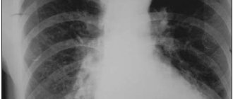
Fig.1 Coronary angiography - x-ray examination of the condition of the arteries of the heart
- What is coronary angiography or cardiac catheterization?
- In what situations is coronary angiography necessary?
- How dangerous is coronary angiography?
- How should a patient prepare for coronary angiography?
- What happens during coronary angiography and how is coronary angiography performed?
What is coronary angiography?
Coronary angiography (also called cardiac catheterization or coronary angiography) is an invasive diagnostic procedure that can detect damage to the vessels (arteries) of the heart and see how well the patient's heart is functioning. The essence and principle of coronary angiography is that a special diagnostic catheter is placed at the origin of the coronary arteries (vessels supplying the heart), through the lumen of which a contrast agent is injected, staining the arteries of the heart. An X-ray tube is used to record images of the coronary arteries, and the internal lumen of the vessel is recorded. A diagnostic catheter is a long, thin plastic elastic tube, thanks to which contrast is supplied to the arteries of the heart through a puncture of a peripheral artery (femoral or axillary). It is also possible to “paint” the cavities of the heart when a catheter is inserted into their lumen.
In what situations is coronary angiography necessary?
Coronary angiography is an invasive procedure, since during its implementation there is minimal, but still traumatization of body tissues (at the stage of access to the vessel for insertion of catheters). Taking this into account, the use of angiographic examination for diagnostic purposes should be carried out according to strict indications, when other additional less traumatic research methods have not made it possible to establish an accurate diagnosis. Coronary angiography is also necessary when planning surgical treatment of a particular cardiovascular pathology. Below are the main indications for using this diagnostic method:
- The need for accurate confirmation of heart and vascular disease (for example, coronary heart disease, heart valve pathology or aortic pathology).
- Assess cardiac and myocardial function.
- Determine the indications and type of proposed surgical treatment (for example, it will be endovascular intervention or bypass surgery).
One of the undeniable advantages of coronary angiography, as well as any angiographic diagnostic study, is the ability to move from a diagnostic procedure to a therapeutic one and perform various endovascular treatment procedures, such as balloon angioplasty and stenting of the coronary arteries.
Fig.2 Stenting of coronary arteries with balloon angioplasty
How dangerous is coronary angiography?
In general, problems when performing coronary angiography are very rare, no more than 1 case per 100,000 examinations. However, given that angiographic examination itself is traumatic, the risks of certain complications are always present. There are always measures to prevent such complications and it is always necessary to discuss this with the doctor who will perform coronary angiography. The following are the most common complications of angiographic studies:
- Bleeding from the area of arterial puncture
- Heart rhythm disturbance
- Formation of blood clots and hematomas
- Infection and inflammation of the wound in the puncture area
- Allergic reaction to contrast agent
- Stroke
- Myocardial infarction
- Perforation of a blood vessel (forming a hole in the vessel) or its rupture
- Decompression sickness (air entering the lumen of a blood vessel, which can be an extremely life-threatening complication)
- Death
How should a patient prepare for coronary angiography?
Before performing coronary angiography, the patient is recommended to complete a standard list of necessary examinations. These include a chest x-ray, blood tests for infections, biochemical and complete blood tests, coagulation tests, platelet aggregation tests, echocardiography, electrocardiogram and urinalysis.
No special preparations are required to perform coronary angiography. The patient may be asked to leave jewelry or jewelry at home. It is recommended to stop taking blood thinning medications, such as warfarin or aspirin, 10 days before the test. The cardiologist will definitely discuss with the patient the need to take certain medications that the patient was taking before hospitalization for the study.
If the patient is diabetic, then he needs to continue taking antidiabetic medications or consult an endocrinologist the day before. The patient must inform the doctor about the presence of allergic reactions to a particular medication, for example, a reaction to iodine and iodine-containing drugs, X-ray contrast agents, alcohol, novocaine, other medications, products containing rubber (rubber gloves), antibiotics, etc. It is necessary to take personal hygiene items with you (toothbrush, slippers, towel, etc.), since your stay in the clinic can last 2-3 days and their presence will make your stay in the hospital more comfortable. On the evening before the study and in the morning, the patient will have their intestines prepared using an enema; they are also advised not to eat or drink in the morning. In the morning, the area where the artery will be punctured for access will be shaved, usually the groin or axilla.
If patients have serious pathologies of other organs and systems of the body, further examination is necessary to determine the extent of their disorders. For example, in the presence of kidney pathology, it is important to assess the filtration function of the renal tissue, since the urinary system is the only place for removal of the contrast agent from the body. If high risks of developing renal dysfunction are identified after coronary angiography, the question of refusing the study or the need for renal replacement therapy (hemodialysis) is usually raised. However, modern contrast agents that are used in coronary angiography have less and less damaging effects and more often coronary angiography, like any other contrast study, proceeds without complications.
The main reason why a doctor may refuse to conduct a coronary angiography study on a patient is the instability of the patient’s condition, shock, or exacerbation of a chronic disease, including infectious etiology.
What happens during coronary angiography and how is coronary angiography performed?
Before the coronary angiography, the patient will be asked to wear special underwear. The nurse will place a peripheral venous catheter in the cubital vein, which is needed to administer fluids and medications during the procedure.
The room in which the angiographic examination is performed is called the cath lab and will be somewhat cool and shaded when coronary angiography is performed. This is necessary for the convenience of the doctor’s work and good visibility of the resulting image on the monitors. The patient will also see the image obtained during the diagnosis. The subject will be placed on a special movable table, around which the X-ray tube will move in different planes.
The operating nurse will cover the area of the artery puncture with sterile linen, which is done to prevent the spread and addition of infection, that is, for the purpose of asepsis. Electrodes of an electrocardiograph, a device with which the activity of the heart will be monitored, will be placed on the chest area. The basic principle of the coronary angiography procedure is presented in the following animation: Coronary angiography in 3D reconstruction.
To monitor the state of kidney function and the rate of urine separation, a catheter will be installed in the bladder. For the purpose of relaxation, the patient is administered sedatives on the eve of coronary angiography. To numb the area through which the catheter will be installed and manipulations will be performed, the x-ray angiosurgeon will administer novocaine.
After anesthetizing the intended artery puncture site, a small skin incision is made necessary to install a special vascular port (intraducer) into the lumen of the artery. Through it, various catheters and devices are inserted. After properly administered anesthesia, pain and discomfort in the puncture hole area rarely occur. Most often, the femoral artery is used as access to the arteries, but it is possible to pass a catheter through the arteries of the arm.
After inserting and positioning the diagnostic catheter in the place required for contrasting, the lights are dimmed and after tests for the reaction to the contrast agent (small doses of contrast are administered), contrasting of the coronary arteries is performed. It is also possible to assess the condition of the valves and chambers (atria and ventricles) of the heart.
When contrast is administered, you may feel a feeling of heat in your chest. This is an absolutely normal reaction of the body to the introduction of almost any drug into the vascular bed and it goes away within a few seconds. If you experience unusual sensations, such as itching of the skin, a feeling of choking or difficulty breathing, swelling in the throat or difficulty swallowing, hoarseness in the throat, chest discomfort and other symptoms of a possible allergic reaction, be sure to notify the doctor who is performing the coronary angiography.
After placing the X-ray tube in the desired position, the x-ray surgeon may ask the patient not to breathe, hold his breath and not cough, after which he will take a series of images. Holding your breath is necessary to obtain a high-quality image. Then, after receiving all the necessary information about the condition of the coronary arteries and the function of the heart, the catheters are removed, and a pressure bandage is applied to the puncture site or special occluders (mechanical systems for closing the arteries) are used.
Upon completion of the angiographic examination, if there were no complications of the procedure, the patient is usually transferred to the department from which he was transferred for coronary angiography. If, however, certain complications of the intervention do arise, then these complications are corrected and the patient is placed in the intensive care ward or in the intensive care unit under the supervision of a resuscitator. The patient is transferred to a specialized department after stabilization of his condition. During the day, until the next morning, the patient must remain with his leg straightened (the one through which the puncture was performed) and in a lying position. It is advisable not to lift your head from the pillow, since this movement causes an increase in pressure in the groin and abdomen, and this can provoke the development of bleeding from the area of access to the artery. If the puncture was performed through the artery of the arm, the restriction of movement concerns only the shoulder girdle. Sitting or standing up immediately after the examination is strictly prohibited. If you experience a feeling of numbness or tingling in your legs or arms, or if the bandage gets wet with blood, you must inform the doctor who is monitoring the patient after the study. There may be a desire to urinate after angiography. A ship is usually used for these purposes. Gradual activation of the patient is carried out the next day. First, they usually ask you to sit for a while, after which you are allowed to stand up. This is due to the fact that when one remains in a horizontal position for 24 hours and then abruptly moves to a vertical position, collapse may develop. Immediately after the study, it is recommended to take a significant amount of fluid. It is usually drunk or administered intravenously through a previously installed catheter in the cubital vein. This is necessary for the rapid removal of the contrast agent, which has a number of toxic properties, from the body. Further treatment and review of the test results are usually carried out by the attending physician. Tactics and possible treatment options, if necessary, are usually discussed with him.
How it's made
Angiography of the heart vessels is performed in a hospital; the patient remains in the hospital for several days for careful preparation and supervision after the procedure. All manipulations are carried out in the sterile conditions of the X-ray operating room - this is an office equipped specifically for such purposes. Before the invasion, the patient signs consent documents.
The patient then changes into hospital gowns. He is placed on the operating table and the position is fixed so that sudden movement does not disrupt the procedure. A cardiac monitor is connected to monitor the heartbeat and blood pressure.
The anesthesiologist administers local anesthesia; the patient will not feel pain, but will remain conscious. A catheter is inserted into a vein in the thigh or arm.
The puncture site is selected depending on the clinical picture. More often the radial artery in the arm is punctured - this is the most painless and safe, less often - the femoral artery in the groin area.
A conductor (thin wire) is inserted through the needle into the lumen of the vessel, and the needle is removed. A catheter (hollow tube) is passed through the guidewire. The catheter is advanced through the vessels to the coronary arteries themselves. There are no nerve endings in the vessels, so the patient will not feel anything. Contrast is fed into the catheter, which fills the vessels and is clearly visualized by fluoroscopy. On the screen, the doctor sees the advancement of the catheter.
When the substance fills the arteries, they become clearly visible on the monitor. To obtain high-quality images, the radiologist may ask you to hold your breath. Data from the screen can be recorded on a digital medium so that doctors can then assess the patient’s condition.
If the patency of the vessels is impaired, a mesh stent can be installed or balloon dilatation can be performed. This is done directly during coronary angiography; such manipulations restore the lumen of the vessel and normal blood flow. After the procedure, the instruments are removed, the puncture site is disinfected and a tight bandage is applied.
The patient remains on bed rest for another day. Some private clinics offer this service on an outpatient basis, the patient rests for a few hours and goes home. The whole process will take no more than an hour.
Reviews from patients confirm that fluoroscopy of blood vessels is quick and without severe pain. When the catheter is inserted, discomfort is felt, and when the contrast enters, a feeling of warmth is felt in the chest.
Why is coronary angiography performed?
Coronary angiography is the most common of a group of procedures known as catheterization. It helps diagnose and treat various heart diseases. It is also called cardiac angiography.
A special contrast agent is injected into the blood vessels of the heart, which can be seen in an angiography unit using X-rays. It takes a series of images (angiograms), providing detailed information about the condition of the veins and arteries.
Contraindications for the procedure
Coronary angiography has only relative contraindications. There is a list of diseases that increase the risk of complications. But even against the background of such diseases, fluoroscopy of blood vessels can be performed if the patient’s life depends on it. Coronary angiography is performed with extreme caution when:
- impaired blood clotting;
- decompensated diabetes mellitus;
- allergies to contrast agent;
- arterial hypertension;
- severe renal failure - the contrast agent has a bad effect on diseased kidneys;
- internal bleeding - postponed until this problem is resolved.
All possible contraindications must be taken into account when prescribing coronary angiography. Absolute contraindications include only a severe allergic reaction to contrast. In most cases, the study is simply postponed.
Procedure
- You lie on your back on the angiography table. The X-ray imaging device moves around the head and chest and records from different angles (views).
- A special catheter is placed intravenously in your arm. They will administer sedatives to make you relax. During the angiography, you will be awake and able to communicate with the doctor and follow his instructions. During the coronary examination, you may be asked to take a deep breath, hold your breath, cough, or place your hands in various positions.
- Electrodes on the chest allow the doctor to monitor cardiac activity throughout the entire period. The cuff on the arm measures blood pressure, and the pulse oximeter shows the oxygen content in the blood. You will be given special medications (anticoagulants) to prevent blood clots from forming in your catheters and arteries.
- The area of the body (on the arm or leg) where the catheter will be installed is treated with a special solution, then local anesthesia is performed. The doctor makes a small incision (about 3 mm) at the access site and inserts a small plastic tube (introducer) into the artery. The catheter for coronary angiography of the heart vessels is inserted through the tube and carefully delivered to the coronary arteries.
- Passing the catheter does not cause pain and you will not feel its movement throughout the body. Make sure of this by reading patient reviews. Tell your doctor if you experience any discomfort during the angiography.
- The contrast agent is injected through the catheter. When this happens, patients notice warmth. During the operation, “jumping” heart contractions (extrasystoles) are a common occurrence; there is no need to worry about this.
- The contrast agent is visible on X-ray images, and by the way it passes through the blood vessels, the doctor will determine the exact location of the stenosis or occlusion and the severity of the affected area. Watch a video of coronary angiography here.
At the end of coronary angiography, the sheath is removed from the arm or leg and the incision is closed using manual pressure or a special device. You will be transferred back to the ward for further observation. You will lie down for several hours to prevent bleeding from the access site.
You will go home the same day if you had an outpatient coronary angiography. If the study was performed through the femoral artery or complications arose during outpatient coronary angiography, you will have to stay in the cardiology clinic until the morning or longer. It is recommended to drink more than usual (about 2 liters of water), unless there are contraindications, so that the contrast is eliminated from the body faster. The doctor will tell you when to resume taking medications and return to work and normal lifestyle. Avoid excessive strain and lifting heavy objects for several days.
Is this examination dangerous?
Everyone who is about to undergo this procedure is interested in what the consequences and complications may be. This type of diagnosis is considered a safe manipulation; the process is controlled using an X-ray or computed tomograph. Nevertheless, the risk of complications is still present; the most dangerous consequence is damage to blood vessels by the catheter.
The likelihood of such a complication is very low; it can occur from sudden movements or carelessness of the doctor. If the patient has not undergone proper preparation, an allergic reaction or bleeding may occur (with poor clotting). All these risks can be reduced if you carefully prepare and relax before the procedure.
If you are overcome by fear and anxiety in the X-ray operating room, you can warn the doctor about this and he will prescribe sedatives.
Possible complications after the procedure
Like any other surgical intervention and violation of the integrity of the vessel, coronary angiography can have a number of complications, even if it is performed by an experienced specialist. Possible complications include:
- heart attack;
- rupture of an artery or heart;
- heart attack or stroke due to a blood clot breaking off from the wall of a vessel;
- arrhythmia;
- bleeding;
- allergies.
Of course, complications do not always arise and are often limited to small hematomas or swelling at the puncture site. However, the possibility of serious consequences cannot be excluded, so before the procedure you need to carefully study the contraindications to it.
Contraindications for the study
Contraindications for cardiac vascular examination with contrast are:
- various respiratory diseases that are accompanied by elevated body temperature;
- anemia;
- open bleeding of various locations;
- low potassium levels in the blood;
- poor blood clotting;
- diabetes;
- obesity or underweight;
- renal, heart failure;
- lung damage.
Interpretation of coronary angiography
How coronary angiography of the heart vessels is performed and what it is is clear. The main thing is not just to take a picture, but to interpret it correctly, that is, decipher it. For the most informative and accurate result, several specialists are involved in decoding: a radiologist, a cardiologist, a vascular surgeon and others.
The thickness of the vessels, their length and width are assessed from the images. Compactions and darkening are revealed. An alarming signal is a violation of the boundaries of blood vessels - blurriness and indistinctness. The pathology of the vessels will also be indicated by their incorrect location.