Breathing is a process without which a person cannot exist. Anyone who cares about their health should know where a person’s lungs are, how they function and what structure they have. The organ is vital because it supplies the entire body with oxygen.
Where are the lungs located?
The lungs are located on either side of the heart and fill the chest. Each lung of an adult weighs a little more than 400 g
.
The right lung is slightly heavier than the left, since the latter has to share space in the chest with the heart. The lungs are protected by the rib cage
. Between her ribs there are small muscles involved in the breathing process.
Check your lungs
If you smoke, be sure to check your lungs with a simple test. Try blowing out a candle at a distance of a meter.
Beneath the lungs is
the diaphragm
, a dome-shaped muscle formation that separates the chest from the abdominal cavity and is also involved in breathing.
Location of the lungs in humans
The lungs are located in the chest cavity, occupying its main part (90%). The location and structure of the lungs makes it possible to unite all the important (main) vessels.
Occupying almost the entire chest cavity, the lungs are located below with their base on the dome of the diaphragm. The right lower pulmonary section is separated by the diaphragm from the liver, the left – from the stomach, spleen, and part of the intestine. The median region is closely adjacent to the heart on both sides. The upper base is 4–5 cm above the collarbone.
The lungs are covered on the outside with a serous protective membrane - the pleura. On one side it passes into the pulmonary tissue, and on the other into the mediastinum and chest cavity. The resulting pleural cavity is filled with fluid. Due to this and due to the effect of negative pressure inside the cavity, the lung tissue is in a straightened state. The pleura located on the surface also protects the lungs from friction against the ribs during breathing.
How does breathing happen?
Before birth, the baby receives oxygen directly from its mother's blood, so its lungs are filled with fluid and do not work. At the moment of birth, the baby takes his first breath, and from that moment his lungs work without rest. The respiratory center of the brain constantly receives signals about how much oxygen the body requires at any given moment. For example, if a person is sleeping, he needs much less oxygen than when he is running for the bus. The brain sends messages along nerves to the breathing muscles, which help regulate the amount of air entering the lungs. As soon as this signal is received, the diaphragm expands and the muscles stretch the chest outward and upward. This maximizes the volume that the lungs can occupy in the chest. When you exhale, the diaphragm and intercostal muscles relax, reducing the volume of the chest. Due to this, air is pushed out of the lungs.
Human lungs: brief characteristics of the organ
The functioning of the brain, the release of energy, and the breakdown of useful substances with the help of oxygen directly depend on the functioning of the respiratory system. The respiratory organs include the nose, mouth, larynx, trachea, and bronchi, which are part of the lungs.
The lung is one of the paired, most voluminous human organs. In a healthy state, it is pink-red in color, the structure is soft, spongy.
The average volume of the organ is 3 liters, but 450-500 ml is enough for inhalation and exhalation. In people who are actively involved in swimming, the lung volume reaches 5 liters.
For a deep breath, about two liters of atmospheric air are used. With such breathing patterns, a reserve remains in the organ, thanks to which the level of oxygen in the alveoli is maintained at the required level.
In the tissues of each lung, venous and arterial blood constantly moves, and lymphatic vessels are present. It also contains nerve cells that are closely connected with blood vessels and bronchi.
In the first month of pregnancy, the rudiments of the main organ of the respiratory system are already formed in the fetus, and by the fifth month its constituent elements are designated. The organ continues to grow until the age of 25, as the growing organism constantly needs oxygen.
How does oxygen get into the blood?
Through the thin walls of the alveoli, oxygen enters the blood vessels. Here it is picked up by “transport” - hemoglobin
, which is contained in red blood cells. At the same time, carbon dioxide enters the opposite direction - into the alveoli - from the blood, which is removed from the body when exhaling. Oxygenated blood is sent from the lungs to the left side of the heart, from where it is distributed throughout the body through arteries. As soon as the oxygen in the blood is used up, the blood flows through the veins to the right side of the heart and from there back to the lungs.
Features of the external structure of the lungs
The lungs are shaped like a cone, divided vertically into two parts. In this case, a convex surface and two concave ones are clearly visible. The convex pulmonary area (costal) is so close to the ribs that sometimes even the lung tissue has traces of them on the surface. One concave surface is located in the middle part of the body, and the second borders the diaphragm. In turn, each of them is also divided into interlobar sections.
In appearance, healthy lung tissue looks like a pink, finely porous sponge. Under the influence of unfavorable factors, its color changes - it becomes darker with age-related changes, pathologies, bad habits (smoking).
According to the anatomical structure, the lungs have different sizes, the right one is larger by about 10% than the left one, and they also differ in shape. The left one is smaller due to its “neighborhood” with the heart, which is closer to it, as if slightly displacing this area, called the cardiac notch. In this place, part of the pericardium remains unclosed, and there is a protrusion below, called the “pulmonary uvula”. The right lung is slightly higher than the left, due to the fact that the liver underneath it pushes it up a little.
On the medial side of each of them there is a “gate”. Important metabolic processes take place through them: the pulmonary artery, bronchi, and nerve plexuses enter the lungs, and the pulmonary veins and lymphatic vessels exit. Together, all this makes up the root of the lung. On the right, the pulmonary root is located behind the atrium and the superior vena cava, below the azygos vein, on the left - under the aortic arch.
What else do the lungs do?
Every day, the lungs of an adult person pump about ten thousand liters of air
.
Consequences of smoking
Chronic obstructive pulmonary disease (COPD) is rightfully considered the most obvious consequence of smoking. Moreover, it is incurable. Find out how to prevent the disease.
With each breath, not only oxygen enters them, but also dust, germs and other foreign objects.
Therefore, the lungs also perform the function of physical and chemical defense against unwanted objects from the air. On the walls of the bronchi there are tiny villi that trap dust and germs
. In the walls of the airways, special cells produce mucus that helps clean and lubricate these villi. Contaminated mucus is removed through the bronchi to the outside and coughed up.
Components of the lungs
The structure of the lungs is a complex structure, which consists of:
- bronchi;
- bronchioles;
- acini.
An important part of the respiratory system is the bronchi. These are tubular branches of the trachea that connect it to the lungs. Their main task is air circulation. In shape they resemble the crown of a tree, due to the many branches and are called the “bronchial tree”. The bifurcation of the trachea into the left and right bronchi occurs in the area of the fifth thoracic vertebra. Then they enter the lung tissue and branch into lobar, then into segmental, and finally into the smallest channels - bronchioles.
Each pulmonary bronchus with the largest diameter has three membranes:
- external;
- fibrinous-muscular, having cartilaginous tissue;
- inside them there is a mucous layer with ciliated epithelium.
As the diameters of the branches of the bronchi decrease, their cartilage tissue and mucous membrane gradually decrease. They are no longer present in the bronchioles, but cuboidal epithelium (a thin layer) is formed.
The framework of the lungs is the bronchial system, which has a branched structure. Many lobules measuring 15x25 mm make up each lung. The apices of the lobules include bronchioles (branches of the bronchi), at the ends of which there are acini - special formations covered with a large number of alveoli.
Acini got their name because of their appearance, reminiscent of a bunch of grapes. Translated from Latin, Acinus means “bunch”. This is the main structural unit of lung tissue, which includes bronchioles, alveolar ducts, and primary pulmonary lobes in the form of small sacs.
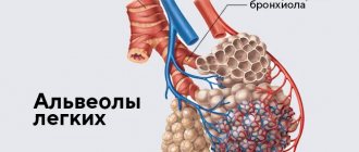
Alveoli
The most important pulmonary elements are the alveoli, which ensure normal exchange of oxygen and carbon dioxide in the body. They are small thin-walled bubbles, tightly enveloped in a network of capillaries. Through the alveolar ducts, blood vessels are continuously supplied with oxygen and cleared of carbon dioxide. The tissue of each lung has 300 million alveoli. Oxygen is supplied to them by arterial capillaries, and carbon dioxide is taken in by venous vessels.
The alveoli themselves are microscopic in size - 0.3 mm. But, thanks to their huge number, the average respiratory surface area when exhaling is 35 square meters, and when inhaling it can reach up to 100 square meters. Of course, the indicators depend on the person’s constitution - height, weight, fitness. Athletes have the highest marks.
How do lungs stay healthy?
The alveoli remain inflated, like balloons, even when you exhale air. Your lungs produce a fluid called a surfactant (surfactant) to help the alveoli stay open. The surfactant also contains fatty proteins that help maintain lung health.
Your lungs cleanse themselves.
They create mucus that traps germs and particles. The mucus is then swept up by cilia, the small hairs that line the airways. Usually you swallow this mucus without noticing it. If you have a respiratory illness, your lungs may produce too much mucus.
The alveoli also contain immune cells called macrophages. These cells "eat" germs and irritants before they can cause infection in the lungs.
Lobes and segments of the lungs
From the small structures of the acini, lobules are formed, then larger segments, which make up the largest pulmonary area - the lobe. The structure of the left and right lungs is different.
The right lung consists of three lobes:
- the upper of the three segments;
- the middle of the two segments;
- lower lobe of five segments.
The left lung consists of two lobes:
- top of five segments;
- the lower of the five segments.
Division into lobes occurs through grooves. One of them (oblique) begins at each lung below 6–7 cm from their apices and extends to the diaphragm, separating the upper lobe from the lower. In the right lung, in the region of the IV rib, there is a horizontal groove separating the wedge-shaped pulmonary area - the middle lobe.
The bronchopulmonary segments do not have clearly defined divisions. The pulmonary segment is a separate area into which blood flows from one artery and ventilation is provided by one bronchus (third order). Lung tissue is divided into segments of different shapes, which differ not only in the right and left lungs, but are also located individually in each person.
Main functions
In addition to the main respiratory function - ensuring gas exchange in the body, the lungs perform several other important missions:
- They normalize the pH composition in the blood, taking part in water, lipid, salt metabolism, and regulating the chlorine balance.
- They protect the body from respiratory infections because they produce antimicrobial substances, immunoglobulins.
- Provide thermoregulation.
- Helps maintain normal water balance in the body.
- Participate in the creation of vocal sounds.
- They serve as a kind of blood storage (contain approximately 9% of the total volume).
- Protect the heart from mechanical influences.
- Promote the removal of toxins, essential and other compounds.
- Participate in coagulation (blood clotting).
Symptoms that require you to see a doctor
Contact your doctor if you have severe symptoms of diseased lungs. According to the American Lung Association, dangerous warning signs of the disease include:
- chronic cough that lasts a month or longer
- shortness of breath, which occurs in the absence of exertion or after little effort
- wheezing or noisy breathing
- chronic mucus or phlegm in the lungs that persists for a month or longer
- chronic chest pain that lasts for a month or longer
- coughing up blood
What X-Rays Don't See: Pulmonary Function Testing
Once in the hospital, during educational supervision, a patient told me: “The doctor ordered some kind of test - the function of external respiration. Is this an exhalation to explore, or what? Why external? Let's figure out what kind of breathing happens, what external breathing is and why study it.
What is a "function"?
Respiratory processes in the body are divided into two large groups. The first is internal respiration, which includes tissue (transfer of oxygen from blood to cells) and cellular (utilization of oxygen inside the cell). The second is external respiration, which combines the processes of air entering the pulmonary alveoli and gas exchange in them: the entry of oxygen into the blood and the extraction of carbon dioxide from it.
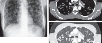
The main function of the respiratory system is to provide the body with oxygen in accordance with the current need. It is obvious that if a person has structural damage to the respiratory apparatus (anything - an abscess in the lung tissue, a foreign body in the bronchus, broken ribs, paralyzed respiratory muscles), then this function will be impaired. Such damage is clearly visible on X-ray, CT or MRI.
But there are situations when the x-ray picture is normal, and the patient complains, for example, of difficulty breathing. That is, there is a violation of the respiratory function, but without structural damage to the respiratory apparatus. What to do in this case?
Volumes and speeds
Here doctors have to turn practically into physicists - to establish a diagnosis, they need to study the movement of air through the system of tubes that form the respiratory system: the trachea and bronchi of various sizes. What matters is both the volume of air that a person can inhale or exhale and the speed at which he does it.
All these indicators are studied using methods such as pneumotachometry, peak flowmetry and spirography. The pneumotachometer measures the volumetric velocity (i.e. liters per minute) of air exhaled during so-called forced expiration. The patient is asked to inhale as deeply as possible and exhale strongly and sharply into the “neck” of the pneumotachometer. To ensure that the air comes out only through the mouth, they put... a clothespin on the nose.
With peak flowmetry, the same thing happens, but, unlike pneumotachometry, you can immediately find out not only the maximum expiratory flow achieved, but also the expiration time, the volume of exhaled air and the volume of air exhaled in the first second (a parameter that is one of the key ones in making a diagnosis "bronchial asthma").
Spirographic research is the most voluminous. The patient is asked to take a series of calm inhalations and exhalations, then a maximally deep inhalation, ending with a maximally sharp exhalation, followed by several more calm inhale-exhale cycles. But here the most data is obtained: in addition to the indicators already listed, the doctor finds out the vital capacity of the lungs, reserve volumes of inhalation and exhalation (they reflect the functional reserves of the respiratory system) and much more.
Types of respiratory dysfunction
There are only three of them, and you know one for sure: the phrase “bronchial obstruction” is very popular with local doctors, especially pediatricians. Obstruction means “blockage, narrowing.” The term refers specifically to the bronchial tree, or more precisely, to a decrease in its patency. Obstructive type disorders are characteristic primarily of bronchial asthma, as well as obstructive bronchitis, a number of chronic diseases and certain stages of pneumonia and ARVI. The study will show a decrease in speed indicators, especially expiratory speed, and the changes will vary depending on what caliber bronchi are affected: large, medium or small (terminal).