Harm of X-rays for pregnant women
Sometimes, while expecting a child, women need to sanitize their oral cavity. And in order to properly plan therapy, an orthopantomogram is often required. “Is it possible to take dental x-rays during pregnancy? What are the consequences? - expectant mothers often worry.Indeed, irradiation is undesirable during this crucial period. But in reality, not everything is as scary as you might think at first glance.
Dental X-ray during early pregnancy
Directly in the first trimester, the risk of undesirable consequences is the highest, since this is when the baby’s organs are formed. For this reason, doctors do not recommend taking dental x-rays during early pregnancy. After all, cells during active division are very sensitive. And if you take an X-ray of a tooth during early pregnancy, they can begin to mutate.
However, many doctors believe that, unlike an X-ray of the back or pelvis, a dental X-ray during pregnancy is not aimed directly at the area where the embryo is located. Therefore, do not worry if you did it before you found out about the joyful event.
In addition, dental x-rays during early pregnancy are allowed in emergency cases when, for example, there is a threat to life. In other situations, it is better to postpone dental x-rays during pregnancy to a more successful period, namely, to the second trimester.
Questions about the intricacies of screening
One of the most pressing issues in pregnancy management is screening, which can determine genetic abnormalities in the child. Screening also determines the degree of risk of abnormalities that are incompatible with life. For many mothers, this can be an alarming moment. We asked our expert some particularly concerning questions about screening.
Article on the topic
CT, MRI, ultrasound, x-ray: what types of studies are there and why are they needed?
— The screening procedure consists of a blood test and ultrasound examination (ultrasound). Analysis of the results of these two studies determines the risk of developing genetic abnormalities in the baby (for example, Down syndrome). However, screening sometimes gives false results. More than one such case has been described when a mother was recommended to have an abortion, but she refused and gave birth to a normal child. What causes the errors? How to avoid them?
— There is not a single 100% diagnostic method, so in the first trimester they conduct a blood test and an ultrasound to determine the degree of risk, after which women at high risk undergo a consultation. It is attended by an obstetrician, a geneticist and an ultrasound diagnostician, whose task is to determine the indications for chromosomal analysis (karyotyping) of the fetus. If chromosomal abnormalities are detected, the doctor discusses the possibility of terminating the pregnancy with the woman and her family, who make the final decision. Only parents can make the final decision about the fate of a potentially sick child, in accordance with their moral, religious beliefs, family circumstances or financial capabilities.
Unfortunately, errors are possible. They can be objective and subjective. The first include individual characteristics of the development of the embryo and fetus and some “dark spots” of our general knowledge, as well as inappropriate equipment and working conditions of the doctor, mainly the limitation of research time. The second includes insufficient competence of the doctor.
— Is there any way to check the screening results? Go through it again?
- Of course, if the diagnosis is unfavorable, the mother can (and is probably to some extent obliged) to check it and undergo additional research. The main method of verification is expert ultrasound examination by a specialist of a higher professional level, as well as karyotyping. For some abnormalities, MRI may be used.
Article on the topic
Nuances of tomography. What is more effective: CT or MRI?
— From a human point of view: is it worth the risk of carrying a potentially sick child?
It all depends on the disease. Now the attitude towards children with Down syndrome has changed a lot, they are successfully socialized. But there are defects that are incompatible with life or that significantly limit physical and mental capabilities, which parents should be aware of. Everyone decides for themselves.
Is it possible to take dental x-rays during pregnancy?
To answer the pressing question “Is it possible to have a dental x-ray during pregnancy?” dentists answer that it is not an absolute contraindication for its implementation. Therefore, if such a study is extremely necessary for the expectant mother, it can be done. The main thing is to follow simple rules:
- So, you can take a dental x-ray during pregnancy, preferably in the second trimester. At earlier or later dates, it is better to refrain from carrying it out.
- To obtain minimal radiation exposure, you can take dental x-rays during pregnancy not with a film fluorograph, but with a computer visiograph.
- During pregnancy, you need to tell the radiologist about it and indicate the due date.
- It is necessary to cover the chest and abdomen with a special protective apron before the examination.
You should also discuss whether it is possible to have a dental x-ray during pregnancy specifically in your case with the gynecologist you are seeing. Perhaps, in order to avoid undesirable consequences, he will have his own opinion.
Visiograph - an alternative to x-ray
A visiograph is a modern analogue of an x-ray machine. Its advantages:
- Allows you to take an image instantly, high-quality data is immediately displayed on your computer monitor.
- A narrow beam of rays is directed exclusively at the diseased tooth, does not affect adjacent tissues, and certainly cannot “touch” the abdomen.
- The duration of exposure (irradiation) is 10 times lower than when using old-style X-ray machines and is only 0.05–0.3 seconds. Radiation exposure is only 2 microsieverts (when using old devices - from 7 to 80 microsieverts).
For comparison, just sitting in front of the TV for three hours and enjoying your favorite series, a mother receives a radiation dose of 5 microsieverts. The safe radiation dose per year is 1,000 microsieverts (that’s 500 shots). Of course, you shouldn’t take the procedure lightly, but you shouldn’t panic and overestimate the danger to the baby either.
Tips for pregnant women on taking x-rays
There are several tips that will help remove the effects of radiation from the body. Red wine and milk are considered effective against rays.
Of course, you will have to give up red wine in a delicate situation, but milk will not harm a pregnant woman in any way and will help remove free radicals. Doctors give the following advice:
- Drink milk immediately after the procedure, and for a few more days after it. Research has proven that milk can remove toxins, so it is often used for detoxification purposes, including after X-ray examination.
- After irradiation, fresh juices will also be beneficial. Only juices should not be purchased, but freshly squeezed. Pomegranate and grape juices are recognized as the best antioxidants. They are able to restore molecular integrity and neutralize free radicals that are released during X-ray examination.
- A good remedy for removing the effects of radiation is iodine. To speed up this process, foods containing iodine are added to food - seaweed, products with iodized salt, etc.
- To alleviate intoxication, take Polyphepan. During pregnancy, this is an approved drug; it is even recommended as a remedy against toxicosis. The drug has a natural composition, it includes wood lignin, which is capable of removing radionuclides from the body.
Indications for the purpose of the study
Mainly, radiography of the paranasal sinuses is prescribed to confirm or exclude sinusitis, sinusitis and other inflammatory processes in the nasal cavity, as well as if cysts or neoplasms are suspected, bleeding, the cause of which has not been identified. X-rays of the paranasal sinuses are taken throughout treatment to monitor the disease, before and after nasal surgery. This is a mandatory procedure for injuries to the nose and face, as well as foreign objects entering the nasal cavity. Typically, x-rays of the paranasal sinuses reveal problems hidden from the doctor’s traditional equipment, so its importance cannot be overestimated.
What can diagnostics show?
X-ray of the paranasal sinuses reveals defects and anomalies in the structure of the bones and cartilage of the face, as well as the nature of their damage, if any. The x-ray clearly shows fluids and their quantity, which most often turn out to be purulent inflammation of the paranasal sinus, which invariably accompanies many diseases of the nose. An X-ray examination of the nose reveals tumors and cysts, which can be distinguished by their outlines. This is an indispensable diagnostic method when foreign objects enter the nasal cavity - to identify their location.
Radiography at the planning stage
As for radiography when planning pregnancy, it is not capable of harming the egg and subsequently causing abnormalities in the child (even if the study was carried out multiple times).
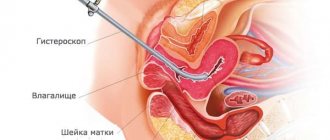
There is a procedure called HSG (hysterosalpingography) - an x-ray of the fallopian tubes with contrast. Its goal is to find out their permeability. It is performed after menstruation, and doctors after such manipulation usually advise the patient to try to conceive a child in this cycle, because after exposure to them the pipes become more passable for a certain time.
Doctors advise women planning to conceive a child to undergo x-rays and fluorography in order to identify possible pathologies in time. After all, they can make themselves felt when carrying a child.
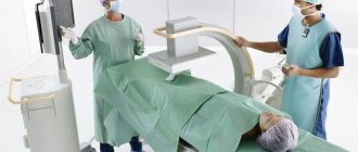
How to reduce the dangers of x-rays
If a pregnant woman needs an x-ray due to indications, doctors take measures to reduce the potential risk to the fetus. This is mandatory shielding of the abdomen and pelvis area with a special lead apron and the use of the diaphragm technique (the spread of rays is limited by special barriers).
When performing x-rays, patients must wear a lead apron.
In addition, pregnant women are strictly prohibited from helping their older child or other relative during the procedure. If a woman herself works as a radiologist, then she will definitely be transferred to another, safer job.
What is an x-ray of the paranasal sinuses?
X-ray of the paranasal sinuses is one of the types of X-ray examination that allows you to clarify the condition of the organ and identify the causes of unpleasant symptoms. X-rays of the paranasal sinuses are taken in two projections: chin and nasomental, which allows visualization of all structures of the paranasal sinuses. For already diagnosed tumors in the sinus cavity and paranasal sinus, this research method is not suitable, since the structure and size of the tumors are poorly captured by x-rays. Therefore, a study is ordered to confirm the occurrence of pathology.
How many times can an x-ray be taken?
If we are talking about analog devices, then experts recommend a break between irradiations of 3 weeks and take one photo per visit.
. However, it happens that it is necessary to increase the number of studies, then they are carried out every couple of days, reducing the negative impact as much as possible. Several x-rays on an analog device in one day can have a bad effect on your health.
The invention of digital equipment has made it possible to greatly reduce risks and allow for more frequent x-ray examinations. There is no longer any need to make compromises between harm and health benefits; doctors prescribe as many procedures as necessary to effectively monitor the progress of treatment.