The computed tomography method is often used to diagnose respiratory pathologies. This is a safe X-ray examination that takes 15-30 minutes and allows you to assess the condition of the lungs. The images are formed from 1000 layer-by-layer images of tissues. Any deviations from the norm are visible on them. Based on the images, the doctor can accurately diagnose and prescribe effective treatment. The method is used to clarify pneumonia, tuberculosis, chronic diseases, neoplasms, and assess lung function.
Operating principle of CT
The technology is based on the use of low-dose X-ray radiation, which can be absorbed by body tissues with varying degrees of intensity. The degree of reflection of the ionizing field from hard and soft fibers is recorded by equipment sensors, displaying them in photographs in black and white. Despite the monochrome images, the equipment is capable of distinguishing more than 500 shades of gray inherent in different tissues and organs. Any deviations from the norm in their structure, density, or size are immediately recorded by equipment.
Scans are made layer by layer with a minimum offset of 1 mm. Thus, up to 1000 sections can be obtained in one procedure without white spots or blind spots. From these scans, a multidimensional model of the organ is compiled, each section of which can be viewed from different angles and with maximum approximation. Since computed tomography involves X-ray radiation (radiation exposure is present), it can only be performed in consultation with the attending physician and no more than 5-8 times a year for an adult patient. Changing the frequency of repeated procedures is available only as prescribed by a specialist.
Where can I get a CT scan?
Sign up for a computed tomography scan at the MRI Center by phone or through the feedback form on the website. Doctors of the highest category, polite staff, and the latest generation tomographs - Philips, Siemens, Toshiba - are waiting for you. You will receive high-quality images, an expert opinion and a chance for a speedy recovery.
There are no queues at our diagnostic centers - studies are carried out by appointment for a specific time. The air in the rooms is disinfected after each patient, all surfaces are thoroughly treated with antiseptic solutions.
What does a computed tomography scan of the lungs reveal?
In pulmonology, CT is used very actively, as it allows one to detect numerous diseases at any stage of their development. The study is prescribed if the following pathologies are suspected:
- chronic obstruction of the lungs and bronchi (asthmatic syndrome, pneumonia, emphysema, etc.);
- infection by viruses and bacteria (viral pneumonia caused by coronavirus, other types of pneumonia, tuberculosis);
- inflammation of different parts of the pleura, pleural adhesions;
- cystic formations of the mediastinum;
- tumors of different quality;
- congenital anomalies of the structure of the respiratory system.
Often the standard computer scanning program is supplemented with an angiography mode. In this case, contrast is added, which better “highlights” the vascular network in the images. This is how thromboembolism, dissection of the aortic walls, stenoses, and the initial stages of the growth of cancerous formations entangled with blood vessels are detected.
MRI or CT scan of the chest
Often patients are concerned with the question: “Which is better - CT or MRI?” Experts believe that these two studies complement each other. A CT scan uses X-rays, while an MRI uses a magnetic field to produce images. Radiation exposure to the body is undesirable during gestation and in childhood under 14 years of age, and magnetic resonance examination cannot be performed on patients with installed implants (pacemakers, insulin pumps, orthoprostheses, etc.). CT scan lasts several minutes, which makes it indispensable in urgent situations - in case of injuries, bleeding, cardiovascular accidents.
A chest CT scan includes assessment of the following organs and structures:
- trachea, bronchi, lungs and pleura;
- heart and blood vessels;
- sternum, ribs and thoracic spine;
- mammary glands;
- esophagus;
- thymus gland.
The same structures are visible on MRI scans, but in diagnosing bone tissue pathologies, CT remains the study of choice.
Can CT scans be used for coronavirus?
According to WHO, the most common complication of COVID-19 is infectious pneumonia associated with coronavirus. This condition is determined on CT scan images of the lungs with an accuracy of 98%. X-ray is also used to visualize pneumonia, but it is not as informative as computer screening. Foci of fibrosis can be seen on x-rays only with a protracted course of the disease, starting from stage 3. The first two degrees of damage are not displayed at all or are controversial. If X-ray scans show signs of fibrosis, specialists still send for a CT scan to get a more complete picture. To avoid increased radiation exposure if you suspect COVID-19, it is better to immediately undergo a lung tomography and get tested for Covid.
What to take with you to the examination
If a CT scan is performed for the first time, the patient needs to take with him a referral, passport and compulsory medical insurance policy. But if a person has previously undergone lung diagnostics, then it is necessary to show the doctor the results of past studies. It can be:
- X-ray;
- fluorogram;
- bronchoscopy results;
- discs with MRI or CT recordings.
Any available data can help diagnose the patient's condition. Therefore, even if only extracts from the medical record remain from the last examination, it is better to take them with you and show them to the doctor.
What can be seen on CT scans for coronavirus?
If the disease has descended into the lungs, the scans will immediately identify foci of inflammation.
The term “ground glass” is used to refer to specific compactions in pneumonia. This means that the lung tissue contains fluid-filled alveoli, which are normally filled only with air. Most often, small areas of “ground glass” (lightened pattern) are found in the lower lateral lobes on each side. If they spread and merge, this indicates a worsening of the disease.
The following changes in tissues are also detected:
- increased density of partitions;
- parenchyma pattern resembling a patchwork quilt;
- hard rims around the lesion (reverse halo syndrome);
- air inclusions in isolated lumens of the bronchi.
In addition to structural changes in the parenchyma, the tomogram shows cavitation bubbles, enlarged lymph nodes, and the presence of free fluid in the pleural cavity. In severe cases of the disease, zones of fibrosis are visible - replacement of the alveolar network with adhesive fibers. They are not capable of breathing or gas exchange, which is why the overall “useful” volume of the lungs suffers. To differentiate viral pneumonia from other diseases with similar symptoms, it is necessary, along with a CT scan, to undergo a PCR test in the clinic.
CT scan of the lungs after coronavirus
A CT scan of the lungs for coronavirus is done not only to assess lung damage, but also to monitor the recovery process as part of therapy. The first is done three days after the start of treatment, if it does not produce results and the patient does not recover. The next tomography can be repeated after a week if the patient’s condition does not improve.
With favorable treatment during the rehabilitation period, a CT scan of the lungs can be performed twice (interval - 2-3 weeks) to monitor the dynamics of lung recovery after coronavirus. In total, it is recommended to do no more than 5 CT scans per year.
Lung tissue is elastic and capable of regeneration. If the pathology is detected in time and treatment measures are taken, the patient’s body can cope with the infection within 1 month, and after rehabilitation, the functionality of the lungs will be completely restored.
If a patient was admitted to a medical facility with more than 50% lung damage, suffered severe pneumonia or acute respiratory distress syndrome, then fibrosis may develop. The consequences of pulmonary fibrosis resemble scars, and such pathological changes may be irreversible. However, if small areas are affected, then from a functional point of view they are easily compensated by healthy ones and are not felt throughout life. The feasibility and number of repeated computed tomography scans during the rehabilitation period are determined by the doctor.
Main indications for the study
It is better to go for a CT scan with a direct referral from a doctor, but if no contraindications to tomography have previously been identified, you can come for the procedure on your own initiative, without a referral. Screening is required for the following conditions:
- persistent cough not associated with a previous acute respiratory viral infection;
- stable temperature 37-39 degrees;
- soreness in the sternum against the background of previously discovered cancer in other areas of the body;
- inhalation of a foreign object;
- dry cough, shortness of breath and difficulty breathing due to spastic phenomena;
- copious sputum production for a long time;
- signs of arterial pulmonary thromboembolism (pain in the heart area, sweating, pallor, rapid heartbeat);
- complications of systemic diseases affecting the respiratory system.
In the current situation with a high risk of contracting Covid, you should not do a tomography just for the sake of interest, since later, in case of a real illness, multiple repeat computer examinations may be required. In order not to expose the body to additional radiation load, at the first suspicion of coronavirus, it is better to take a test at home. If the result is positive, you must consult a doctor who will decide on computer screening.
MRI and CT with and without contrast - what is the difference?
Check the possibility of treatment in this area at the moment
Magnetic resonance and computed tomography are considered the most informative, safe and fast diagnostic methods in modern medicine. With their help, pathologies of various organs and systems are identified, the condition of the body is monitored during the rehabilitation period, and congenital anomalies are found. At CDC-24 you can undergo examinations using new generation tomographs, including MRI and CT with and without contrast
. The difference lies in the need to use medications that are administered intravenously or orally. Enhanced examinations are safe, but require certain preparation and are done exclusively as prescribed by a specialist: cardiologist, neurologist, gastroenterologist, oncologist, traumatologist, etc.
Why is contrast used in MRI?
In most cases, diagnostics using radio waves and magnetic fields is sufficient to detect abnormalities in internal organs, joints, brain structures and the spine. But sometimes there is a need to obtain the most accurate and detailed image, and then the doctor recommends a contrast method. The injected substance influences the properties of water molecules in the area under study, thereby improving visualization and making it possible to find the smallest anomalies due to the amplified signal.
Some people think that the main difference between an MRI with and without contrast is
is that enhanced diagnostics are harmful, but conventional magnetic resonance imaging is not. In fact, both procedures are safe, and the substances used do not affect the body in any way; their purpose is to “illuminate” the picture. They are quickly eliminated from the body and do not cause a hypersensitivity reaction (with rare exceptions). As for adverse reactions, they occur infrequently and disappear within 1-2 hours - as a rule, they are a slight headache, rashes and dizziness.
Depending on the area under study, enhancer substances may differ in composition (with iron oxide, manganese compounds, gadolinium, etc.) and method of use. Most drugs are administered by intravenous injection, but when studying the state of the gastrointestinal tract, oral varieties are used.
Does MRI without contrast show tumors?
It is possible to detect neoplasms without introducing special drugs into the patient’s body. However, in some cases, for example, with a small tumor or its complex location, amplification is required in order to most accurately determine the type and stage of the pathology. The study is also prescribed to determine malignancy and when deciding on surgical intervention.
Indications and contraindications
Enhanced MRI is performed on patients with suspected cancer, aneurysms, pituitary gland pathologies, as well as nerve damage and disorders of the cardiovascular system. Diagnostics allows us to identify necrotic lesions of soft tissues of any location, injuries of the musculo-ligamentous apparatus, diseases of the reproductive organs, lungs, liver, gallbladder and spleen, as well as degenerative changes in the central nervous system.
Indications for the examination include:
- convulsions, migraines, frequent fainting;
- decrease or loss of hearing, vision;
- palpable neoplasms in the neck area;
- arthrosis of joints, hernias, protrusions;
- multiple sclerosis;
- joint pain;
- muscle atrophy, paralysis, paresis of the lower and upper extremities;
- infectious lesions of the brain or spinal cord;
- neoplasms in the chest, pelvic organs and gastrointestinal tract;
- infertility in men and women.
There are no restrictions on the frequency of the procedure - it is prescribed as many times as required for effective treatment of the patient. Before conducting diagnostics at CDC-24, it is mandatory to check for contraindications, which include:
- pregnancy, regardless of trimester;
- individual intolerance to contrast agent;
- chronic heart failure;
- serious diseases of the kidneys and other organs of the urinary system;
- the presence of metal objects and devices in the body;
- fear of closed spaces.
MRI with and without contrast: which is better?
Both methods are effective and safe, and their differences are due to the use of drugs that enhance the signal. Compared to conventional magnetic resonance imaging, enhanced MRI:
- Lasts longer - the patient will have to stay 30-40 minutes longer inside the tomograph.
- Allows you to get more detailed pictures.
- May cause minor allergic reactions.
- Provides additional information about the state of the structures under study.
- Requires large financial costs.
- Assumes compliance with additional training recommendations.
PET CT, CT with and without contrast: what studies show, what are their differences
If we are talking about injuries and diseases of dense structures, then patients are prescribed computed tomography and PET CT, and CT can also be performed with the introduction of a contrast agent. In the first case, the radiologist studies the anatomical features of organs and tissues, using signal-enhancing drugs if necessary, and in the second, he determines deviations in dynamics by examining functional activity. Positron emission tomography is considered the most advanced method and provides more information than CT with contrast. The disadvantages of the examination include its duration (and the patient must remain motionless during the entire diagnosis) and high cost.
Conclusion
To summarize, we can say that the difference between MRI, CT with and without contrast
lies in the quality of visualization. Enhanced examination makes it possible to detect pathologies at the initial stage and is indispensable in differential diagnosis. Most often, the procedure is performed on patients with cardiac, neurological symptoms, as well as malignant and benign neoplasms. When contrast is administered, the examination takes longer and in some cases may be accompanied by side effects.
When scanning is prohibited
The main danger of the technology is the presence of an X-ray field during the examination. It is minimized to low-dose levels, which is considered safe for relatively healthy people. However, this type of diagnosis is not allowed:
- young children;
- women at any stage of pregnancy;
- cancer patients who have recently undergone radiation therapy;
- patients living or working in places with increased background radiation, often requiring radiography.
In some cases, limitations may be associated with the use of contrast. To enhance the clarity of the images, an iodine-based staining agent is injected intravenously. For more information about contrast enhancement, please follow the link: Lung CT with Contrast.
Preparing for a CT scan of the chest
A thoracic CT scan can be performed immediately on the day of your visit.
Preliminary preparation includes:
- Questioning the patient to identify possible contraindications;
- Making entries in the outpatient card, signing voluntary informed consent for the study;
- Mandatory removal by the patient of all metal objects that may affect the quality of the study;
- Explaining to the patient the basic rules of diagnostics, as well as explanations of actions in the event of a sudden deterioration of the condition.
Carrying out a CT scan of the lungs using contrast does not cause negative reactions in most patients, with the exception of individual intolerance to the components of the substance.
A CT scan takes about 2 minutes, and up to 10 minutes when using contrast. During the entire procedure, the radiologist monitors the progress of the procedure and the patient’s condition.
Do I need to prepare for the procedure?
There is no need to prepare specifically or for a long time for a tomography session, but when going for a scan, it is important to consider the following points:
- Interaction with the device occurs in a lying position, so it is better to wear loose, comfortable clothes that do not restrict movement when lying on a high couch.
- All foreign objects, metal jewelry, and small change must be removed from the body and pockets, as distortions and artifacts may appear in the photographs.
- Tell the specialist about chronic pathologies, possible pregnancy, the presence of built-in implants, electronic stimulators.
- To register or conclude an agreement for the service, bring your passport, as well as the results of other studies and a referral from the treating doctor.
Contraindications
Computed tomography is not a preferred test for pregnant women and children under 14 years of age. They try not to perform CT with contrast in this category of people and in patients with a complicated medical history, which includes:
- hyperfunction of the thyroid gland;
- allergy to iodine-containing substances;
- urological and nephrological pathology with impaired renal function;
- cardiovascular diseases in the stage of decompensation.
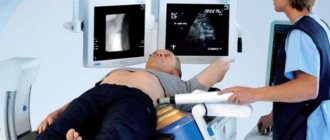
How does the MSCT procedure of the lungs work?
The visitor is escorted to the room with the equipment and helped to sit on the retractable part of the apparatus while lying on his back.
Your hands need to be raised behind your head and held motionless. The ring part of the scanner will slowly move along the study area, remotely taking layer-by-layer images. At the right time, the doctor will ask you to hold your breath several times. The entire process takes no more than 15 minutes. The session takes place without penetration into the tissue, without pain. Upon completion of the scan, the patient does not need rehabilitation; he can go about his remaining business.
What may be in the results
The scans produced by the equipment are immediately sent to the doctor’s computer, who studies them in detail after the procedure. The interpretation takes from half an hour to a day, so it is not necessary to wait for the results in the clinic. It is enough to leave your contacts at the reception so that you will be notified of readiness or a digital version of the conclusion and photographs will be sent to your email address.
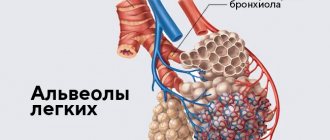
In the photographs you can see clear boundaries of the internal organs, blood vessels and bones. The denser the fabric, the lighter it appears in the images. The pulmonary parenchyma is normally homogeneous, gray, but with the development of inflammation, fibrosis, and filling of the alveoli with fluid, the pattern changes. Foci of compaction are displayed in the form of a lightened network, “frosted glass”. Tumors and other neoplasms are also visible with precise sizes and configurations; they are indicated by a developed vascular network around the pathological object.
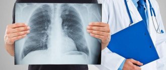
Chest tomography photo
The CT result of the OGK will be ready in 90-120 minutes; at the Magnit diagnostic center in St. Petersburg, the data obtained in doubtful situations are assessed collectively. Let us present several tomograms that demonstrate pathological processes in the chest:
CT scan of the chest demonstrates a mediastinal tumor (the boundaries are highlighted with markers)
CT scan of the mediastinum. Red arrows indicate lipoma.
Signs of peripheral lung cancer on CT (highlighted in red)
Advantages over other technologies
Despite the large number of diagnostic procedures, CT scans of the lungs and bronchi are more often chosen. This is due to the following advantages:
- comprehensive visualization of the study area;
- Possibility of shooting from different angles;
- no blind spots;
- obtaining complete results in just one procedure;
- high accuracy, maximum scan resolution;
- detection of early stages of serious diseases;
- the presence of metal and electronic stimulators is not a serious contraindication to the study;
- construction of three-dimensional models of the organ with subsequent mathematical analysis of the data.
It should be noted that there is increased accessibility for all categories of patients. In comparison with alternative types of tomography, computer screening is one and a half or more times cheaper than MRI or PET, while in terms of information content regarding the lungs it is in no way inferior to them, and sometimes even surpasses them in certain indicators.
CT scan of the chest for tuberculosis
Focal changes on CT scan of the lungs, suspicious for tuberculous lesions (highlighted in red)
Tuberculosis affects patients of any age and gender; the lungs are most often involved in the process. The disease is considered socially dangerous, since if the open form is not diagnosed in a timely manner (release of mycobacteria into the environment when coughing), there is a high risk of infection of surrounding people. Chest CT for tuberculosis is prescribed in the following situations:
- screening radiography showed changes suspicious for the pathology in question;
- the patient has positive results of laboratory tests for tuberculosis (Mantoux test, Diaskintest) with ambiguous signs on the fluorogram;
- tracking dynamics after specific therapy;
- as a preoperative diagnosis to identify the anatomical features of blood supply and innervation of lesions, localization of pathology.
How much does a CT scan of the lungs cost in St. Petersburg and what does the price depend on?
In St. Petersburg, the cost of lung tomography starts from 2800 rubles and depends on the following factors:
- type, quality and year of manufacture of the tomograph;
- price level in the medical center (economy, average or premium);
- time of day at which the study is carried out (often it can be done cheaper at night, for more details - CT scan of the lungs at night);
- the need to obtain film with a photograph (usually pictures are recorded on disk, and film costs extra);
- the need to administer contrast, read more here - CT scan of the lungs with contrast;
CT scan of the chest with contrast
Bolus contrast allows obtaining high-quality tomograms, ensuring the delivery of the drug to the required phases of the study
The introduction of contrast during CT is a necessary measure if it is planned to examine blood vessels, soft tissues, find out the location of the tumor and its relationship with nearby structures, or metastasis. MRI is preferable for diagnosing neoplastic processes, but computed tomography helps to establish the cause when magnetic scanning is contraindicated. Chest CT with contrast helps to obtain more informative tomograms for assessing vascular pathologies. It is easier to perform a differential diagnosis of diseases by analyzing the results of images obtained after enhancement. Doctors often have to use CT scan data to distinguish changes typical of a tuberculous cavity from a cancerous tumor, a bronchopleural fistula from a lung abscess, emphysema from a tense cyst, etc.
Preparation for a chest CT scan with contrast:
- taking a blood test for creatinine level (can be done using the express method for an additional fee before the test at the Magnit diagnostic center);
- a break in taking glucose-lowering drugs (Metformin and its analogues) after permission from the endocrinologist;
- refusal of lactation for 12 hours after performing a CT scan of the chest with the introduction of contrast and a preliminary supply of milk for the next two feedings.
What to do if you need to undergo a test urgently
To urgently undergo the study, call: +7 (812) 385-77-56. Our operators see a summary table of available places in most diagnostic clinics in St. Petersburg, so they can make an appointment in the near future.
Sources used:
- Gedymin L.E., Filippov V.P., Evgushchenko G.V., et al. The role of biopsy in the diagnosis of pulmonary diseases at the prehospital level of observation // Clinical Medicine. - 2009. - No. 4. — P.41-44.
- Ivanova E.V., Bilichenko T.N., Chuchalin A.G. Morbidity and mortality of the working age population in Russia due to respiratory diseases in 2010 -2012. // Pulmonology. - 2015. - T. 25. - No. 3. - P.291-297.
- Karashchuk N.P., Kiseleva M.V. Lung cancer and tuberculosis // Scientific Medical Bulletin of Ugra. -2014. — No. 1-2(5-6). — P.71-73.
- Kartashov M.V., Kartashova O.M., Kotlyarov P.M. First experience of using diffusion-weighted magnetic resonance imaging for small cell lung cancer // Medical visualization. – 2011. – No. 4. – P.28-33.
- Kotlyarov P.M. Post-processing of multislice computed tomography data in the refined diagnosis of pathological changes in diffuse lung diseases // Pulmonology. - 2021. - No. 4. — P.472-477.
- Kotlyarov P.M., Sergeev N.I. Radiation research methods in the differential diagnosis of parasitic and tumor lesions of the lungs // Siberian Journal of Oncology. – 2021. – No. 15(4). – P.33-39.
- Ternovoy S.K., Nasnikova I.Yu., Morozov S.P. Modern computed tomography in clinical medicine // Kremlin Medicine. Clinical Bulletin. - 2008. - No. 2. — P.9-13.
- Tyurin I.E. Differential diagnosis of single lesions in the lungs // Polyclinic. - 2014. - No. 3-1. — P.28-32.
- Yudin A.L., Afanasyeva N.I., Abovich Yu.A., et al. High-resolution computed tomography in the diagnosis of interstitial pneumonia // Medical visualization. -2002. - No. 4. — P.40-48.
Text approved by therapist Laeva Alina Vadimovna
CT lungs Admiralty CT lungs Bucharest CT lungs Vasileostrovskaya CT lungs Volkovskaya CT lungs Gorky CT lungs Gostiny Dvor CT lungs Dostoevskaya CT lungs Elizarovskaya CT lungs Zvenigorodskaya CT lungs Zvezdny CT lungs Komendantsky Prospekt CT lungs Krestovsky Island CT lungs Kupchino CT lungs Ladozhskaya CT lungs Ligovsky Prospect CT lungs Lomonosov CT lungs Mayakovskaya CT lungs International CT lungs Moscow CT lungs Moscow Gate CT lungs Nevsky Prospekt CT lungs Novocherkasskaya CT lungs Bypass channel CT lungs Obukhovo CT lungs Pobeda Park CT lungs Alexander Nevsky Square CT lungs Primorskaya CT lungs Proletarskaya CT lungs Bolshevikov Avenue CT lungs Rybatskoe CT lungs Sadovaya CT lungs Sennaya Square CT lungs Spasskaya CT lungs Sports CT lungs Staraya Derevnya CT lungs Technological Institute CT lungs Dybenko Street CT lungs Frunzenskaya CT lungs Chkalovskaya CT lungs Elektrosila show all metro show more
How is a chest CT scan done?
The Magnit clinic has installed a modern and safe Siemens Somatom Emotion tomograph (16-slice)
To get a CT scan of the chest organs at the Magnit diagnostic center in St. Petersburg, you must first fill out an application form on the website or make an appointment by phone: +7, it is possible to undergo diagnostics for emergency indications on the day of treatment.
At the appointed time, the patient is placed on the tomograph table in a horizontal position. To ensure immobility, fixing belts and rollers can be used; this is necessary to prevent artifacts (defects) on the films, which complicate diagnosis. If a chest CT (CT) is performed with contrast, the pharmaceutical is administered intravenously at once or as a bolus (using an injector that supplies an amplifier during certain phases of the study).
The table moves the patient deep into the tomograph, inside the ring of which sensors begin to rotate around the area under study, capturing the response of organs and systems to the ionizing stream of particles emitted by the beam tube. The data is transferred to a computer, and a special program builds the images. Modern MSCT (multispiral computed tomography), using multiple detectors, allows minimizing radiation exposure, which makes the examination safe for most patients. There is no pain, you can use headphones to level out the mechanical noise from the operation of the equipment. Communication with staff takes place via speakerphone, and there is a panic button at hand. The examination takes 15-20 minutes; after a CT scan of the chest organs, the patient can do his usual activities.