The clavicle is a supporting support for the shoulder girdle, ensuring the function of the upper limb and its position. It is vulnerable to damage because it performs a special function, is located under the skin and has an unusual geometry.
Clavicle fractures are the most common injury among adults and account for 5% of the total number of fractures. The incidence is 29 cases per year per 100,000 population with a mean age of 33 years and a male to female ratio of 2.6:1. In men, the peak occurs between the ages of 13 and 20 years.
The main mechanism of injury is a fall during sports.
Fractures of the middle third of the clavicle are the most common and occur in 69-81% of cases, while injuries to the distal third occur in 16-28%.
Clinical examination
The evaluation of patients should take into account the mechanism of injury and possible associated injuries. When assessing a patient with high-energy trauma, the ATLS protocol should be followed. In case of open fractures, fractures of the middle third, concomitant fractures of the scapula, it is necessary to use additional imaging methods to exclude concomitant damage to the chest organs.
Patient complaints
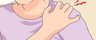
The main complaint of patients with all clavicle fractures is pain in the shoulder joint , however, to obtain a complete picture it is necessary to examine the cervical spine, chest and evaluate the neurological status to exclude possible combined injuries.
Clinical examination
The examination should begin with an examination of the shoulder areas on both sides. A displaced fracture is characterized by the presence of a typical deformity with protrusion of the medial fragment as a result of the action of the weight of the limb and traction of the sternocleidomastoid muscle.
Shortening of a broken clavicle is manifested by a decrease in the width of the shoulder girdle as a result of protraction of the scapula. During examination, it is necessary to evaluate the condition of the skin, since in case of open fractures or significant skin tension, surgical treatment is indicated. With a direct fall on the shoulder, there are abrasions, which are often located in the projection of the deltoid muscle and acromion process.
Palpation should examine the sternoclavicular joint, subcutaneous surface of the clavicle, acromioclavicular joint, acromial process and spine of the scapula. Detection of specific areas of tenderness determines the need for imaging techniques.
Due to the pain syndrome, there is a restriction of movement, however, with an isolated fracture of the clavicle, passive internal and external rotation is possible. The presence of full passive rotational movements indicates the absence of dislocation in the shoulder joint. The proximity of the brachial plexus and the vessels of the subclavian region requires an assessment of the neurovascular status (examination of the pulse and sensory-motor characteristics of the limb distal to the site of injury).
Visualization
Minimal x-ray examination
should include a direct view with full coverage of the clavicle and acromioclavicular joint. Additional radiography with a radiator tilted 20° radially helps assess clavicle displacement and shortening.
If damage to the shoulder joint is suspected, radiography should be performed in direct and axial projection.
Performing a CT
with reconstruction gives a complete picture of the configuration of the fracture, displacement and shortening
of the clavicle .
Classification
There are several classifications of clavicle fractures . A more practical and constructive classification was proposed by Robinson. This Edinburgh system considers the displacement and comminuted nature of fractures as key features of therapeutic and prognostic significance, as well as inter-rater reliability.
Type I – Medial 1/5 (fracture medial to the first rib)
- IA: no offset
- IA1: extra-articular
- IA2: intra-articular
- IB: offset
- IV1: extra-articular
- IV2: intra-articular
Type II – Moderate 3/5 (Damage to the central 3/5 of the clavicle)
- IIA: without disruption of the cortical layer
- IIA1: no offset
- IIA2: angulation
- IIB: with offset
- IIВ1: simple
- IIВ2: splintered/segmented
Type III – Lateral 1/3 (Injury to the lateral part of the clavicle)
- IIIA: without disruption of the cortical layer
- IIIA1: extra-articular
- IIIA2: intra-articular
- IIIB: offset
- IIIВ1: extra-articular
- IIIB2: intra-articular
All types are further subdivided depending on the displacement, where subtype A is fractures with a displacement of less than 100%, subtype B is more than 100%. Groups are also distinguished depending on the splintered nature of the damage and the relationship of the fracture line to the structure of the joints.
Anatomy
Rice.
1. Right clavicle (a - top view, b - bottom view): 1 - acromial end; 2 - sternal articular surface; 3 - sternal end; 4 - acromial articular surface; 5 - coracoid tuberosity; 6 - nutrient opening; 7 - costal tuberosity. The clavicle has the shape of an elongated letter S (Fig. 1); the length of the adult human body is 12-15 cm. The medial part of the body (about 2/3 of the length) is curved anteriorly, the lateral third is curved posteriorly and ends with a plate of spongy structure, flattened from top to bottom. The middle section of the kidney is cylindrical in shape and has a bone marrow canal. The sternal end is thickened, has the shape of a triangular prism with blunt edges and a pronounced rough impression on the lower side, caused by the attachment of the costoclavicular ligament. On the lower side of the acromial end of the cone there is a cone-shaped tubercle (tuberculum conoideum) and a trapezoidal line (linea trapezoidea)—the attachment points of the coracoclavicular ligament. The deltoid muscle (m. deltoideus) is attached to the acromial end from above and in front, and the trapezius muscle (m. trapezius) from above and behind.
The acromial end of the joint articulates with the acromial process of the scapula, forming the acromial clavicular joint. Mobility in it is due to weak tension of the articular capsule and the fibrocartilaginous layer between the bones; the edges are often formed into a more or less isolated disc. The joint is strengthened by two ligaments: acromioclavicular (lig. acromioclaviculare) and coracoclavicular (lig. coracoclaviculare).
The sternal end of the joint articulates with the sternum (sternoclavicular joint). The joint is divided into two cavities by a cartilaginous articular disc. The stability of the joint is due to powerful ligaments. It is connected to the first rib by a two-layer costoclavicular ligament (lig. costoclaviculare). In front and behind, the sternoclavicular joint is strengthened by the anterior and posterior sternoclavicular ligaments (lig. sternoclaviculare ant. et post.). The interclavicular ligament (lig. interclaviculare) passes across the jugular notch.
The sternocleidomastoid muscle (m. sternocleidomastoideus) is attached to the sternal end of the muscle at its posterior edge, and the sternohyoid muscle (m. sternohyoideus) is attached below. The pectoralis major muscle (m. pectoralis major) is attached to the medial two-thirds of the chest in front. The subclavian muscle (m. subclavius) runs along the lower surface of the muscle between the first rib and its acromial end. The nutrient opening (foramen nutricium) is also located here, through which vessels penetrate into the bone.
The sternoclavicular and acromioclavicular joints are involved in active movements of the upper limb. With their participation, it rises above the horizontal line, moves forward and to the side. The functional significance of the shoulder is that it is a “spacer” between the bones of the torso (sternum) and the shoulder joint (see Shoulder girdle). This stabilizes the joint and increases its range of motion.
At the level of the middle third of the chest, behind it, between the attachment of the deltoid and pectoralis major muscles, in front of the first rib, the subclavian artery and vein and the brachial plexus pass.
Age characteristics
Clavicles: the first ossification nuclei appear in the body of the body at 5-6 weeks. intrauterine life and merge at the 7th week. The ossification nucleus in the cartilaginous sternal end of the cartilage appears at 16-20 years and merges with the body of the cartilage by 20-25 years.
X-ray anatomy
For X-ray examination of the clavicle, radiography is used in the anterior and posterior projections, sometimes in the anterior bilateral projection - to obtain a symmetrical image of both C., as well as in the so-called. superior projection - to obtain: images of K. outside the background of the ribs (when the rays move from below to the front, along the anterior wall of the chest). If there are indications, use: tomography (see). All pictures of K. are taken with held breathing.
Rice. 2. Anterior radiograph of a normal right clavicle.
In direct projection photographs, the S-shaped curvature of the knee in the horizontal plane is concealed (Fig. 2). The wide flat acromial end of the K. is projected against the background of thin integument and therefore appears sparse; it has a small articular surface. On the lower contour of the K., near the sternal end, a depression is visible at the site of attachment of the costoclavicular ligament, sometimes mistakenly taken for a focus of destruction. In the diaphysis of K. the cortical layer and the medullary cavity are expressed, and at the ends there is a spongy structure and a thin cortical layer. Near the upper contour of the K. in the diaphysis, an oblong clearing of diameter is sometimes found. up to 3 mm - canal for the lateral branch of the middle supraclavicular nerve.
Functions
The main functions are:
- transport function (movement of physical impulses to other bones of the skeleton);
- protective (protection of blood and lymphatic vessels that are located under this anatomical segment);
- provides range of movement of the arm (this fact eliminates bone friction, since the bone is distant from the chest).
It is noteworthy that in our primitive ancestors and intermediate forms of apes, this bone was much shorter than in modern humans. In the process of evolution, it changed, as man began to use his hands to perform various work activities.
Pathology
Congenital deformities
Congenital deformities are rare and are divided into two groups: 1) violations of the size and shape of the joint—additional coracoclavicular, costoclavicular joint, bifurcation of the joint; 2) so-called defective anomalies - absence of part or all of the blood, perforated blood, clavicular-cranial dysostosis (see).
For congenital deformities of the clavicle, in case of functional disorders, surgical treatment (bone grafting) is indicated.
Damage
Clavicle dislocations
, according to a number of authors, constitute from 3 to 19% of all dislocations. There are dislocations of the sternal and acromial ends of the joint.
Dislocations of the acromial end of the shoulder are more common and usually occur when there is a blow to the acromial process of the scapula or a fall on the adducted shoulder; they can be incomplete (subluxations due to rupture or tear of one acromiocleidoclavicular ligament) and complete (if the coracoclavicular ligament is also ruptured). When the acromial end of the joint is dislocated, its protrusion is visible under the skin, and when pressure is applied from above, mobility is determined (“key symptom”). If a dislocation of the acromial end of the joint is suspected, bilateral photographs are taken (for comparison with the opposite side) with the patient in a vertical position, since in a horizontal position the displacement can be eliminated on its own. A radiograph showing dislocation or subluxation shows an upward displacement of the acromial end of the joint.
Dislocation of the sternal end of the K. can be incomplete (subluxation) or complete. There are presternal, suprasternal and retrosternal dislocations. These dislocations are difficult to diagnose, since the deformation is not significant, and on the anterior bilateral radiograph, the asymmetry of the sternal ends of the joint is revealed only with suprasternal dislocation.
To reduce dislocations of the acromial end of the joint, under local anesthesia, the shoulder is raised, retracted and pressed from above on the outer end of the joint. To hold the joint in this position, there are a large number of plaster casts and splints, the general principle of which is pressure from above on the acromial end of the joint. Good results are achieved by using staged belt-type bandages, the main advantage of which is constant pressure on the acromial end of the shoulder, the reliability of its immobilization and the possibility of early restoration of the function of the shoulder joint. The pressure element of these dressings is used in the first 3 weeks. replaceable plaster splint, and then rubber honey. bandage folded in several layers. Complete and chronic (3 weeks or more after injury) dislocations of the acromial end of the clavicle are usually treated surgically. Fixation of the joint to the acromial and coracoid processes using a silk thread has become widespread; a combined method involves connecting the joint to the coracoid process with Mylar tape, to the acromial process with Kirschner wires, and osteosynthesis of the joint with metal clamps. After the operation, a Deso-type plaster cast with a roller is applied in the armpit for 3-4 weeks. (see Desmurgy). With fresh dislocations of the sternal end of the joint, it is easy to perform a closed reduction, but it is extremely difficult to hold the reduced end of the joint. Therefore, open reduction of the dislocation with fixation with one or more wires is often performed. Immobilization after surgery - up to 6 weeks.
Clavicle fractures
amount to approx.
3% of all fractures. Direct blows to the knee are relatively rarely the causes of fractures. The latter are most often caused by an indirect effect on the knee: a fall on an outstretched arm, a blow to the shoulder joint, compression of the body in the sagittal plane, etc. Fig .
3. Schematic representation of a typical displacement of fragments in a clavicle fracture: 1 - sternocleidomastoid muscle; 2 - internal fragment; 3 - subclavian muscle; 4 - external fragment. The most common localization of fractures of the joint is the border of its middle and outer thirds, which is due to its physiological bending and the lowest mechanical strength in this place. In adults, fractures are transverse, oblique, comminuted, often with significant displacement. Subperiosteal greenstick fractures are more common in children. Under the influence of muscle traction when a fracture occurs, its lateral end moves downward and anteriorly, and the medial end moves upward and inward (Fig. 3). Occasionally, K.'s fractures are fraught with the danger of breaking the skin with a sharp fragment, injury to the subclavian vessels and parietal pleura.
Fractures of the Clavicle are characterized by all the classic signs of fractures (see): deformation in the form of swelling and often protrusion of the ends of the fragments under the skin, subcutaneous hematoma, severe pain, palpation of bone fragments and their crepitus in the area of K., dysfunction of the corresponding upper limb. The shoulder joint on the side of the fracture is displaced downwards and slightly forward, the head is tilted towards the side of the injury. With subperiosteal fractures in children, symptoms are limited to swelling, pain on palpation and during movements of the upper limb. The X-ray image clearly shows the fracture line of the joint. In doubtful cases (for example, non-displaced fractures, “greenstick” fractures in children), an x-ray of both joints is needed on the same film.
K.'s combat injuries are accompanied by damage to the neurovascular formations, the lung, nearby large bones and a large mass of soft tissue (see Wounds, wounds) and, in connection with this, massive bleeding (see) and traumatic shock (see Shock).
Treatment of K.'s fractures can be conservative and surgical. The conservative method is used most often, especially in children.
Rice. 4. Figure-of-eight bandage applied after reposition for a clavicle fracture: 1 - application stage; 2 — front view; 3 - rear view
For immediate reposition of the fracture, K., performed under local anesthesia with 2% novocaine solution (15-20 ml), the patient is seated on a stool, and an assistant with both hands, standing behind, spreads the patient’s shoulders so as to bring the inner edges of the shoulder blades closer together . At this time, the surgeon uses his thumb to reposition the K. fragments, followed by immobilization (see). Retaining fragments presents significant difficulties, which explains the existence of more than 250 different types of bandages and splints for the treatment of fractures. The most widely used are Kuzminsky, Kaplan, Chaklin splints, a figure-of-eight bandage (Fig. 4), plaster casts such as Dezo, Sitenko, etc. Consolidation of the fracture K. in the overwhelming majority of cases occurs with immobilization for 4-6 weeks, often with some displacement of fragments. However, hand function is usually not affected. Non-union of a fracture is extremely rare; it is associated with significant interposition of soft tissues in the fracture zone or with gross errors in immobilization.
Rice. 5. Intramedullary osteosynthesis of the clavicle: 1 - acromial end of the clavicle; 2 - sternal articular surface; 3—sternal end of the clavicle; The dotted line shows a metal rod inserted into the bone and passing through the fracture line.
Indications for surgical treatment are only fractures with significant displacement of fragments and interposition of soft tissues, the threat of skin breakout by a fragment of the bone and damage to blood vessels, false joints of the knee with impaired limb function. Various options for intramedullary fixation of K fragments are used (Fig. 5). Regardless of the method of osteosynthesis (OS), after surgery a plaster cast is applied for 6-10 weeks, and sometimes for a longer period.
Diseases
Osteomyelitis (see) of the clavicle occurs more often in children, usually affecting the sternal end of the joint. Surgical treatment is necrectomy (see), sequestrectomy (see), resection of the joint.
The diagnosis of specific lesions (tuberculosis, syphilis, actinomycosis) of K. is made on the basis of a characteristic clinical picture, radiological and laboratory data. Treatment is surgical in combination with specific antibacterial therapy (see Actinomycosis, Syphilis, Tuberculosis of bones and joints).
In adolescence, there is osteochondropathy of the sternal end of the blood, which consists of aseptic subchondral necrosis (see Friedrich's syndrome).
Tumors
The most common are sarcoma (see), osteoblastoclastoma (see), osteoma (see) and angioma (see). Treatment depends on the type and nature of the tumor.
Bibliography:
Kaplan A. V. Closed injuries of bones and joints, M., 1967, bibliogr.; Krupko I. L. Guide to traumatology and orthopedics, book. 1, L., 1974, bibliogr.; Multi-volume guide to orthopedics and traumatology, ed. N. P. Novachenko, vol. 2, p. 480, vol. 3, p. 381, M., 1968; Nagy D. X-ray anatomy, trans. from Hungarian, p. 56, Budapest, 1961; Reinberg S. A. X-ray diagnosis of diseases of bones and joints, t. 1-2, M., 1964; Watson-Jones R. Bone fractures and joint damage, trans. from English, M., 1972; Burnell S. Surgery of the hand, Montreal - Philadelphia, 1964; Herman S. Congenital bilateral pseudarthrosis of the clavicles, Clin. Orthop. related Res., No. 91, p. 162, 1973; Kohler A.u. Zimmer EA Grenzen des Normalen und Anfange des Pathologischen im Rontgenbilde des Skelettes, S. 168, Stuttgart, 1956; Sycamore LK Common congenital anomalies of bony thorax, Amer. J. Roentgenol., v. 51, p. 593, 1944.
E. P. Mattis; S. I. Finkelshtein (rent.).
Diagnosis of pain in the collarbone
Specialists at the CELT Pain Clinic do not recommend self-medication, as it can cause irreparable harm to your body. In order for the treatment to be successful, it is necessary to identify the reason why the collarbone hurts and direct all efforts to eliminate it.
In addition to being examined by a doctor and taking a medical history, diagnosing collarbone pain includes the following:
- radiography of the collarbone;
- CT scan;
- Magnetic resonance imaging.
Arthritis
The most vulnerable part of the skeleton is the shoulder joint and collarbone, especially when a person is diagnosed with arthritis. This disease can vary depending on the species and occurs as a result of injury, as well as infectious or autoimmune diseases.
To get rid of arthritis, patients are prescribed special exercises and physical therapy. Treatment is selected individually in each case. When such a diagnosis is made, the patient should be provided with constant care and rest. Physical stress on the limb should be completely avoided. If the above treatment methods do not help, then surgical intervention is preferred.
First aid for acromial dislocation
When providing first aid to a victim who has suffered a dislocation of the collarbone, first of all, one should ensure fixation and calmness of the injured limb. This can be done using a bandage or a rag scarf. The arm is suspended in a bent state, and a small soft cushion is placed in the armpit.
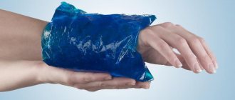
Since the injury is very painful, it is permissible to apply a cold compress to the painful area. Ice should be wrapped in cloth to prevent the skin from freezing. This kind of compress can be applied for no more than a quarter of an hour with an interval of thirty minutes between procedures.
It is important to know!
Attempting to straighten a dislocation on your own is strictly prohibited, since inexperienced actions can lead to negative consequences. Also, until the ambulance arrives, you should not give the patient strong painkillers, which can change the clinical picture.
Histogenesis of bone tissue
During the embryonic period of development, such types of cells as fibroblasts, chondroblasts and osteoblasts are formed from the middle germ layer. From them, tissues that form the organs of the musculoskeletal system, such as connective, cartilaginous and bone, subsequently develop. To answer the question: what is the clavicle from the point of view of its internal structure, let us remember that in the human skeleton there are bones that are called mixed. They are formed by elements having different origins. For example, the clavicle is formed partly from connective tissue that has undergone osteogenesis without the formation of cartilage, and partly from chondrocytes in which the process of ossification has taken place.
Symptoms of types of dislocations
At the same time, there are symptoms characteristic of each individual type of dislocation.
Prosternal type of dislocation
Thus, the first, prosternal type of dislocation is the most common and is easily determined by the protrusion of the inner end of the clavicle forward.
Suprasternal type of dislocation
In the case of a suprasternal dislocation, the collarbone protrudes forward and upward.
Substernal type of dislocation
For the third type of dislocation, according to its name, there is a retraction of the inner end of the clavicle. Retrosternal dislocation of the inner end of the clavicle is considered to be the most dangerous, since in this case there is a serious risk of damage to important anatomical structures.
Also helping to avoid confusion is the fact that in the event of a fracture, in addition to swelling and deformation, there is limited mobility of the shoulder, bruising and rupture of soft tissues from fragments of the broken bone. In addition, displacement during a fracture, unlike a dislocation, usually occurs forward and downward. However, an x-ray should be taken to rule out the presence of a bone fracture.
Attention!
In people who are obese, the outward signs of a dislocated collarbone may be less noticeable.
Possible injuries
When it has been possible to determine what the clavicle is, its features and structure, as well as how it is formed during human development, it is necessary to find out what anomalies it is susceptible to.
One of the most common injuries to the collarbone and neck is a dislocation. It can be caused by a bad fall. As a result of a dislocation, the limb becomes deformed, lengthens and protrudes from the body, forming swelling on the skin.
Another serious collarbone injury is a fracture. The causes of this condition may be birth trauma, which mainly occurs during the baby’s passage through the birth canal, a concentrated blow from a heavy object, or a fall. Most people are diagnosed with a fracture of the right or left clavicle, and in extremely serious cases - on both sides. The following symptoms indicate a fracture:
Diagnosing a fracture is not difficult, since the fragments protrude under the skin and can be easily felt. Fractures of the clavicle of the shoulder are a very painful injury. It fundamentally limits the mobility of the skeleton and brings a lot of inconvenience to the victim. In addition, minor cracks belong to fractures. But, as practice shows, they heal much faster, especially in childhood.
Chondroma
The pathology is classified as a benign neoplasm of the clavicle. When such a disease develops, the patient experiences only one single symptom. This is pain in the bone when moving. Most often, this disease is diagnosed in patients during adolescence, since rapid skeletal development occurs during this period of time.