First symptoms
A pulmonary infarction does not manifest itself immediately. For the first two to three days after a vessel is blocked, a person may not be aware of the problems. Many people confuse the first symptoms with the discomfort that occurs with angina pectoris. Sometimes pain is possible, as with an acute abdomen. You should be alert to the following symptoms:
- acute chest pain;
- increased pain when coughing, rapid breathing, bending the body;
- shortness of breath that occurs out of the blue, without physical activity;
- when coughing, blood is coughed up (not in everyone, in about half of the cases), complete pulmonary hemorrhage rarely occurs;
- the nasolabial triangle and fingers of the limbs become bluish;
- cold sticky sweat during painful attacks;
- hiccups;
- general malaise, possible fever;
- Some patients vomit.
People suffering from thrombophlebitis and thrombosis, pulmonary hypertension, and frequent relapses of pulmonary embolism are predisposed to pulmonary infarction. Risk factors include age over 60 years, excess weight, pregnancy and childbirth (or caesarean section).
Myocardial infarction
Diabetes
Atherosclerosis
7485 16 February
IMPORTANT!
The information in this section cannot be used for self-diagnosis and self-treatment.
In case of pain or other exacerbation of the disease, diagnostic tests should be prescribed only by the attending physician. To make a diagnosis and properly prescribe treatment, you should contact your doctor. Myocardial infarction: causes, symptoms, diagnosis and treatment methods.
Definition
Myocardial infarction (MI) is the necrosis of an area of the heart muscle due to insufficient blood supply with the development of a characteristic clinical picture.
Causes of myocardial infarction
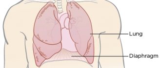
The heart is a hollow muscular organ shaped like a cone. The walls of the heart consist of three layers. The inner layer - the endocardium - lines the cavities of the heart from the inside, and its outgrowths form the heart valves. The middle layer, the myocardium, consists of cardiac muscle tissue. The outer layer is the pericardium. The human heart has four chambers: two atria and two ventricles. The right atrium receives blood from the tissues of the heart itself and all parts of the body (through the superior and inferior vena cava). Four pulmonary veins flow into the left atrium, carrying arterial blood from the lungs. The pulmonary trunk emerges from the right ventricle, through which venous blood enters the lungs. The aorta emerges from the left ventricle, carrying arterial blood to the vessels of the systemic circulation. Oxygen is delivered to the myocardium of the heart through the coronary arteries.
The heart is very sensitive to lack of blood supply (oxygen deficiency). In the case of blockage of a large coronary artery and in the absence of effective alternative blood circulation through other vessels, within 30 minutes the death of cardiomyocytes (muscle cells of the heart) begins in the affected area.
Poor circulation due to atherosclerotic lesions of the coronary arteries in 97-98% of cases is of primary importance in the occurrence of myocardial infarction.
Atherosclerosis can affect either one coronary artery or all three. The degree and extent of narrowing of the artery can vary. When blood pressure increases, the sclerotic inner layer of the vessel (endothelium) is easily damaged, blood penetrates into the plaque, the blood clotting process is activated and a blood clot is formed, which can partially or completely block the vessel.
Where a blood clot can form:
- at the site of rupture of a vulnerable (unstable) atherosclerotic plaque;
- on a defect (erosion) of the endothelium of the coronary artery, not necessarily localized on the surface of the atherosclerotic plaque;
- at the site of hemodynamically insignificant narrowing of the coronary artery.
Coronary artery thrombosis occurs with complete or incomplete parietal occlusion. A thrombus located proximally (closer to the center) in the coronary artery tends to be transported with the blood flow more distally (closer to the vessel wall) and lead to the formation of small foci of necrosis and/or contribute to the expansion of the main area of necrosis.
Great importance in the development of myocardial infarction is attached to the development of spasm of the coronary arteries both with atherosclerosis and with unchanged vessels.
Developing myocardial necrosis can be of various sizes, and necrosis passing through all layers of the heart (transmural) can cause myocardial rupture.
The formation of foci of necrosis in the myocardium is accompanied by changes in the size, shape and thickness of the heart wall, and the remaining myocardium experiences increased stress and undergoes hypertrophy with an increase in volume and mass.
Concomitant conditions such as anemia, inflammation, infection, fever, metabolic or endocrine disorders (in particular, hyperthyroidism) can provoke or aggravate myocardial ischemia.
Risk factors for developing myocardial infarction include:
- hyperlipidemia (violation of the normal ratio of blood lipids);
- smoking,
- diabetes,
- arterial hypertension,
- abdominal obesity,
- psychosocial reasons (stress, depression, etc.),
- low physical activity,
- unbalanced diet and alcohol consumption.
Recurrent myocardial infarction occurs within 28 days of the initial myocardial infarction.
If myocardial infarction develops at a later date, they speak of recurrent myocardial infarction. Classification of myocardial infarction
I. Acute myocardial infarction.
- Acute transmural infarction of the anterior myocardial wall.
- Acute transmural infarction of the lower myocardial wall.
- Acute transmural myocardial infarction of other specified locations.
- Acute transmural myocardial infarction of unspecified localization.
- Acute myocardial infarction, unspecified.
II. Repeated myocardial infarction.
- Repeated infarction of the anterior myocardial wall.
- Repeated infarction of the lower myocardial wall.
- Repeated myocardial infarction of another specified location.
- Repeated myocardial infarction of unspecified localization.
III. Some current complications of acute myocardial infarction.
- Hemopericardium.
- Atrial septal defect.
- Ventricular septal defect.
- Rupture of the heart wall without hemopericardium.
- Rupture of the tendinous chord.
- Rupture of the papillary muscle.
- Thrombosis of the atrium, atrial appendage and ventricle of the heart.
- Other current complications of acute myocardial infarction.
IV. Other forms of acute coronary heart disease.
- Coronary thrombosis not leading to myocardial infarction.
- Dressler's syndrome is post-infarction sclerosis.
- Other forms of acute coronary heart disease.
- Acute coronary heart disease, unspecified.
V. Previous myocardial infarction.
Symptoms of myocardial infarction
During myocardial infarction, as a result of circulatory disorders, metabolic products accumulate in the affected area of the heart, which irritate the receptors of the myocardium and coronary vessels, which is manifested by acute pain. A painful attack leads to the release of adrenaline and norepinephrine from the adrenal cortex.
Pain in the typical course of myocardial infarction is its main symptom. It arises behind the sternum, sometimes it can radiate to the left arm, left shoulder, throat, lower jaw, and to the epigastric region.
In intensity and duration, such pain significantly exceeds a regular angina attack. The pain is not relieved by taking nitroglycerin. The duration of the pain syndrome can vary - from 1 hour to several days. Sometimes myocardial infarction is accompanied by severe weakness, dizziness, headache, vomiting, and loss of consciousness. The patient looks pale, lips turn blue, and sweating occurs.
On the first day of myocardial infarction, tachycardia (rapid heartbeat), rhythm disturbance, and temperature rise to 37-38℃ may be recorded.
In 30% of cases, myocardial infarction in the first hours of its development may manifest itself atypically.
The following clinical options are distinguished:
- asthmatic - occurs as an attack of bronchial asthma (there is shortness of breath, difficulty breathing, a feeling of lack of air);
- gastralgic - characterized by pain in the stomach spreading to the retrosternal space, there may be belching, hiccups, nausea, repeated vomiting, bloating;
- arrhythmic - life-threatening heart rhythm disturbances occur;
- cerebral – characterized by impaired cerebral circulation (nausea, dizziness, impaired consciousness with the development of fainting are observed);
- asymptomatic - myocardial infarction without a typical pain attack. Due to non-compliance with bed rest and lack of proper treatment, the course is unfavorable.
Diagnosis of myocardial infarction
There are clear criteria for diagnosing myocardial infarction:
- clinical picture of myocardial infarction;
- picture of myocardial infarction according to ECG data;
- the presence of new areas of the myocardium with reduced blood circulation or impaired myocardial contractility according to instrumental studies;
- detection of coronary artery thrombosis according to angiography.
To confirm the diagnosis, the following laboratory tests are performed:
- determination of the level of biochemical markers of cardiomyocyte damage in the blood;
Why is a pulmonary infarction dangerous?
When a large artery is blocked, the patient's condition quickly deteriorates. Pain and fever intensify, panic attacks appear, and attacks of suffocation occur. If a person is not taken to the hospital, he will face serious consequences, including death.
If there is a problem with a small or medium vessel, the pathological condition develops more slowly, and at first the symptoms are not pronounced. But if treatment is left untreated, complications arise:
- post-infarction pneumonia is associated with congestion in those parts of the lung where the blood supply has been disrupted;
- lung abscess - an advanced stage of the infectious process, purulent melting of tissue, subsequently - the formation of a cavity in the lung;
- necrosis of affected tissues due to developing ischemia and cessation of nutrition of a part of the organ.
How to diagnose
If a pulmonary infarction is suspected, the patient is examined by a pulmonologist and a cardiologist. At the first stage, the doctor interviews the patient and listens to him using a phonendoscope. Small wheezing, pleural flapping noise, systolic murmurs and other specific signs should alert you. The patient experiences rapid breathing, and palpation of the abdominal area reveals an enlarged and painful liver.
An important test is an x-ray of the lungs. Photographs are taken in frontal and lateral projections, effusion in the pleural cavity, deformation and expansion of the lung root are detected. The state of pulmonary circulation is assessed using angiopulmonography - x-ray with contrast injected into the arteries.
To complete the clinical picture, it is necessary to take a general blood test and undergo an ECG. The patient may also be sent for Doppler ultrasound of the lower extremities to assess the condition of the veins and the presence of thrombosis.
Treatment and prevention
To eliminate pain, the patient is prescribed non-narcotic (less often narcotic) analgesics. Coagulants are used to prevent further thrombosis. And to stimulate the dissolution of already formed blood clots - streptokinase and urokinase, tissue plasminogen activator.
If there is a suspicion of post-infarction pneumonia, antibiotics are prescribed. The drugs are prescribed orally and in the form of droppers. In cases where conservative treatment does not give the expected effect, surgical intervention is indicated - pulmonary embolectomy.
To avoid problems in the future, it is recommended not to start and treat thrombophlebitis in a timely manner. Since pulmonary infarction in the vast majority of cases develops precisely against their background. Therefore, for prevention it is necessary:
- wear compression stockings;
- do therapeutic exercises;
- Visit a phlebologist regularly if you are prone to varicose veins and thrombosis.
If you experience symptoms of a pulmonary infarction, consult your doctor immediately. At the medical center you will receive a consultation with a pulmonologist. If necessary, you will be examined by a surgeon and a cardiologist. At the clinic you will also be tested and undergo the necessary examinations. If therapy is started in a timely manner, the prognosis is favorable.
Treatment of heart attack-pneumonia
If the cause of pneumonia after a pulmonary infarction is thromboembolic complications, then therapy begins with fibrinolytic agents and anticoagulants.
Symptomatic treatment involves reducing pain by taking analgesics. Etiotropic treatment involves a course of antibiotics depending on the nature of the pathogen. The principle of therapy after myocardial infarction is based on the use of steroid hormones, which contribute to the rapid improvement of general well-being.
Only an experienced doctor can prescribe treatment, based on the results of the studies obtained, and taking into account the individual characteristics of each patient’s body. Pulmonologists, cardiologists, therapists, neurologists and other doctors at the Yusupov Hospital always prescribe a course of treatment for their patients on an individual basis. In this case, the general condition of the patient, age factor, the presence of concomitant diseases and much more are taken into account. Doctors at the Yusupov Hospital are putting back on their feet even those patients who were abandoned in other medical centers.
You can make an appointment with a doctor at the Yusupov Hospital by phone and by filling out the feedback form on the website.