Varicose veins of the esophagus are a serious pathology caused by diseases of the liver, heart, digestive organs and, much less frequently, venous walls. It is more often observed in the lower sections along with lesions of the gastric veins in men after 50 years.
Massive bleeding may be unexpected and the only sign. A timely diagnosis is necessary to take preventive measures. To eliminate esophageal varicose veins, special surgical approaches have been developed in vascular surgery.
In the International Classification, the disease is taken into account under different codes:
- I85.9 - without bleeding;
- I85.0 - with bleeding;
- I98.2 - against the background of another pathology.
How does blood flow through the veins of the esophagus?
The esophagus is connected with blood supply to many organs of the chest and abdominal cavity. Arterial branches to it come from the thoracic aorta. The venous apparatus is unevenly developed. Blood flows through the veins of the esophagus into the vessels of the azygos and semi-gyzygos veins, then passes through anastomoses through the veins of the diaphragm into the inferior vena cava, and through the venous network of the stomach into the portal vein of the liver.
From the upper parts of the esophagus, venous outflow goes into the vessels of the superior vena cava. The anatomical location and connections form the venous apparatus of the esophagus, as an intermediary between three outflow systems: the portal vein, the inferior and superior cava.
This feature provokes the occurrence of compensatory varicose veins at the level of the esophagus due to the opening of auxiliary vessels (collaterals) in diseases of the spleen and intestines, accompanied by a block of their own veins.
Reasons for expansion
Esophageal varices are caused by two mechanisms. There is either a difficulty in outflow due to a mechanical obstruction in the underlying parts of the venous system (high pressure, thrombosis, phlebitis), or a loss of tone of the venous wall due to impaired synthesis of collagen fibers (varicose veins of the SMV).
The cause of stagnation in the upper sections is often a malignant goiter. In the lower part of the esophagus, venous blood flow is delayed due to:
- portal hypertension caused by liver cirrhosis;
- portal vein thrombosis.
Rare causes of the formation of esophageal varices (EVV) are considered to be a vascular tumor (angioma) and venous changes in Randu-Osler syndrome.
Liver cirrhosis is a long-term chronic disease that complicates hepatitis (in first place is viral hepatitis B), alcoholic disease with fatty degeneration. Pathological changes are expressed in disruption of the structure of the hepatic lobules and the surrounding space.
Dense scar (connective) tissue grows, functioning cells are replaced by bumps with the formation of liver failure. Under these conditions, both arterial and venous vessels are compressed. A decrease in oxygen supply aggravates the situation, causing ischemia of the organ.
Violation of venous outflow leads to an increase in pressure in the portal system (hypertension)
Liver cirrhosis can be caused by:
- medications (Methotrexate, Isoniazid);
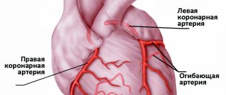
- congestive heart failure due to defects, complications of extensive infarction, myocardial dystrophy, cardiopathy;
- hereditary diseases with metabolic changes (galactosemia, hepatocerebral dystrophy, hemochromatosis);
- fetal hepatitis in newborn babies occurs when the mother has an infection (rubella, herpes, cytomegalovirus), when the pathogen is transmitted to the fetus through the placental barrier.
Varicose veins of the esophagus due to the opening of collaterals can provoke intestinal and liver tumors, peritonitis, any enlargement of the spleen and lymph nodes.
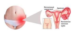
Banti's syndrome is a violation of circulation in the veins of the spleen (splenohepatomegaly) that occurs in young women against the background of anemia, thrombocyto- and leukopenia, congestion in the liver with portal hypertension and cirrhosis. It is caused by infectious diseases (brucellosis, malaria, syphilis, leishmaniasis).
Randu-Osler syndrome (hereditary telangiectasia), in addition to lesions of the skin and mucous membranes, causes multiple angiomatous changes in internal organs with a tendency to bleeding. Localization in the esophagus creates conditions for vein dilation. To prevent bleeding from the dilated venous network of the esophagus, it is necessary to treat the underlying disease.
Course of the disease
The course of the disease can be divided into 4 stages:
Stage I - initial
The disease has already appeared, but cannot yet be diagnosed. It is asymptomatic, or patients feel heaviness in the right hypochondrium and mild malaise.
Stage II - moderate
Pronounced clinical symptoms of the disease appear. Pain in the upper abdomen, flatulence, digestive disorders (belching, heartburn, heaviness in the abdomen, etc.), splenomegaly and hepatomegaly (enlarged liver).
Stage III - pronounced
All the signs and symptoms of portal hypertension are present in a pronounced form.
Stage IV - stage of complications
Intense ascites develops, difficult to treat, repeated bleeding, as well as possible complications.
Current classification
There are several proposed classifications of the disease. Signs are detected during esophagogastroscopic examination. The most acceptable division of esophageal varicose veins according to the degree of change in the veins is considered.
- 1st degree - the maximum diameter of the vessels is 5 mm, they are elongated, localized in the lower part of the esophagus;
- 2nd degree - the tortuosity of the veins is determined, the diameter is increased to 1 cm, reaching the middle third of the organ;
- 3rd degree - draws attention to the thinning and tension of the walls of venous vessels, diameter over 10 mm, they are side by side, on the surface there are characteristic red markers from the smallest capillaries.
The initial stage is characterized by the absence of vein tortuosity
According to another classification (Vytenasoma and Tamulevichute), it is proposed to take into account 4 stages of the disease:
- 1 - vein diameter is allowed 2–3 mm, they are bluish in color and straight in shape;
- 2 - veins become tortuous, nodular, and increase in diameter to more than 3 mm;
- 3 - varicose nodes are clearly visible, significant tortuosity, protrusion appears into the lumen of the esophagus;
- 4 - the nodes grow to a grape-shaped shape, significantly narrow the lumen of the esophagus, and a thin network of small capillaries is visible on the outer surface.
In addition, the diagnosis takes into account:
- congenital form occurring against the background of pathologies of unknown origin;
- acquired - caused by various diseases.
Esophageal varicose veins: symptoms
Depending on the degree of development, the following signs are distinguished:
- First degree. The diameter of the veins is increased by no more than 3 mm, the formation of single nodes is possible. Diagnosis of the disease is possible only by the endoscopic method; there are no clinical signs.
- Second degree. A rather dangerous period of varicose veins of the esophagus - there are no symptoms (even the mucous membrane of the organ still remains unchanged), but at the same time the contour of the veins already becomes unclear, and X-ray diagnostics reveal thickenings on the veins (harbingers of nodules). Already at this stage, bleeding is possible, which in the absence of immediate medical attention often leads to death.
- The third and fourth degrees of varicose veins of the esophagus are characterized by severe symptoms - the patient is often bothered by heartburn, pain occurs in the sternum, shortness of breath is possible, not only during physical activity, but also at rest. Often the disease is diagnosed only in the later stages of its development, when the first bleeding appears; blood is released directly from the esophagus or found in the vomit.
How do esophageal varices manifest?
Symptoms of the disease depend on the pathology that caused esophageal varicose veins. The initial period proceeds without clinical manifestations, patients are unaware of the development of pathology. But there are frequent cases of a progressive course with sudden bleeding.
Why is the test for occult blood in stool positive?
The condition worsens within 4–5 days. Patients feel increasing heaviness behind the sternum, compression. This sign is considered a harbinger of massive bleeding and requires urgent measures, since surgeons' observations associate it with death.
All symptoms of varicose veins are determined by threatened manifestations of blood loss. In a chronic course with a small amount of excreted blood, the body gradually weakens. Hypochromic anemia develops. The patient is pale, losing weight, has difficulty moving, and is troubled by shortness of breath. Sometimes there is liquid black feces.
Harbingers of bleeding and initial signs of varicose veins can be:
- vague pain in the chest;
- severe heartburn;
- belching after eating;
- difficulty swallowing dry food.
Heartburn and belching are explained by dysfunction of the esophageal sphincters, reverse (reflux) reflux from the stomach. Some patients experience a tickling in the throat, soreness, and a salty taste in the mouth before bleeding begins.
In acute bleeding, the following appears:
- increasing pallor of the skin;
- vomiting blood (“coffee grounds”);
- constant dizziness;
- loose, tarry stools;
- darkening of the eyes;
- severe weakness.
Bleeding is provoked by lifting weights, physical work, elevated body temperature, taking anticoagulants, and the fibrogastroscopy procedure. But sometimes it occurs spontaneously against the background of general health. It is necessary to differentiate bleeding from a disintegrating tumor of the esophagus and stomach, tumor germination into a large vessel and its breakthrough, and injury to blood vessels by a foreign body.
Treatment
Treatment of varicose veins of the esophagus begins with finding out the true causes of the development of the disease. If a pathology of the liver, thyroid or pancreas is diagnosed, then it must be treated - the provoking factor must be eliminated or its influence must be weakened. Medications include vitamin complexes, antacids and astringents. If bleeding is diagnosed, there is always a risk of its reoccurrence. In this case, the following is prescribed:
- blood transfusion;
- hemostatic drugs;
- introduction of a special probe that can compress the vessels.
Varicose veins of the esophagus, bleeding in which occurs too often, require surgical treatment.
Surgery is also performed if there is no effect from therapeutic treatment. Can esophageal varicose veins be stopped in this case? The prognosis is more favorable - the survival rate of patients increases 3 times. After any treatment, you must follow the dietary recommendations of doctors, undergo regular examinations and avoid excessive physical activity.
You can find out more about what esophageal varicose veins are and which doctor you should contact on the pages of our website Dobrobut.com.
Diagnostics
The diagnosis can be suspected, but cannot be confirmed without esophagogastroduodenoscopy. This is practically the only way to establish a connection between bleeding and esophageal varices; gastric varices are often simultaneously detected.
An x-ray can reveal inflammation, tumors, spastic contraction with obstruction of patency
The procedure allows you to determine the degree of deformation of the veins, the stage of the disease, visually determine the condition of the vascular walls, and predict rupture. It is almost impossible to conduct research during bleeding.
Contrast radiography of the esophagus is routinely prescribed; before the image, the patient is given a barium mixture to drink. A series of radiographs are used to monitor the movement of the contrast and its spreading in the lumen of the esophagus.
Laboratory way:
- it is necessary to determine the presence of anemia by the content of red blood cells, platelets, and color indicator;
- in case of acute bleeding, the hematocrit is calculated;
- be sure to do an analysis of clotting indicators;
- determine liver function by enzyme tests, protein, glucose, bilirubin levels; deviations in the results make it possible to suspect the influence of liver pathology on changes in the venous system of the esophagus;
- if there are signs of bleeding, the blood type and Rh factor are determined in case of a necessary blood transfusion.
Even minimal excretion of blood in the stool is confirmed by Gregersen's reaction to occult blood.
How is esophageal vein pathology treated?
Treatment of varicose veins of the esophagus is characterized by a planned option and a scheme depending on the occurrence of an emergency problem, life-threatening bleeding.
In the absence of massive bleeding, the patient needs treatment for the underlying disease and increased administration of hemostatic agents. The patient must be hospitalized in a specialized department. Mode: bed rest, the head end of the bed is raised.
Diet Requirements
Therapeutic nutrition provides for the absence of irritating foods (spicy seasonings, fried and smoked meats, coarse vegetables, whole fruits, bread crusts, bones, carbonated water). Alcohol and chocolate are strictly prohibited.
The diet is based on fairly high-calorie, but liquid, cooled food. We recommend lukewarm broths, boiled liquid porridges, milk noodles, cottage cheese, sweet fruit jelly, cooled tea, white bread pulp, and cooked minced meat.
Preference is given to boiled products
Treatment with medications
In order to reduce the activity of cirrhotic changes in the liver, the treatment regimen includes:
- antiviral drugs (for indolent hepatitis);
- steroid hormones;
- antibiotics for bacterial infections;
- diuretics to reduce pressure in the inferior vena cava system;
- cardiac glycosides, if cirrhosis is caused by myocardial decompensation;
- hepatoprotectors;
- vitamin preparations in high doses to restore all types of metabolism.
Particular importance in the treatment of varicose veins is given to vitamins K, C, D, E. Vikasol is administered intramuscularly or intravenously. If the patient is diagnosed with anemia with impaired coagulation, then a transfusion of fresh frozen single-group plasma (1-2 doses), red blood cells or platelets is prescribed.
To stop bleeding, intravenous administration of Octreotide is widely practiced. The drug is able to suppress the release of hormones into the blood that dilate blood vessels. Vasopressin and Terlipressin are also used, but compared to Octreotide they have more side effects. A solution of calcium chloride is administered intravenously.
You should be careful with medications that increase blood pressure, as they increase bleeding.
If bleeding continues, use: rinsing the esophagus with hot water (40–45 degrees) through a probe, installing a rubber inflating probe - there are standard corrugated products (obturator probes) for pressing the bleeding vessel in the esophagus and in the stomach ulcer.
Balloon dilatation of the esophagus is used both to stop bleeding and to treat narrowed areas
How does surgery help?
An unfavorable clinical course is an indication for endoscopic ligation. The technique involves suturing the veins of the esophagus using an endoscope. Surgeons consider it more effective than injecting sclerosing agents into the veins (sclerotherapy), which requires repetition at least four times a year.
Treatment of esophageal varices with bleeding that is not eliminated by therapeutic methods requires emergency surgery. The goal of surgery is to reduce the pressure in the portal vein by creating shunts and dumping them into the inferior vena cava.
Creation of an artificial anastomosis (installation of a metal stent) between the portal and hepatic veins is called transjugular intrahepatic portosystemic shunt. The operation is technically difficult. Experts believe that it can be successfully performed in 95% of cases.
It is accompanied not only by technical difficulties, but also by early relapse of bleeding and inflammation. In 1/3 of patients, re-installation is required, since the stent quickly thromboses, blocking the lumen. Within a month, up to 13% of patients die. This classifies the operation as an emergency measure of choice.
Another method of improving portacaval blood flow is to create an anastomosis between the splenic and left renal veins. The surgical technique is complex and risky for the patient, and is accompanied by high mortality. The devascularization operation involves excision and removal of the affected veins and their replacement with prostheses.
Creating a bypass reduces the pressure in the portal system by half
Is it possible to be treated with folk remedies?
The use of folk remedies in the presence of bleeding is unsuccessful. But they can be used in the treatment of the main cause of varicose veins - liver damage. Long-term use of decoctions is suitable for this:
- from milk thistle;
- chicory root;
- corn silk;
- Japanese Sophora;
- oats;
- rowan fruit;
- rosehip.
Varicose veins of the esophagus grades 1, 2 and 3 - causes
Varicose veins of the esophagus are a dangerous pathology that occurs, like any varicose veins, when the venous outflow is disrupted. Lengthening, dilatation and twisting of the esophageal veins appear in certain diseases of the digestive system. Varicose veins of the esophagus are a concomitant symptom of diseases of the liver, pancreas and stomach.
Causes of pathology
The main factor in the development of pathology is the complexity of the functional structure of the blood supply to the abdominal organs. The veins of the esophagus connect with similar vessels of the spleen, the portal vein and the veins of the abdominal organs.
What normally works, when there are obstacles to blood flow, turns into a pathological formation. It causes a variety of negative symptoms that indicate problems.
Having diagnosed the first signs of the disease, you must immediately consult a doctor.
Conventionally, the factors causing pathology are divided into three large groups: intra- and extrahepatic, and mixed.
Reasons for formation in a particular case:
- Intrahepatic blockage occurs in the portal vein of the liver. It is provoked by cirrhosis, thrombosis, splenomegaly, cancer or purulent disease.
- Extrahepatic blockade is associated with obstruction of the bile ducts and vascular changes. It can often be caused by formations in the esophagus or thyroid gland.
- The mixed form appears if other organs are involved (for example, the heart with cardiovascular failure). It appears when there is already an existing violation of systemic blood flow.
- Modern medicine cannot yet explain the idiopathic form (congenital varicose veins of the esophagus and some others).
The causes of varicose veins of the esophagus most often lie in diseases of the digestive or hepato-biliary systems.
Symptoms of the disease
The main manifestations depend on the severity of the condition. Grade 1 varicose veins of the esophagus are characterized by initial changes:
- VRV extension;
- expansion of vein diameter;
- bleeding;
- decrease in blood pressure;
- tachycardia;
- admixture of blood in vomit and feces.
Other characteristic symptoms: splenomegaly (enlarged spleen), increased bleeding, ascites, yellowing of the skin, itching and rashes - all these symptoms depend on the causes that caused esophageal varices.
Diagnosis of varicose veins of the esophagus
There are several special hardware research methods used to establish grade 1 esophageal varicose veins. Diagnosis of varicose veins is carried out first in the presence of liver cirrhosis and some other severe conditions, because clinical practice suggests its presence.
If parallel studies have not been carried out, varicose veins can be detected using:
- esophagogastroduodenoscopy (diagnosis using an endoscope);
- capsule endoscopy (used much less frequently, because it involves swallowing a video camera; it is quite expensive and therefore inaccessible);
- auxiliary methods, such as vascular Dopplerography, computed tomography and magnetic resonance imaging;
- X-ray of the chest cavity.
In addition to hardware tests, laboratory tests are certainly carried out. Basically, this is a study of the composition of the blood (biochemistry, coagulability, determination of the level of various components); an analysis of the ascites fluid filling the abdominal cavity can also be carried out.
Endoscopic examinations are necessary to correlate the degree of development of the disease with the category, to determine the severity and existing risks according to the classification of esophageal varices.
Main classification
International ICD 10 distinguishes only 2 types of varicose veins of the esophagus: with and without bleeding. In clinical practice, a classification is used that differentiates varicose veins of the esophagus into 3 degrees, according to the pathological changes that occur.
This classification of varicose veins of the esophagus was introduced in 1997, and as the main diagnostic criterion implies the degree of change in the condition of the veins, the expansion of the natural diameter:
- To diagnose grade 1 varicose veins of the esophagus, it is enough that the veins located in the lower section reach 5 mm and are elongated compared to the physiological state.
- The diagnosis of grade 2 varicose veins of the esophagus assumes a wider diameter of the vein, increased under the influence of pathological reasons to 10 mm, the veins begin to twist and are located in the middle part of the organ.
- To determine the third, not only the diameter of the veins is important (it must exceed the centimeter mark). Grade 3 varicose veins of the esophagus are already thinned venous walls, formed twists, fully formed varicose nodes and red marks on the outside, which are considered markers for determining the severity of the condition.
Treatment of varicose veins of the esophagus
Treatment tactics are carried out in two directions, the use of which depends on the degree of development of the disease. Methods of conservative treatment of esophageal varicose veins include treatment with medications, folk remedies and diet.
Disease prognosis
In the initial stages of esophageal varicose veins, with constant treatment, a sufficient functional state of the liver, and compliance with the recommendations for regimen and diet, it is possible to stop bleeding in 80% of patients. In 2/3 of patients, bleeding recurs after a single episode within 1–2 years. They are constantly at high risk. The survival rate of individuals with advanced cirrhosis is low.
Varicose veins of the esophagus are complications of the disease. This in itself is already a sign of severe damage to the body. Support can only be provided by timely detection using endoscopy and monitoring the patient.
Key features of the intervention!
The advantages of vein embolization include:
- High speed of intervention. The operation takes 30-60 minutes. Complex procedures may take longer.
- No significant discomfort. Pain and any discomfort during vein embolization are relieved with special modern medications.
- Speed of recovery after intervention. The patient will have to stay in the clinic for only 1-2 days. Note! In some cases, the length of hospitalization after vein embolization increases. In any case, the patient can return to normal life in the near future.
What recommendations do doctors give to patients?
- Strengthening the drinking regime. In the first 7 days after surgery, you need to drink plenty of clean water.
- Elimination of water procedures for 3-5 days. After this, you can take both a shower and a bath.
- Physical peace. It is usually prescribed for 2-3 weeks. Patients are prohibited from lifting heavy objects or playing sports. Bed rest is not prescribed for this period. Slow walks are even beneficial.
What lifestyle should I lead after embolization of esophageal vessels?
Patients should:
- Follow a specially prescribed diet that includes a large number of foods that help reduce total cholesterol levels.
- Avoid heavy exertion.
- Take various medications. They will be prescribed by your attending physician.
- Eat food often enough, but in as small portions as possible. The daily dose is best divided into 5-6 doses. Eating food is undesirable 2-3 hours before bedtime.
- Give preference to boiled dishes. Food can also be steamed. This way it retains a lot of useful substances, is easily digestible and does not become a source of additional harmful cholesterol.
It is forbidden to eat too hot or cold food. This can damage the esophagus and its veins.