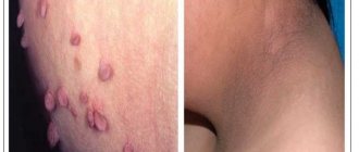
Skin xerosis is a pathological condition accompanied by dry skin, flaking and itching. Dry skin, losing elasticity, ceases to be a barrier to infectious agents and toxic substances. The disease can manifest itself on various parts of the body, but more often exposed areas are affected: face, torso, limbs. Sometimes the process affects the anogenital area, causing discomfort and provoking the development of infectious and inflammatory diseases and allergic reactions in this area.
Medical specialists have developed and successfully used a method for treating xerosis of the skin of the anogenital area using preparations based on hyaluronic acid. Hyaluronic acid allows you to retain water in the upper layers of the skin, thereby maintaining normal water balance and slowing down the process of desquamation of the epidermis. The method allows you to achieve good results in patients with various causes of increased dryness.
Reasons for the development of xerosis
There are several common reasons that contribute to the development of xerosis:
- excessively dry and/or cold air;
- increased solar insolation or frequent visits to the solarium;
- skin diseases accompanied by increased dryness (atopic dermatitis, psoriasis, hypothyroidism);
- decreased sebum production by skin glands;
- abuse of hot baths;
- use of personal hygiene products with aggressive detergent components.
The last three factors are most often involved in the occurrence of xerosis of the anogenital zone.
Navigation
- Symptoms of the disease
For certain reasons, not enough fat is produced in the upper stratum corneum, as a result of which moisture is lost and strong susceptibility to any external irritant appears. As a rule, excessive dry skin is accompanied by noticeable itching, which forces a person to scratch and thereby injure the skin, making it open to infectious agents.
Xerosis can be congenital (or atopic) and acquired. The main factors provoking acquired xerosis of the skin are:
- certain climatic and weather conditions (for example, dry frost, heat, low humidity combined with strong wind, etc.);
- increased air dryness during heating in winter;
- constant long-term water procedures (constant bathing, swimming in a pool with chlorinated water, can lead to disruption of lipid metabolism of skin cells);
- ultraviolet radiation, which penetrates the skin, dries it out, making it flabby and wrinkled;
- household chemicals with aggressive components that destroy the fat layer;
- various dermatological diseases (atopic dermatitis, seborrhea, ichthyosis, eczema, psoriasis);
- age-related skin changes associated with a slowdown in cellular metabolic processes.
Skin xerosis can develop in any person. Therefore, any manifestations of symptoms should be considered as an alarming signal and appropriate measures should be taken immediately. After all, many people consider severe dry skin to be a temporary phenomenon and do not pay due attention to it.
Clinical manifestations of vaginal dryness
The first sign of the development of skin xerosis is increased peeling in a certain area. As the process progresses, patients begin to complain of skin redness, a feeling of tightness, and itching. In some cases, small bleeding cracks may form, accompanied by pain. The skin becomes dull and becomes rough to the touch.
The severity of xerosis manifestations depends on many reasons: age, time of exposure to the provoking factor, lifestyle, nutrition, and general condition of the body.
Symptoms of the disease
First stage: The protective function of the skin is impaired, which is expressed by symptoms such as tightness, dryness, which are especially felt when the facial muscles contract. These symptoms can be easily and quickly eliminated with moisturizers (lotions, creams).
Second phase. Hyperkeratosis is observed, that is, the skin is covered with scales of dead epithelium, and its peeling begins. Moisturizers no longer help, the skin remains tight, itching often occurs, and sometimes sensitivity to external irritation is added to it.
Third stage. The dermis undergoes noticeable changes: it becomes thinner, the upper layer atrophies, while the peeling becomes lamellar (large structures peel off), and expression wrinkles become pronounced. The skin becomes inelastic, hardens, bursts, and reacts to external irritants with redness, itching, and an erythematous rash. Negative processes affect the papillary layer of the dermis.
Fourth stage. The skin layers—epidermal and dermal—are greatly thinned (which is even visually noticeable), and various pronounced trophic changes (to the point of erosion) appear.
Xerosis is not so difficult to notice. And here it is important to begin the fight against abnormal dryness at the very initial stage, when the earliest symptoms appear, which will avoid the development of the disease and its negative consequences.
Treatment method for xerosis of the skin of the anogenital zone
Medical specialists treat dry skin in the intimate area using modern innovative techniques. One of the promising areas of the clinic’s work in this direction is the use of drugs based on hyaluronic acid.
Hyaluronic acid in the human body is part of many tissues: connective, epithelial, nervous, cartilage. It is synthesized in large quantities in the skin, helping to retain fluid in tissues and increase their elasticity. When exposed to unfavorable factors, the skin synthesis of hyaluronic acid may slow down, and the rate of its breakdown may increase. As a result, fabrics lose moisture, their elasticity decreases, and resistance to various external influences decreases.
The essence of the treatment method for skin xerosis is to replenish the missing amount of hyaluronic acid using intradermal injections. Injections are performed by qualified doctors at our clinic who are familiar with the technique of administering hyaluronic acid preparations and have undergone special training. The dose of the drug is selected individually, taking into account the severity of xerosis manifestations and the prevalence of the lesion.
Skin thickening, or hyperkeratosis
Diabetes mellitus
Human papillomavirus (HPV)
Fungus
Menopause
Climax
13717 16 August
IMPORTANT!
The information in this section cannot be used for self-diagnosis and self-treatment.
In case of pain or other exacerbation of the disease, diagnostic tests should be prescribed only by the attending physician. To make a diagnosis and properly prescribe treatment, you should contact your doctor. Thickening of the skin: causes of occurrence, what diseases it occurs with, diagnosis and treatment methods.
Definition
Hyperkeratosis is an abnormal thickening of the top layer of skin (epidermis) as a result of excessive sun exposure, exposure to chemicals, frequent friction or pressure. In addition, hyperkeratosis can occur against the background of certain diseases.
Thickening of the skin occurs in the stratum corneum of the epidermis, which is the end point of the differentiation process of keratinocytes - cells containing the protein keratin. It is in the stratum corneum that keratinocytes lose water and their nucleus and turn into scales of the stratum corneum - corneocytes.
Normally, corneocytes are gradually exfoliated, due to which the skin is renewed.
With hyperkeratosis, accelerated differentiation of keratinocytes occurs, and the physiological exfoliation of horny scales from the skin surface, on the contrary, slows down.
Types of hyperkeratosis
Depending on the origin, acquired and hereditary hyperkeratosis are distinguished.
According to clinical manifestations.
- Calluses are a common type of hyperkeratosis. There are several types of calluses, but they all appear due to thickening of the skin in places most susceptible to mechanical stress. Moreover, such skin changes can be associated both with increased physical activity and with various chronic diseases, which is typical for elderly patients.
- Horny (tilotic) eczema manifests itself as hyperkeratosis of the palms and soles.
- Psoriasis is an autoimmune inflammatory disease in which hyperkeratotic scaly plaques form on the skin.
- Actinic keratosis usually appears as small, reddish, scaly bumps that appear after excessive sun exposure. Actinic keratosis is a serious condition with a high probability of malignancy and requires mandatory consultation with a doctor.
- Seborrheic keratosis is characterized by small brown or black patches, usually located on the face, neck, shoulders and back. This is one of the most common benign skin tumors in adults.
- Follicular hyperkeratosis (“goose bumps”) is characterized by blockage of the mouths of the follicles with keratinized epidermal cells.
- Epidermolytic hyperkeratosis is a rare hereditary disease that appears immediately at birth. Newborns have reddish skin, sometimes covered with small blisters.
Possible causes of skin thickening
Skin hyperkeratosis can occur in people who neglect regular skin care procedures, as a result of which dead cells of the stratum corneum accumulate and form keratomas - benign neoplasms.
Our skin is constantly exposed to adverse external factors, such as chlorinated water and detergents, and UV radiation. As a result, the protective lipid layer of the skin is damaged, and moisture begins to rapidly evaporate from its surface, and corneocytes lose their ability to physiologically exfoliate.
In diabetes mellitus, hyperkeratosis becomes a consequence of metabolic disorders and deterioration of skin microcirculation.
Wearing tight or uncomfortable shoes, especially with flat feet, congenital foot pathologies, and obesity, can cause thickening of the skin on the feet.
The development of cervical hyperkeratosis (leukoplakia) is promoted by the human papillomavirus.
The cause of hyperkeratosis can be chronic fungal infection, as well as herpes zoster.
It is believed that symptoms of thickened and dry skin may be caused by a deficiency of vitamins A, E, D and C.
Hyperkeratosis often results from a lack of the hormone estrogen in women during menopause.
Diseases and conditions in which hyperkeratosis develops
- Diabetes.
- Obesity.
- Flat feet.
- Ichthyosis.
- Psoriasis.
- Eczema.
- Menopause.
- Fungal skin infection.
- Shingles.
- Erythroderma.
- Atopic dermatitis.
- Seborrheic keratosis.
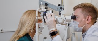
Which doctors to contact for hyperkeratosis
Most often, the first consultation regarding skin thickening is addressed to a dermatologist. After a thorough examination, collecting complaints, finding out the patient’s medical and family history, conducting laboratory and instrumental studies, a consultation with an infectious disease specialist may be required.
Diagnosis and examinations for thickening of the skin
A thorough history taking, taking into account all the patient’s complaints, examination and additional diagnostic methods will help determine the cause of hyperkeratosis.
- Clinical blood test with a detailed leukocyte formula to identify inflammatory processes in the body.
Xerosis in children
The causes of skin xerosis in children are different, and treatment depends on their nature.
So, the causes of skin xerosis in children may be as follows:
- allergy;
- endocrinological disorders;
- weak immunity.
In most cases, all this is transmitted with genes. To identify the cause, it is worth visiting the appropriate specialists.
It is acceptable to use special children's cosmetics. For example, moisturizing and soothing creams. Sometimes the doctor may prescribe medications.
To combat the scourge on their own, parents can be recommended to use “improvised” measures. This will improve the condition of the child’s skin texture (examples of skin xerosis in children photos).
- start hardening (if there are no contraindications);
- instill a love of hygiene;
- improve nutrition (fresh natural foods, more vegetables, eating at the same time);
- establish a routine (wake up and go to bed at the same time every day);
- ensure healthy sleep (8-9 hours a day, in absolute darkness, in a pre-ventilated room);
- get interested in sports (for example, enroll in some sports section).
Regular spending time outdoors will also benefit your growing body. However, it is better to avoid active sun.
In general, skin xerosis in children is similar to that in adults, regardless of the cause, so the approach to therapy is similar.
Treatment
Methods for moisturizing the skin are different and are largely determined by eliminating the source - the catalyst of the problem. If the cause is a specific disease, then treatment of dry skin of the body includes all the necessary measures that can stop the pathology, and hence remove its accompanying symptoms.
If the reason is different, then it is necessary to take measures to protect the skin from the negative influence of external irritants, which can be eliminated independently. It is necessary to keep the skin clean, but you should not overdo it with the amount of washing and taking baths and showers. Cosmetics for washing must be of high quality and match your skin type. You should use only high-quality, non-expired cosmetics. In winter, it is necessary to use protective creams. In everyday life, prefer clothes and shoes made from natural materials. This will protect against the occurrence of allergic reactions to artificial material and protect against thermal overheating of the skin. Anti-inflammatory agents must be used along with moisturizers. It is recommended to use anti-allergy medications or general health-improving medications internally, which will be prescribed by your dermatologist.
Xerosis. Diet
To make dry skin feel better, you should include these products in your menu:
- seafood (contains a lot of polyunsaturated fatty acids);
- vegetables and fruits rich in vitamin C (an antioxidant responsible for skin renewal);
- vegetables and fruits of red and green colors (contain beta-carotene, which is converted into vitamin A. This is a serious antioxidant that does not allow cells to be destroyed);
- clean, fresh water (daily intake for women - from 30 ml per kg of weight, for men - from 40 ml per kg of weight per day).
Traditional recipes:
- Glycerin bath. Restores the epidermal cover. Add 0.5 cups of natural glycerin (non-technical) to a bath of warm water. Take 10-15 minutes.
- Honey-oil bath. A liter of milk is heated without boiling. Add 200 g of natural honey (not store-bought, it is better to purchase it from beekeepers). Mix and gradually add 1 tablespoon of almond oil. The mixture is poured into a bath of warm water. Take - 10-15 minutes. Softens the skin.
- Olive mask. Mix 2 tablespoons each of olive oil and honey. Apply to damp skin after shower or bath. Keep for 20 minutes. Rinse off with warm water. Softens and moisturizes the skin.
Diagnostics
A detailed history taking is of great importance in making the correct diagnosis. The collected data (onset of the disease in early childhood, seasonality of symptoms, high photosensitivity, consanguineous marriages) suggest xeroderma pigmentosum. The diagnosis is confirmed after cytological and histological examination of the affected areas of the skin.
Additional examinations include examination by a dentist, otorhinolaryngologist, ophthalmologist and neurologist to exclude damage to the central nervous system, oral apparatus, visual and hearing organs. For De Sanctis-Cacchione syndrome, an X-ray examination or computed tomography of the skull bones, and magnetic resonance imaging of the brain are also performed. In this case, small sizes of the sella turcica, a decrease in the volume of the cranium and brain, and underdevelopment of the cerebellum and pituitary gland are detected.
Kinds
Depending on the damaged chromosome, there are 8 subtypes of the disease: A, B, C, D, E, FG and Young's pigmented xerodermoid. The first 7 types of xeroderma pigmentosum have a similar clinical course and are determined only after molecular genetic analysis. Young's xerodermoid pigmentosa has a more favorable prognosis - the symptoms of the disease appear later, and the pathological process itself is mild.
An independent clinical form of the disease is De Sanctis-Cacchione syndrome. It is considered the most aggressive type of xeroderma pigmentosum and is characterized by pronounced changes in the central nervous system. The syndrome can develop with any chromosomal variant of the disease, but most often occurs with subtype A.
Restoring the ratio of water and lipids in the stratum corneum
To get rid of dryness, it is not enough to simply saturate it with water. Water must also be retained in sufficient quantity, and for this it is necessary to restore the normal ratio of lipids in the stratum corneum. Therefore, you should select skin care products that contain both moisture-retaining and lipid-replenishing components.
Useful tips
To care for very dry body skin, it is useful to use special cosmetics - emollients. They contain special ingredients that not only moisturize and soften the skin, but also restore its protective function.
The principle of selecting suitable cosmetics should be based on the degree of dryness of the skin. The drier the skin, the more lipids (fats, oils) it needs. When areas of very dry, rough skin appear (for example, on the heels), it is advisable to use creams with a high urea content. DiaDerm Intensive foot cream 10% urea, specially designed for people with diabetes, with constant use, effectively and safely eliminates hyperkeratosis thanks to urea and an increased concentration of lipids in the reverse water-in-oil emulsion.
Softening the skin significantly reduces the itching that often accompanies xerosis. This painful sign of dryness manifests itself especially clearly on the legs, primarily on the legs and thighs. DiaDerm Softening foot cream saturates the skin with nourishing oils, eliminates flaking and restores the protective barrier. The entry gates for triggers that cause irritation and itching are closed.
Useful tips
To care for irritated skin (itching, redness, excessive peeling), you can use cosmetic creams containing 2.5-5% dexpanthenol (or other names: panthenol, D-panthenol, vitamin B5). Preferably without fragrances or dyes!
What is it and what do the signs of the disease look like?
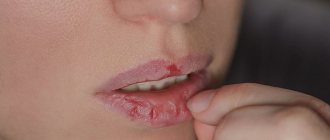
xerosis of the skin in the photo
Xerosis of the skin is a critical drying out of the epithelial layer with the destruction of a large number of its cells. Some scientific dermatologists who deal with the problem of the occurrence of this disease believe that xeroderma refers to the initial stage of skin ichthyosis. Xerosis is also present when the patient has certain types of infectious diseases.
Dry skin occurs when the sebaceous glands stop producing sebum or do so in reduced tone. The first symptoms of the disease are as follows:
- abnormally dry skin that does not show any external signs of hydration;
- as the condition of the skin deteriorates, its surface layer begins to form scales that peel off greatly;
- the affected areas of the epithelium become rough to the touch and it seems that the skin has become one continuous calloused neoplasm;
- mild itching, which gradually becomes simply unbearable, is present day and night, significantly reducing the patient’s quality of life;
- swelling of the epidermal tissues, inflammation and redness, on which xeroderma develops;
- loss of elasticity of diseased skin, as well as the disappearance of pores that cannot be seen during visual inspection;
- very strong drying out of individual parts of the body with the formation of cracks from which lymphatic fluid is released.
In addition to the skin, xerosis can spread to the mucous membrane of the oral cavity, nasal canals and organs of vision. People at risk are between 45 and 80 years old. The danger of the disease lies in the fact that its first signs are often confused with an allergic reaction to external environmental factors or banal dehydration of the surface epithelial layer.
Clinical picture
Newborn children have no symptoms of the disease. The first signs of xeroderma pigmentosum begin to be noticed between the ages of 3 months and 4 years. In rare cases, the onset of the disease is possible in adulthood. The literature contains data on the appearance of symptoms in patients aged 25-30 years.
Xeroderma pigmentosum is characterized by seasonality and stages of progression. An exacerbation of the disease occurs in the sunny season (spring - early autumn). The first stage is called inflammatory and is manifested by a specific triad of symptoms:
- photosensitivity - intolerance to direct sunlight;
- erythema - redness of the skin of exposed areas of the body after exposure to the sun;
- foci of hyperpigmentation - the appearance of areas of excess color.
Erythema of the skin is persistent. It is often accompanied by severe swelling, peeling and the formation of small bubbles with transparent contents. After the inflammatory processes subside, at the site of the erythema, foci of hyperpigmentation appear in the form of light or dark brown spots of different sizes. In most cases, they resemble a scattering of freckles, but can be large (lentigines).
The second stage is poikilodermic. It begins at the age of 3-7 years and is characterized by pronounced changes in the skin in the form of telangiectasia (spider veins), areas of hyperkeratosis with abundant peeling, areas of atrophy and thinning of the skin. A typical manifestation of the second stage is also the formation of small, smooth, whitish scars with a shiny surface. Upon careful examination, areas of skin atrophy have an uneven, rough appearance and resemble a burn surface.
Pronounced changes in the skin lead to a number of serious consequences:
- deformation of the external nose and nasal passages, resulting in difficulty breathing;
- curvature of the mouth;
- deformation of the ears due to cartilage atrophy;
- eyelash loss;
- eversion or inversion of the eyelids.
In patients with xeroderma pigmentosum, areas of hyperpigmentation persist, and as the disease progresses, hypopigmented spots appear. As a rule, they are localized on the back and wings of the nose, on the chin.
In 75-80% of patients at the second stage, signs of damage to the organ of vision are also determined: lacrimation and photophobia, blepharitis, ulceration of the conjunctiva and its inflammation, clouding of the cornea and the appearance of spots on it. In 20% of patients, symptoms of dysfunction of the central nervous system are detected in the form of decreased sensitivity, mental retardation, changes in coordinated muscle function (ataxia) and decreased or absent unconditioned reflexes (for example, tendon reflexes).
The disease enters the third (tumor-like) stage in adolescence. It is characterized by the appearance of benign or malignant neoplasms of various shapes and sizes on the affected areas. Benign tumors can be represented by papillomas, nevi, fibromas, angiomas and have a high risk of oncological transformation. Often, neoplasms are also detected on the mucous membrane of the mouth and nose.
De Sanctis-Cacchione syndrome has a severe clinical course. Its main symptoms are impaired mental development (idiocy) due to a decrease in the size of the brain and skull, dwarfism and typical skin manifestations of xeroderma pigmentosum. Additional signs of the syndrome are delayed sexual development and severe disturbances of the nervous system (paresis, paralysis, ataxia). Skin manifestations appear early in patients with De Sanctis-Cacchione syndrome. They are pronounced, rapidly progress and develop into oncological neoplasms already in childhood.
Cleansing plus moisturizing
Proper care for dry skin is cleansing and moisturizing. You should never forget about the first stage: you should not apply the cream to dirty skin! First you need to prepare it, clean it, that is, wash it. You can't just wash away all the impurities on your skin with water. Still, most of them are located in a water-fat film on the surface, and various detergents are used to remove them (soaps, bathing gels and foams, shampoos, etc.).
If you have xerosis, you should be especially careful when choosing a foam detergent, paying first of all to its acidity or pH value. Normally, human skin is slightly acidified (pH from 4.1 to 5.8)3. In case of skin diseases (as well as diabetes), its pH shifts to the alkaline region, which provokes the growth of pathogenic microorganisms on its surface.
Useful tips
Do not use alkaline soap (hard, bar) with pH 7.0 -12.0.
Choice - liquid products with pH 4.5-5.7
Moreover, any, even the mildest detergent, affects the structure of the lipids of the stratum corneum to one degree or another. After water procedures, it is advisable to apply an appropriate moisturizer to the skin; for legs - yours, for the body, face or hands - yours. DiaDerm hand and nail cream nourishes the skin with healthy oils, is quickly absorbed and has a pleasant sweet orange scent.
Useful tips
The high content of nourishing oils (shea butter, avocado oil, coconut oil) in hand creams will help the skin recover faster from exposure to aggressive environmental factors.
Creams with vitamins A, E and F are well suited for strengthening brittle, peeling nails. And, of course, the most important factor in restoring skin in diabetes, reducing its dryness, is normalizing glucose levels, maintaining them as close to the physiological norm as possible.
The editors thank the specialists of the AVANTA company for their assistance in preparing the material.