The tumor differs from all other tissues in its ability to constantly create new vessels that support its unprecedented ability to grow without limit. Based on the volume of the vascular network, one can quite clearly determine the boundaries of the tumor with normal tissues. The tumor is able to invade any tissue and large vessels, pushing them aside and involving them in the cancer process, leading to a narrowing of the lumen of the vessel. When a cancerous tumor appears in an anatomical area where large vessels supplying organs lie, the possibility of surgical removal depends on the degree of involvement of the artery in the tumor process.
A diagnostic method that allows one to find out the location of a tumor by the branching of the vascular network, its connection with large vessels and at the same time resolve the issue of the possibility of radical removal of cancer is called angiography.
The method involves introducing an X-ray contrast agent into the vascular bed in order to use snapshots to record the filling of tumor vessels and all nearby arteries with contrast. The passage of a contrast agent can be recorded not only with x-rays and its variant - computed tomography, but also with magnetic resonance imaging.
X-ray angiography
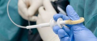
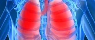
Angiography is divided according to the organ in which the vessels are contrasted. For example, angiocardiography visualizes the atria and ventricles with large vessels, and angiopulmonography visualizes the vascular network of the lungs. The procedure requires access to a vessel as close as possible to the affected area, the continuation of which is the artery feeding the tumor. A catheter is inserted into the artery and guided under X-ray control to the desired vessel. A contrast agent is passed through the catheter and pictures are taken at a certain time - the phase of passage of the contrast containing iodine.
With an X-ray examination, it is possible to contrast not only arteries, but also veins, and, if necessary, obtain an image of lymphatic vessels, therefore, in principle, this study is called vasography - an image of blood vessels. The study of arteries is called arteriography, venography or phlebography is the image of veins and lymphography, respectively, contrasting the lymphatic vessels. The study not only provides a picture of the vessels, but based on the “picture” of its blood supply, it allows us to identify pathological conditions of the organ and its functions that affect the state of the blood supply.
When deciding on the surgical treatment of cancer, the surgeon’s preliminary understanding of the state of the arterial bed in the area of the tumor is very important, since it allows one to determine the boundaries of tumor growth and the involvement of large-diameter arteries with damage. Blood flows through the arteries under high pressure, so during the operation, a fairly large volume of blood will be lost from the vessel damaged by the tumor until the bleeding is completely stopped. Blood flows through the veins at low speed and low pressure; a large vein damaged by a tumor can easily be compressed without any significant blood loss.
Angiographic information obtained before surgery allows you to prepare for the unexpected, for example, the installation of a prosthesis instead of a vessel affected by cancer. With X-ray angiography, you can immediately carry out a therapeutic measure - install a stent in the vessel narrowed by the tumor. Malignant tumors induce increased blood clotting with the formation of blood clots, the length of which reaches tens of centimeters. With angiography, the thrombus is clearly visible and can be removed immediately.
Book a consultation around the clock +7+7+78
Indications for use
The main tasks of angiography are to identify pathologies of the circulatory system and determine their nature. And this is really possible to do, since with the help of this research method it is possible to achieve ideal visualization of large veins of the extremities, arteries, aortas and pulmonary arteries. With the help of angiography, doctors have the opportunity to detect diseases such as:
- cerebral vascular sclerosis;
- congenital malformations of veins and arteries;
- hypertension of various etiologies.
There are many indications for angiography. Among the most common are:
- deep vein thrombosis with identification of the nature of the changes;
- thromboembolism (that is, blood clots in the pulmonary artery duct);
- defects of arteries and veins;
- lesions of the renal arteries;
- lesions and diseases of the aorta;
- aneurysms.
A useful trend of our time
Today, during angiography, they not only take X-rays, but also subject the images to computer processing in order to improve the quality of the image and highlight certain vessels from the general mass. And most importantly, digital processing makes it possible to avoid introducing a catheter with contrast into an artery, limiting it to a vein, which is much simpler for the surgeon performing the study and easier for the patient, and even reduce the amount of contrast agent, which is very harmful to the body. This enhanced angiography is called digital subtraction angiography.
Computer processing techniques make it possible to obtain a 3D image, where the organ is visible in three dimensions and all stages of the passage of blood through it. You can divide the blood flow into arterial and venous, all in the appropriate color, and see the entire process of blood supply to the entire organ. The X-ray angiography method is invasive, that is, with penetration into the body, so the study is performed in the operating room with all precautions to exclude any infection of the patient and the development of dangerous bleeding.
But the main advantage of X-ray angiography is the possibility of simultaneous therapeutic manipulation in vessels, which is impossible with more “advanced” angiographic studies performed on CT and MRI equipment. Today, X-ray contrast angiography is the “gold” standard for vascular examination in general and is completely integral to modern high-tech oncological care.
How is the procedure done?
How angiography is done can be described as follows:
- administration of antihistamines (even if a preliminary test showed that there is no allergy to iodine);
- antiseptic treatment of the area of the body where the puncture will be made to introduce contrast;
- anesthesia (usually lidocaine is used);
- incision of the skin to provide access to a vein or artery;
- installation of an introducer (hollow tube) to facilitate further insertion of the catheter;
- administration of a drug that prevents vascular spasm;
- inserting a catheter into a hollow tube and moving it to the beginning of the vessel to be examined;
- administration of contrast;
- shooting.
After the shooting is completed, the catheter is removed, hemostatic measures are carried out (if there is bleeding), and then a tight bandage is applied to the puncture site.
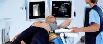
Computed tomography angiography (CTA)
This version of angiography does not require introduction into the body, there is no need for special preparation or an operating room; you just need to inject a contrast agent, also containing iodine, into the patient’s vein and take pictures with a computed tomograph with the necessary software. The study is fast; not only visualization of the vascular bed is possible, but also various reconstructions of the blood supply at any time. Only with CTA can one obtain an image of the lumen of the vessel “in its natural form” and evaluate its narrowing at 100%. Difficulties arise when calcium is deposited in an atherosclerotic vessel, since calcium is not permeable to x-rays.
Computer tomographs are classified by the number of slices - x-ray images in one projection, which the device takes in one second. Today, 16-slice, 32- and 64-slice tomographs, and even 256- and 320-slice tomographs are used. Each picture is taken at a certain distance; the more slices, the more accurate the final “picture” will be. With an increase in the number of slices, the image improves, the negative effect of the patient’s breathing on the image is “extinguished,” and the amount of contrast agent is reduced, which is important for health. But a very large number of sections is required for research work; in clinical practice, 16 sections are quite sufficient.
Limitations for tomographic angiography:
- Contraindications include intolerance to iodine preparations and severe renal impairment, since the contrast agent is toxic to the kidneys. In a typical 16-slice CT scan, approximately 100 ml of contrast is injected.
- After administration of contrast to a nursing mother, milk should not be used for feeding during the day.
- It is not possible to perform CT angiography on a very obese or very active patient.
- X-ray exposure is dangerous for children and pregnant women, and they should be offered alternative vascular testing.
During a CTA examination, therapeutic manipulations with blood vessels are impossible; a simple visual picture of the vascular lesion is obtained. Therefore, CTA could not replace conventional radiocontrast angiography.
Preliminary preparation
Angiography requires special preparation:
- 2 weeks before the procedure you should not drink alcoholic beverages.
- A week before the procedure, you need to exclude all blood thinners (for example, aspirin) from your medications (if any).
- Five days before the procedure, the patient must undergo additional examinations: fluorography, ultrasound of the heart
, ECG, coagulogram (blood test for clotting), blood biochemistry, blood test for Rh factor, test for HIV, hepatitis, syphilis. - 2 days before angiography, a test is performed to determine the patient’s tolerance to the components of the contrast agent (in particular iodine). If the test shows the presence of an allergic reaction, this is a contraindication and the procedure must be cancelled.
- 1 day before the examination, in the evening, you need to remove hair from the area where the puncture is expected to be made.
- At night (immediately before the procedure) you need to take a sedative. The doctor will recommend which one.
- On the morning of the procedure, the patient is given a cleansing enema for the intestines.
- On the day of the procedure, you should refrain from eating and drinking to avoid nausea and vomiting after contrast is injected into your body.
- Before starting the procedure, you must empty your bladder.
Magnetic resonance angiography (MRA)
Like CTA, magnetic resonance angiography is a non-invasive study, that is, it does not penetrate the body. But it allows you to obtain a highly accurate image of the vascular network of the organ under study. There is no need for an operating room or any preliminary preparation of the patient, everything is simple: come and do it. For greater accuracy of the study, and when studying the vascular bed of a tumor without fail, the manipulation is carried out with the introduction of a contrast agent, which highlights the vessels against the general background. The contrast agent for MRA does not contain iodine, but it can also damage the kidneys, although to a lesser extent.
The power of a magnetic resonance imaging scanner and, consequently, the accuracy of the study is determined by the magnetic field created by the device. The higher the magnetic field voltage, measured in Tesla units, the better the image. The simplest “low-field” MRI scanners correspond to 0.5 Tesla, the most accurate are “ultra-high-field” 3 Tesla, but in clinical practice “high-field” 1.5 Tesla are quite sufficient. MRA is more accurate than all other angiographic techniques when examining calcified vessels, but the degree of vessel narrowing may exaggerate.
There are limitations in the use of magnetic resonance angiography in the form of the presence of pacemakers and defibrillators in the patient’s body, which go astray from the program, which threatens the patient with irreparable consequences. It is impossible to study obese patients and those who are terrified of closed spaces - claustrophobia; a magnetic resonance imaging scanner is a tube of limited diameter where the patient is located. The main difference between MRA and CTA is the absence of ionizing radiation - radiation, so it is possible to study children and pregnant women.
Book a consultation 24 hours a day
+7+7+78
Recovery after the procedure
How quickly a patient recovers after angiography depends largely on how extensive the study was. There are several recommendations that will help the patient recover faster:
- Follow bed rest and diet for several days after the procedure;
- eliminate stress and anxiety;
- exclude physical activity;
- Take antihistamines to minimize the likelihood of an allergic reaction.
As a rule, if the study was performed correctly, the patient's recovery takes approximately 2-3 days.
Decoding the results
There is a certain technique for deciphering angiography results. Markers are identified for each vascular pathology. For example, if the vessels have intermittent outlines in the images, we can talk about the presence of pathologies such as thrombosis or atherosclerosis. The detection of protrusions in the walls of blood vessels in the images indicates that there are either congenital changes, an aneurysm, or the same thrombosis. We can also talk about congenital pathologies in cases where the contrast agent does not enter the capillaries, but passes directly into the vein.
If, upon administration of contrast, there is a lack of its spread, this indicates the presence of thrombophlebitis, thrombosis or thromboembolism.