For a more detailed diagnosis of diseases of internal organs, medicine practices the use of a puncture to take their contents for analysis. In addition, punctures enable doctors to “deliver” medications directly to the diseased organ and, if necessary, remove excess fluid or air from it.
The most common procedure in thoracic surgery is puncture of the pleural cavity, the types and algorithm of which will be discussed in this article. Its essence boils down to a puncture of the chest and pleura in order to carry out diagnostics, establish the characteristics of the course of the disease and ensure the necessary medical procedures.
Performing a pleural puncture is vitally important in cases of disruption of the correct outflow of plasma (the liquid component of blood) from the vessels of the pleura, which causes the accumulation of fluid in the cavity (pleural effusion). Pleural puncture helps doctors determine the cause of the disease and take measures to eliminate its symptoms.
A little anatomy
Content:
- A little anatomy
- Indications for manipulation
- Contraindications for puncture
- Technique of pleural puncture
- Features of the procedure for different types of effusion
- Pleural puncture in children
- Laboratory test results
- Complications of pleural puncture
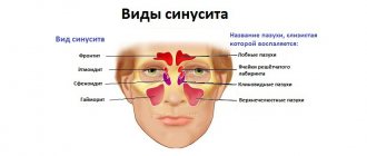
The serous membrane that lines the lungs and the surface of the chest is called the pleura. In the normal state, between its two sheets there is from one to two milligrams of straw-yellow liquid, which is odorless and viscosity, and is necessary to ensure good sliding of the pleural sheets. During physical activity, the amount of fluid increases tens of times, reaching 20 ml.
At the same time, some diseases can also lead to changes in the composition and increase in the contents of the pleural cavity. Diseases of the cardiovascular system, post-infarction syndrome, cancer, lung diseases, including tuberculosis, and even injuries can cause disruption of the outflow of pleural fluid, which provokes the so-called pleural effusion.
An increase in the volume of fluid in the pleural cavity (effusion), the accumulation of air in it that does not escape due to a mechanical obstruction (pneumothorax), as well as the appearance of blood caused by various types of injuries, tumors or tuberculosis (hemothorax) can lead to respiratory or heart failure. In order to clarify the diagnosis and in cases where the patient’s condition is rapidly deteriorating and there is no time left for a detailed examination to save his life, doctors make the only right decision - performing a pleural puncture.
How should you prepare for the procedure?
Usually, before treatment, a series of blood tests are carried out to assess the function of the liver and kidneys, as well as the functioning of the blood coagulation system.
It is very important to inform the doctor about all medications the patient is taking, including those of herbal origin, as well as any allergies, especially to local anesthetics, anesthesia drugs, or iodinated contrast materials. Some time before the procedure, you should stop taking aspirin or other blood thinners, as well as non-steroidal anti-inflammatory drugs.
You should also tell your doctor if you have any recent illnesses or other conditions.
The doctor must be aware of the following:
- A blood clotting disorder or taking blood thinning medications such as aspirin, warfarin (Coumadin), etc.
- Previous lung surgeries.
- Lung diseases such as emphysema, etc.
Women should always inform their doctor and radiologist of any possibility of pregnancy. As a rule, studies using x-rays are not carried out during pregnancy to avoid negative effects on the fetus. If x-rays are necessary, every effort should be made to minimize the effects of radiation on the developing child.
The doctor should provide the patient with detailed instructions for preparing for the procedure, including any necessary changes to the usual drug regimen.
During the procedure, you will need to remove some or all of your clothing and wear a special hospital gown. In addition, you should remove all jewelry, glasses, and any metal or clothing that could interfere with the x-ray image.
It is advisable to come to the hospital with a relative or friend who will help the patient get home after the procedure.
Up
Indications for manipulation
A pleural puncture can be performed for both diagnostic and therapeutic indications. Firstly, the reason for diagnosis is effusion, an increase in the amount of fluid in the pleural cavity to 3-4 ml, as well as taking a tissue sample for examination if a tumor is suspected.
Symptoms of effusion include:
- The appearance of pain when coughing and taking a deep breath.
- Feeling of fullness.
- The appearance of shortness of breath.
- Constant dry reflex cough.
- Asymmetry of the chest.
- Changes in percussion sound during tapping in specific areas.
- Weak breathing and voice tremors.
- Darkening on an x-ray.
- Changes in the location of the anatomical space in the middle parts of the chest (mediastinum).
Secondly, pleural puncture is indicated for collecting contents from the cavity for bacteriological and cytological analysis in order to identify and confirm pathologies such as:
- Congestive effusion.
- Inflammatory process due to fluid stagnation (inflammatory exudate).
- Accumulation of air and gases in the pleural cavity (spontaneous or traumatic pneumothorax).
- Collection of blood (hemothorax).
- The presence of pus in the pleura (pleural empyema).
- Purulent melting of lung tissue (lung abscess).
- Accumulation of non-inflammatory fluid in the pleura (hydrothorax).
In some cases, diagnostic pleural puncture can simultaneously become therapeutic. The therapeutic indication for pleural puncture is the need to carry out a number of therapeutic manipulations, such as:
- Extraction of contents from the cavity in the form of blood, air, pus, etc.
- Drainage of a lung abscess found in close proximity to the chest wall.
- Introduction of antibacterial or antitumor drugs into the pleural cavity directly into the lesion.
- Lavage (therapeutic bronchoscopy) of the cavity for certain inflammations.
Benefits and risks of chest interventions
Advantages:
- In general, thoracentesis and other chest interventions are a safe procedure.
- No surgical incisions are required.
Risks:
- Any procedure that involves violating the integrity of the skin carries a risk of developing infection. However, in this case, the chance of developing an infection that requires antibiotic treatment is less than 1 in 1000 cases.
- The following complications may develop: Pneumothorax or partial collapse of the lung caused by injury to the lung tissue and air entering the pleural cavity.
- Pulmonary edema, which can occur when too much fluid is removed.
- Infection and bleeding.
- Serious difficulty breathing.
Up
Contraindications for puncture
Despite numerous indications, puncture of the chest wall is not recommended in some cases. However, most of the contraindications are relative. For example, regardless of the high risks for the patient in the case of valvular pneumothorax, pleural puncture is performed to save his life.
The following are the circumstances in which physicians will have to decide whether to perform a thoracentesis on an individual basis:
- High risks of serious complications during and after puncture.
- Instability in the patient's condition (myocardial infarction, angina, acute heart failure or hypoxia, arrhythmia).
- Pathology of blood clotting.
- Continuous cough.
- Bullous emphysema.
- Features in the anatomy of the chest.
- The presence of fused layers of the pleura with obliteration of the pleural cavity.
- High degree of obesity.
Technique of pleural puncture
A pleural puncture is performed in a treatment room or operating room. For bedridden patients, doctors can perform a similar procedure directly in the ward. Depending on the specific circumstances, the puncture of the chest wall is performed in a lying or sitting position.
During the manipulation, the following set of tools is used:
- Tweezers.
- Clamp.
- Syringes.
- Needles for administering anesthetic and drainage.
- Electric suction.
- Disposable drainage system.
The algorithm for performing the procedure includes the following steps:
- Local anesthesia.
- Treating the future puncture site with an antiseptic.
- Puncture of the sternum and advancement of the needle deeper as the anesthetic infiltrates the tissue.
- Replacing the needle with a puncture needle and taking a sample for visual assessment.
- Replacing the syringe with a disposable system for removing fluid from the pleural cavity.
After treating the manipulation site twice with iodine and then with ethyl alcohol and drying it with a sterile napkin, the patient, who sits leaning forward and leaning on his hands, is given local anesthesia, most often with novocaine.
To avoid pain during puncture, it is recommended to use a small volume syringe with a thin needle. The puncture site chosen in advance is usually located where the thickness of the effusion is greatest: in the 7-8 or 8-9 intercostal space from the scapular to the posterior axillary line. It is established after analyzing tapping data (percussion data), ultrasound results and x-rays of the lungs in two projections.
The doctor inserts a needle under the skin, into the tissue and muscle tissue gradually to achieve infiltration of the puncture site with a solution of novocaine until complete anesthesia. To avoid excessive bleeding due to possible injuries to the nerve and intercostal artery, the puncture needle is inserted in a clearly defined area: along the upper edge of the underlying rib.
Once the needle reaches the pleural cavity, the feeling of elasticity and resistance when inserting the needle into the soft tissue is replaced by a dip into emptiness. Air bubbles or pleural contents in the syringe indicate that the needle has reached the puncture site. The pulmonary surgeon uses a syringe to aspirate a small amount of effusion (blood, pus, or lymph) for visual analysis.
Having determined the nature of the contents, the doctor changes the thin needle in the syringe to a reusable one with a larger diameter. Having connected an electric suction hose to the syringe, he inserts a new needle into the pleural cavity through the previously anesthetized tissue and pumps out its contents.
Another option for the procedure is to use a thick needle for puncturing. This approach subsequently requires replacing the syringe with a special drainage system.
At the end of the procedure, the puncture site is treated with an antiseptic and a sterile bandage or patch is applied. The patient must be under the supervision of a doctor for 24 hours. After the procedure, an X-ray examination is performed.
Who studies the results of the procedure and where can they be obtained?
The results of the procedure are analyzed by an interventional radiology specialist, who draws up and signs a report for the attending physician.
After completion of the procedure or other treatment, the specialist may recommend that the patient undergo a dynamic follow-up examination, during which an objective examination, blood tests or other tests, and instrumental examination are performed. During this examination, the patient can discuss with the doctor any changes or side effects that have appeared after the treatment.
Up
Features of the procedure for different types of effusion
The volume of fluid in the pleural cavity is determined using ultrasound data, which is performed immediately before the procedure. If there is a small amount of exudate in the pleural cavity, the effusion is removed directly with a syringe, without connecting an electric suction. In such cases, a rubber tube is installed between the syringe and the needle, which the doctor squeezes whenever the syringe with liquid is removed to empty it.
After evacuating liquid effusion from the pleural cavity and measuring its volume, the doctor compares the information received with ultrasound data. To ensure that there are no adverse effects, in particular air entering the pleural cavity, a control x-ray is performed.
Puncture for hydrothorax
If there is a significant volume of fluid and blood in the pleural cavity, the blood is first completely removed. After this, in order to avoid displacement of the mediastinal organs and in order not to provoke cardiovascular failure, liquid effusion is removed in a volume of no more than a liter.
Samples of the material obtained as a result of the procedure are sent for bacteriological and histological examination. If there is evidence indicating the presence of non-inflammatory fluid, in particular hydrothorax, the gradual accumulation of fluid after puncture in patients with congestive heart failure does not require repeated puncture. Such effusion is not life-threatening.
Puncture for hemothorax
This type of procedure is carried out in accordance with the established procedure. However, more research is needed to choose the right treatment for hemothorax (collection of blood). The puncture material is used for the Revilois-Gregoire test, which can be used to determine whether bleeding has stopped or is still ongoing. Its continuation is indicated by the presence of clots in the blood.
Puncture for pneumothorax
This procedure can be performed either sitting or lying down. Depending on the position of the patient during the procedure, the puncture site is selected. If the puncture is performed in a supine position, the patient lies on the healthy side of the body and raises the arm held behind the head. The puncture is performed in the 5th-6th intercostal space along the midaxillary line of the chest. If the procedure is performed in a sitting position, the puncture is made in the second intercostal space along the midclavicular line. This type of puncture does not require pain relief.
Puncture for cleansing pathological contents
Large volumes of blood, pus and other effusions in cases of injury and the development of complications after punctures are removed using drainage. To clean the pleural cavity from pathological contents, it is drained according to Bulau. This cleansing method is based on outflow based on the principle of communicating vessels.
Indications for the use of this type of puncture are as follows:
- Pneumothorax, treatment of which by other methods has not given a positive result.
- Tension pneumothorax.
- Purulent inflammation of the pleura as a result of injury.
A similar technique is also known as passive Bulau aspiration. The place for drainage in case of gas accumulation is in the 2-3 intercostal space along the midclavicular line, and the liquid content is along the posterior axillary line in the 5-6 intercostal space. After treatment with iodine, a 1.5-centimeter incision is made with a scalpel, into which a special instrument for puncture, a trocar, is inserted.
A drainage tube is inserted into the hollow outer part of the instrument, through the hole in which the pathological contents are removed. Instead of a trocar, a clamp and a rubber drainage tube are sometimes used. The drainage system is attached to the skin with silk threads, its peripheral part is lowered into a vessel with furatsilin. A rubber valve at the distal end of the tube prevents air from entering the cavity.
What does the equipment for the procedure look like?
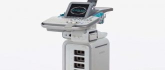
For interventions on the chest organs, X-ray or ultrasound equipment, a CT scanner and a biopsy needle can be used. As a rule, procedures are carried out under ultrasound control, but in some cases X-ray or CT control is used.
An ultrasound scanner consists of a console that includes a computer and electronic equipment, a video display and an ultrasound transducer used for scanning. The sensor is a small, portable device that resembles a microphone and is attached to the scanner using an electrical cord. The ultrasound sensor sends high-frequency sound signals and picks up echoes reflected from the internal structures of the body. The principle of operation of the device is similar to sonars that are used on submarines.
In this case, an image instantly appears on a monitor resembling a television or computer screen. Its appearance depends on the amplitude (strength), frequency and time it takes for the sound signal to return from the patient's body to the transducer, as well as the type and structure of tissue through which the sound travels.
A CT scanner is a massive rectangular machine with a hole, or short tunnel, in the middle. During the procedure, the patient is placed on a narrow table that slides inside the tunnel. The X-ray tube and electronic X-ray detectors are located opposite each other inside a ring-shaped structure called a gantry. In a separate office there is a computer workstation where the resulting image is processed. There is also a doctor or technologist here who monitors the operation of the tomograph and the progress of the examination.
The biopsy needle is usually several centimeters long, and the syringe barrel is the diameter of a wide paper clip. The needle is hollow inside, allowing the tissue sample to be grasped and removed.
When performing interventions on the chest organs, two types of needles are used:
- A thin diameter needle attached to a syringe or vial. Its diameter does not exceed the size of the needles that are used to draw blood from a vein.
- Automatic Cutting Needle with Spring Mechanism: Consists of an inner “thick” needle that is inserted into a locking cell covered with a cutting sheath and attached to a spring mechanism.
Up
Pleural puncture in children
In childhood, the procedure for therapeutic purposes is indicated:
- For aspiration of liquid or gas components from the pleural cavity in order to facilitate breathing.
- With exudative pleurisy and pleural ampyema.
- For tumor diseases in the chest.
- In case of hemothorax and pneumothorax.
For diagnostic purposes, a puncture is performed to obtain analysis from the pleural cavity.
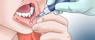
The procedure is carried out directly in the manipulation rooms. The child should lie on his side (back) or sit on a chair. The puncture site is the 5th-6th intercostal space (nipple level) or the deepest point of the effusion. First, local anesthesia is performed with a solution of novocaine (0.25%). A “lemon peel” is made with a thin needle, after which it is changed to a needle with a large lumen, which first pierces the skin, and then the subcutaneous fat and muscles. Having moved the needle to the level of the upper edge of the underlying rib, the surgeon punctures the chest wall and infiltrates the tissue with novocaine. Puncture of the pleura gives the feeling of the needle falling into emptiness.
The pleural cavity is anesthetized with two to three milliliters of novocaine, after which liquid is sucked out of it with a syringe for testing. If there is blood, pus or air in it, the doctor connects the needle to the adapter tube and aspirates the contents of the cavity. The contents are removed from the syringe into a previously prepared container, and the syringe is disconnected from the tube with a special clamp. After evacuating the contents, the cavity is washed with antiseptics. The procedure ends with the introduction of an antibiotic, but only after it has been possible to achieve maximum vacuum in the pleural cavity (“collapse” of the rubber tube).
If the first puncture has a positive effect, the manipulations are repeated until complete recovery. If the result of the procedure is unsuccessful (thick pus or an unsuccessful puncture site), one-time punctures are performed in other places until a positive result is obtained.
In the absence of positive results, passive drainage according to Bulau, or active, is indicated by creating a vacuum when connecting the drainage tube to a water jet or electric suction. Also in modern medicine, microdrainage is increasingly being practiced - the use of a venous polyethylene catheter with a diameter of 0.8-1.0 mm, inserted after removing the needle. Its advantages: elimination of organ injury and the possibility of repeated lavages of the pleural cavity with the administration of antibiotics.
To protect the child from a state of shock due to the loss of a large volume of fluid, as well as to prevent the development of infection and the formation of a fistula at the site of the canal, special care is required for him. Upon completion of the manipulation, the patient is placed on the punctured side and, in order to facilitate breathing, the upper part of the body is placed in an elevated position. Basic vital processes are monitored, in particular, respiratory function is monitored first every quarter of an hour, then every half hour, and then every 2-4 hours. Also make sure that there is no bleeding.
Laboratory test results
Best materials of the month
- Coronaviruses: SARS-CoV-2 (COVID-19)
- Antibiotics for the prevention and treatment of COVID-19: how effective are they?
- The most common "office" diseases
- Does vodka kill coronavirus?
- How to stay alive on our roads?
The puncture material is examined for tumor cells and pathogenic microorganisms. It also determines the amount of protein, enzymes and blood components.
The accumulation of excess proteins in the pleural cavity indicates the inflammatory nature of the fluid as a result of pneumonia, tuberculosis, pulmonary embolism, lung cancer or diseases of the digestive tract, as well as rheumatoid arthritis or lupus erythematosus.
The cause of insufficient protein content in the effusion can be heart failure and a number of other diseases, including sarcoidosis, myxedema, glomerulonephritis.
Blood cells in the effusion are a consequence of injuries or tumors of the pulmonary artery. The detection of tumor cells indicates the presence of metastases and new malignant formations.
Bacteriological analysis of effusion allows us to identify the causative agents of infectious pleurisy.