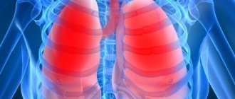
Pulmonary edema is a severe pathological condition associated with the massive release of non-inflammatory transudate from the capillaries into the interstitium of the lungs, then into the alveoli. The process leads to a decrease in the functions of the alveoli and disruption of gas exchange, and hypoxia develops. The gas composition of the blood changes significantly, and the concentration of carbon dioxide increases. Along with hypoxia, severe depression of central nervous system functions occurs. Exceeding the normal (physiological) level of interstitial fluid leads to edema.
The interstitium contains: lymphatic vessels, connective tissue elements, intercellular fluid, blood vessels. The entire system is covered by the visceral pleura. The branched hollow tubes and tubes are the complex that makes up the lungs. The entire complex is immersed in the interstitium. The interstitium is formed by plasma leaving the blood vessels. The plasma is then absorbed back into the lymphatic vessels that drain into the vena cava. Through this mechanism, the intercellular fluid delivers oxygen and necessary nutrients to the cells and removes metabolic products.
Violation of the amount and outflow of intercellular fluid leads to pulmonary edema:
- when an increase in hydrostatic pressure in the blood vessels of the lungs causes an increase in intercellular fluid, hydrostatic edema occurs;
- the increase occurred due to excessive filtration of plasma (for example: with the activity of inflammatory mediators), membrane edema occurs.
Condition assessment
Depending on the rate of transition from the interstitial stage of edema to the alveolar stage, the patient’s condition is assessed. In the case of chronic diseases, edema develops more gradually, more often at night. This swelling can be easily controlled with medications. Edema associated with mitral valve defects, myocardial infarction, and damage to the pulmonary parenchyma increases rapidly. The condition is rapidly deteriorating. Acute swelling leaves very little time to respond.
Disease prognosis
The prognosis for pulmonary edema is unfavorable. Depends on the reasons that actually caused the swelling. If the edema is non-cardiogenic, it responds well to treatment. Cardiogenic edema is difficult to treat. After long-term treatment after cardiogenic edema, the 1-year survival rate is 50%. In the lightning-fast form, it is often impossible to save a person.
With toxic edema, the prognosis is very serious. Favorable prognosis when taking large doses of diuretics. Depends on the individual reaction of the body.
Diagnostics
The picture of any type of pulmonary edema is bright. Therefore, diagnosis is simple. For adequate therapy, it is necessary to determine the reasons that caused the swelling. Symptoms depend on the form of edema. The fulminant form is characterized by rapidly increasing suffocation and respiratory arrest. The acute form has more pronounced symptoms, in contrast to the subacute and protracted form.
Symptoms of pulmonary edema
The main symptoms of pulmonary edema include:
- frequent coughing;
- increasing hoarseness;
- cyanosis (the face and mucous membranes become bluish);
- increasing suffocation;
- tightness in the chest, pressing pain;
- increased breathing;
- dizziness, weakness;
- bubbling wheezing is heard;
- with increasing cough - foamy pink sputum;
- as the condition worsens, sputum is released from the nose;
- the person is scared, consciousness may be confused;
- sweating, cold and sticky sweat;
- fear of death;
- increased heart rate up to 200 beats per minute. Can easily develop into life-threatening bradycardia;
- drop or surge in blood pressure.
Pulmonary edema itself is a disease that does not occur on its own. Many pathologies can lead to edema, sometimes completely unrelated to diseases of the bronchopulmonary and other systems.
Medical assistance
Pulmonary edema in elderly people, treatment of this syndrome includes the following manipulations:
- Use of a special mask that provides oxygen supply. In emergency situations, oxygen is replaced by forced ventilation.
- Administration of painkillers and sedatives.
- Administration of Aminophylline, which promotes dilation of the bronchi and improves blood circulation in the kidneys.
- Blood pressure control. If it is higher than normal values, the patient is injected with sodium nitroprusside. For low blood pressure - dobutamine.
Subsequent treatment consists of taking medications. These can be hormonal medications, antibiotics, hepatoprotectors, antihistamines.
The treatment regimen is determined by the doctor based on the patient’s condition, his age, and the form of the disease. If there is the slightest chance of pulmonary edema, you need to contact a medical facility to avoid the development of complications. The latter can be so serious that they will lead to death if the patient does not receive timely medical attention. Such a serious disease cannot be joked about, especially when it comes to older people. Pulmonary edema progresses quickly, and measures must be taken without delay. A medical examination in such cases is mandatory.
Causes of pulmonary edema
Causes of pulmonary edema include:
- Sepsis. Usually is the penetration into the bloodstream of exogenous or endogenous toxins;
- Pneumonia;
- Overdose of certain (NSAIDs, cytostatics) drugs;
- Radiation damage to the lungs;
- Drug overdose;
- Myocardial infarction, heart disease, ischemia, hypertension, any heart disease in the decompensation stage;
- Congestion in the right circulation that occurs with bronchial asthma, emphysema and other pulmonary diseases;
- A sharp or chronic decrease in protein in the blood. Hypoalbuminemia occurs with liver cirrhosis, nephrotic syndrome and other kidney pathologies;
- Infusions in large volumes without forced diuresis;
- Poisoning by toxic gases;
- Poisons;
- Gastric aspiration;
- Shock due to serious injuries;
- Enteropathies;
- Being at high altitude;
- Acute hemorrhagic pancreatitis.
Types of pulmonary edema
There are two types of pulmonary edema: cardiogenic and non-cardiogenic. There is also a 3rd group of pulmonary edema (non-cardiogenic) - toxic edema.
Cardiogenic edema (cardiac edema)
Cardiogenic edema is always caused by acute left ventricular failure and obligatory stagnation of blood in the lungs. Myocardial infarction, heart defects, angina pectoris, arterial hypertension, left ventricular failure are the main causes of cardiogenic edema. To relate pulmonary edema to chronic or acute heart failure, pulmonary capillary pressure is measured. In the case of cardiogenic type of edema, the pressure rises above 30 mmHg. Art. Cardiogenic edema provokes transudation of fluid into the interstitial space, then into the alveoli. Attacks of interstitial edema are observed at night (paroxysmal dyspnea). The patient does not have enough air. Auscultation detects harsh breathing. Breathing is increased during exhalation. Choking is the main symptom of alveolar edema.
The following symptoms are characteristic of cardiogenic edema:
- suffocation;
- increasing cough;
- inspiratory dyspnea. The patient is characterized by a sitting position; in the lying position, shortness of breath increases;
- tissue hyperhydration (swelling);
- dry whistling, turning into wet gurgling wheezing;
- separation of pink frothy sputum;
- acrocyanosis;
- unstable blood pressure. It is difficult to reduce it to normal. A decrease below normal can lead to bradycardia and death;
- tachycardia;
- severe pain behind the sternum or in the chest area;
- fear of death;
- The electrocardiogram shows hypertrophy of the left atrium and ventricle, sometimes blockade of the left bundle branch.
Hemodynamic conditions of cardiogenic edema
- violation of left ventricular systole;
- diastolic dysfunction;
- systolic dysfunction.
The leading cause of cardiogenic edema is left ventricular dysfunction.
Cardiogenic edema must be differentiated from non-cardiogenic edema. With non-cardiogenic edema, changes in the cardiogram are less pronounced. Cardiogenic edema occurs more quickly. There is less time for emergency care than for other types of edema. Death is most often caused by cardiogenic edema.
Toxic pulmonary edema
Toxic edema has certain specific features that contribute to differentiation. There is a period here when there is no swelling itself, there are only reflex reactions of the body to irritation. A burn of lung tissue or a burn of the respiratory tract causes a reflex spasm. This is a combination of symptoms of damage to the respiratory organs and the resorptive effects of toxic substances (poisons). Toxic edema can develop regardless of the dose of the drug that caused it.
Medicines that can cause pulmonary edema:
- narcotic analgesics;
- many cytostatics;
- diuretics;
- X-ray contrast agents;
- non-steroidal anti-inflammatory drugs.
Risk factors for the occurrence of toxic edema are old age and long-term smoking.
It has 2 forms, developed and abortive. There is a so-called “silent” edema. It can be detected by X-ray examination of the lungs. There is practically no definite clinical picture for such edema.
Characterized by periodicity. Has 4 periods:
- Reflex disorders. Characterized by symptoms of irritation of the mucous membranes: lacrimation, cough, shortness of breath. The period is dangerous due to respiratory and cardiac arrest;
- Hidden period of subsidence of irritation. May last 4-24 hours. Characterized by clinical well-being. A thorough examination may show signs of impending edema: bradycardia, pulmonary emphysema;
- Direct pulmonary edema. The course is sometimes slow, reaching 24 hours. Most often, symptoms increase within 4-6 hours. During this period, the temperature rises, the blood count shows neutrophilic leukocytosis, and there is a danger of collapse. The developed form of toxic edema has a fourth period of completed edema. The completed period has “blue hypoxemia”. Cyanosis of the skin and mucous membranes. The completed period increases the breathing rate to 50-60 times per minute. Bubbling breathing can be heard from a distance, sputum mixed with blood. Blood clotting increases. Gas acidosis develops. “Gray” hypoxemia is characterized by a more severe course. Vascular complications are added. The skin takes on a pale grayish tint. The limbs are cold. Thread-like pulse and drop in blood pressure to critical values. This condition is promoted by physical activity or improper transportation of the patient;
- Complications. When leaving the period of immediate pulmonary edema, there is a risk of developing secondary edema. This is due to left ventricular failure. Pneumonia, pneumosclerosis, emphysema are common complications caused by drugs and toxic edema. At the end of the 3rd week, “secondary” edema may occur due to acute heart failure. Rarely does an exacerbation of latent tuberculosis and other chronic diseases occur. Depression, drowsiness, asthenia.
With quick and effective therapy, a period of reverse development of edema begins. It does not belong to the main periods of toxic edema. Here everything depends only on the quality of the assistance provided. Cough and shortness of breath decrease, cyanosis decreases, wheezing in the lungs disappears. The X-ray shows the disappearance of large, then small lesions. The peripheral blood picture is normalizing. The recovery period from toxic edema can be several weeks.
In rare cases, toxic edema can be caused by taking tocolytics. The catalyst for edema can be: large volumes of intravenously administered fluid, recent treatment with glucocorticoids, multiple pregnancy, anemia, unstable hemodynamics in a woman.
Clinical manifestations of the disease:
- The key symptom is respiratory failure;
- Severe shortness of breath;
- Cough;
- Severe chest pain;
- Cyanosis of the skin and mucous membranes;
- Arterial hypotension in combination with tachycardia.
Toxic edema differs from cardiogenic edema by its protracted course and the content of a small amount of protein in the fluid. The size of the heart does not change (it rarely changes). Venous pressure is often within normal limits.
Diagnosis of toxic edema is not difficult. An exception is bronchorrhea due to FOS poisoning.
Non-cardiogenic pulmonary edema
Occurs due to increased vascular permeability and high filtration of fluid through the wall of the pulmonary capillaries. With a large amount of fluid, the functioning of blood vessels worsens. Liquid begins to fill the alveoli and gas exchange is disrupted.
Causes of non-cardiogenic edema:
- renal artery stenosis;
- pheochromocytoma;
- massive renal failure, hyperalbuminemia;
- exudative enteropathy;
- pneumothorax can cause unilateral non-cardiogenic pulmonary edema;
- severe attack of bronchial asthma;
- inflammatory lung diseases;
- pneumosclerosis;
- sepsis;
- aspiration of gastric contents;
- cancerous lymphangitis;
- shock, especially with sepsis, aspiration and pancreatic necrosis;
- cirrhosis of the liver;
- radiation;
- inhalation of toxic substances;
- lungs' cancer;
- large transfusions of drug solutions;
- in elderly patients taking acetylsalicylic acid for a long time;
- drug addict.
To clearly differentiate edema, the following measures should be taken:
- study the patient's history;
- apply methods of direct measurement of central hemodynamics;
- radiography;
- assess the affected area during myocardial ischemia (enzyme tests, ECG).
To differentiate non-cardiogenic edema, the main indicator will be the measurement of wedge pressure. Normal cardiac output and positive wedge pressure results indicate a non-cardiogenic nature of the edema.
Diagnosis of pathology
The diagnosis of cardiopulmonary failure is established based on the results of a comprehensive examination. During the initial examination, the doctor collects anamnesis (listens about symptoms, chronic pathologies in the patient and immediate family), performs a visual examination (possible swelling of the feet and legs), conducts a physical examination and standard measures (measuring blood pressure, listening to breathing using a phonendoscope). Upon palpation, a cardiac impulse is detected, and upon percussion, an expansion of the boundaries of the region of the muscular organ, which is partially covered by the lungs, is detected.
Among the laboratory diagnostic methods for making a diagnosis, an analysis of the gas composition of arterial blood is required. If there are problems, patients experience a decrease in P02 and SAO2 P02, and an increase in PC02. Additionally, a general blood test is prescribed to detect an infectious process in the body (by increasing the level of leukocytes). The most informative ways to detect cardiopulmonary failure are instrumental diagnostic methods. Depending on the symptoms, the patient may be prescribed:
- study of respiratory function (external respiration function). With the help of this examination, it is possible to establish the nature and severity of pulmonary ventilation impairment;
- electrocardiography (ECG). This is an accessible method that does not require additional preparation from the patient. The essence of electrocardiography is to record the electrical potentials of a muscle organ, which makes it possible to detect changes in rhythm and electrolyte deficiency. If the disease occurs in an acute form, it is possible to detect signs of overload in the right parts of the muscular organ. If the disease is chronic, it is possible to detect markers (both direct and indirect) of right ventricular hypertrophy;
- echocardiography (EchoCG). It is often prescribed in addition to an ECG, since the information content of this examination regarding cardiopulmonary failure is very high. This is the main non-invasive method with which it is possible to assess intracardiac hemodynamics, determine the size of the cavities of the muscular organ and the degree of pulmonary hypertension. EchoCG allows you to detect a number of possible disturbances in the functioning of the heart, differentiate it from heart disease and other pathologies;
- chest x-ray. This is a simple technique with which it is possible to assess the condition of the respiratory system (detect damage to the internal organ and notice signs of pulmonary hypertension);
- transbronchial lung biopsy. These methods are rarely resorted to, in particularly advanced clinical cases. A puncture biopsy of the lung is performed under ultrasound or X-ray guidance using a local anesthetic.
Consequences of pulmonary edema
When the swelling is stopped, it is too early to end the treatment. After an extremely severe condition of pulmonary edema, serious complications often arise:
- addition of a secondary infection. Pneumonia most often develops. Against the background of reduced immunity, even bronchitis can lead to adverse complications. Pneumonia associated with pulmonary edema is difficult to treat;
- hypoxia, characteristic of pulmonary edema, affects vital organs. The most serious consequences can affect the brain and cardiovascular system - the effects of swelling may be irreversible. Cerebral circulation disorders, cardiosclerosis, heart failure without powerful pharmacological support lead to death;
- ischemic damage to many organs and systems of the body;
- pneumofibrosis, segmental atelectasis.
Emergency care for pulmonary edema
Required for every patient with signs of pulmonary edema. Emergency care highlights:
- the patient must be placed in a semi-sitting position;
- aspiration (removal) of foam from the upper respiratory tract. Aspiration is performed by inhaling oxygen through 33% ethanol;
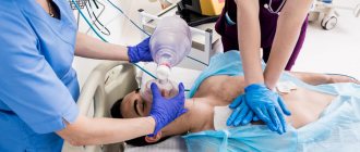
- urgent oxygen inhalation (oxygen therapy);
- elimination of acute pain syndrome with the help of antipsychotics;
- restoration of heart rhythm;
- correction of electrolyte balance;
- normalization of acid-base balance;
- normalization of hydrostatic pressure in the pulmonary circulation. Narcotic analgesics “Omnopon” and “Promedol” are used. They depress the respiratory center, relieve tachycardia, reduce venous blood flow, lower blood pressure, reduce anxiety and fear of death;
- vasodilators (Nitromint aerosol). The drugs reduce vascular tone and intrathoracic blood volume. Nitroglycerin preparations facilitate the outflow of blood from the lungs, acting on peripheral vascular resistance;
- application of venous tourniquets to the lower extremities. The procedure is necessary to reduce CTC - an old effective method. Currently, 40 mg of Lasix intravenously is used to dehydrate the lung parenchyma. The action of furosemide (Lasix) develops within a few minutes and lasts up to 3 hours. The drug is capable of removing 2 liters of urine in a short period of time. A reduced plasma volume with increased colloid osmotic pressure contributes to the transition of edematous fluid into the bloodstream. Filtration pressure decreases. For low blood pressure, diuretics can be used only after it has normalized;
- prescription of diuretics for lung dehydration (Lasix 80 mg intravenously);
- administration of cardiac glycosides to increase myocardial contractility;
- immediate hospitalization.
Cardiogenic form
Cardiogenic OB occurs as a manifestation of left ventricular heart failure. The following diseases can lead to the development of pathology:
- myocardial infarction, post-infarction conditions;
- acquired heart defects (both aortic and mitral);
- hypertensive crisis;
- disturbances of heart rhythm and conduction (arrhythmias, blockades).
The mechanism of development of the cardiogenic form of edema is associated with an increase in pressure in the pulmonary capillaries. This occurs due to increased diastolic pressure in the left ventricle.
The left ventricle cannot contract enough to push all the blood out of the cavity into the aorta. This leads to an increase in pressure first in the left ventricle and then in the left atrium. Since the pulmonary veins flow into the left atrium, over time the pressure also increases in the pulmonary circulation. The fluid sweats first into the interstitial tissue and then into the alveoli.
By the same principle, the lungs swell due to hypervolemia (increased blood volume).
Hypervolemia can develop as a complication of infusion therapy.
Main complications after emergency care
Such complications include:
- development of a fulminant form of edema;
- intensive foam production can cause airway obstruction;
- depression (depression) of breathing;
- tachyarrhythmia, asystole;
- angiotic pain. Such pain is characterized by an unbearable pain syndrome; the patient may experience pain shock, worsening the prognosis;
- impossibility of stabilizing blood pressure. Pulmonary edema often occurs against a background of low and high blood pressure, which can alternate within a large amplitude. The vessels will not be able to withstand such a load for long and the patient’s condition worsens;
- increase in pulmonary edema against the background of high blood pressure.
Complications
The main thing that threatens pulmonary edema in an elderly person is the lack of oxygen in the tissues. Even if the disease has been stopped, the brain will experience serious changes, and the tissues of the heart and lungs will suffer.
Also among other serious consequences of suffering from pulmonary edema are:
- formation of congestion in the lungs;
- organ ischemia;
- emphysema.
Due to oxygen starvation, the patient’s memory will deteriorate, during the daytime he will constantly feel sleepy, general lethargy will be felt, and his mood will begin to deteriorate. You will have to carefully monitor your own condition in order to notice serious deterioration in time and consult a doctor.
Pulmonary edema in old age is a serious pathology. Even if the disease manifests itself in a protracted or subacute form, the risk of complications after therapy is high. An immediate or acute syndrome practically does not allow saving the patient. So at the first symptoms indicating the development of the disease, you need to undergo an examination and consult a doctor so that it is not too late.
Treatment of pulmonary edema
It comes down to one thing - the swelling should be removed as soon as possible. Then, after intensive treatment of the pulmonary edema itself, medications are prescribed to treat the disease that caused the edema.
So, means for relieving swelling and subsequent therapy:
- Morphine hydrochloride. A vital drug for the treatment of cardiogenic type and other edema in case of hyperventilation. The administration of morphine hydrochloride requires readiness to transfer the patient to controlled breathing;
- Nitrate preparations in infusion form (glycerol trinitrate, isosorbitol dinitrate) are used for any edema, excluding edema with hypovolemia due to pulmonary embolism;
- The introduction of loop diuretics (“Furosemide”, “Torasemide”) in the first minutes of edema saves the lives of many patients;
- In the case of cardiogenic pulmonary edema as a result of myocardial infarction, administration of tissue plasminogen activator is mandatory;
- With atrial fibrillation. Only if the effectiveness of electropulse therapy is low. Often, against the background of even a slight decrease in rhythm, the patient’s condition can significantly worsen. When amiodarone is prescribed, dobutamine infusion is sometimes required to increase the rate;
- Corticosteroids are used only for non-cardiogenic edema. The most commonly used is dexamethasone. It is actively absorbed into the systemic bloodstream and negatively affects the immune system. Modern medicine now recommends the use of methylprednisolone. Its elimination period is much shorter, side effects are less pronounced, and its activity is higher than that of dexamethasone;
- For inotropic rhythm support in case of overdose of beta-blockers, dopamine is used;
- Cardiac glycosides (digoxin) are necessary for constant atrial fibrillation, sodium thiopental is necessary for short-term anesthesia, to relieve pain;
- "Diazepam" with ketamine is used for premedication;
- For heroin-induced pulmonary edema or iatrogenic complications, muscle relaxants (naloxone) are prescribed;
- In conditions of high-altitude pulmonary edema, Nifedipine is needed; it quickly lowers blood pressure;
- At the inpatient stage of treatment, loading doses of antibiotics are prescribed to prevent infection. In first place are drugs from the group of fluoroquinolones: Tavanik, Tsifran, levofloxacin;
- To facilitate the removal of accumulated fluid, large doses of ambroxol are prescribed;
- Prescribing a surfactant is mandatory. It reduces tension in the alveolus and has a protective effect. Surfactant improves oxygen absorption by the lungs, reduces hypoxia;
- Sedatives for pulmonary edema. In the treatment of patients who have suffered pulmonary edema, the leading role is played by the normalization of the emotional background. Often, severe stress itself can trigger swelling. The stress trigger often causes pancreatic necrosis and myocardial infarction. Sedatives can, in combination with other drugs, normalize the content of catecholamines. Due to this, spasm of peripheral vessels is reduced, blood flow is significantly reduced, and the load on the heart is removed. Normal heart function improves the outflow of blood from the pulmonary circle. The calming effect of sedatives can relieve vegetative-vascular manifestations of edema. With the help of sedatives, it is possible to reduce the filtration of tissue fluid through the alveolar-capillary membrane. Means that can influence the emotional background can reduce blood pressure, tachycardia, reduce shortness of breath, vegetative-vascular manifestations, and reduce the intensity of metabolic processes - this facilitates the course of hypoxia. Not counting the morphine solution - the first, most effective aid for pulmonary edema, 4 ml of a solution of droperidol 0.25% or Relanium 0.5% - 2 ml is prescribed. Unlike morphine, these drugs are used for all types of pulmonary edema;
- Ganglion blockers: Arfonad, pentamin, benzohexonium. They allow you to quickly relieve pulmonary edema with high blood pressure (from 180 mm Hg). Improvement comes quickly. 20 minutes after the first administration of the drugs, shortness of breath and wheezing decrease, and breathing becomes calmer. With the help of these drugs, pulmonary edema can be stopped completely.
Treatment of heart failure
For drug therapy of heart failure, the following groups of drugs are used: diuretics, cardiac glycosides, vasodilators (nitrates), calcium channel blockers, beta blockers and others. In particularly severe cases, surgical treatment is performed.
Diuretics have been used since the 50s of the 20th century. The drugs help the heart function by stimulating the excretion of excess salt and water in the urine. As a result, the volume of circulating blood decreases, blood pressure decreases, and blood flow improves.
The most important for heart failure is a group of drugs derived from the digitalis plant or “cardiac glycosides”. These medicinal substances were first discovered in the 18th century and are widely used to this day. Cardiac glycosides affect internal metabolic processes inside heart cells, increasing the force of heart contractions. Thanks to this, blood supply to internal organs significantly improves.
Recently, new classes of drugs, such as vasodilators (vasodilators), have been used to treat heart failure. These drugs primarily affect peripheral arteries, stimulating their dilation. As a result, by facilitating blood flow through the vessels, heart function improves. Vasodilators include nitrates, angiotensin-converting enzyme blockers, and calcium channel blockers.
In emergency cases, surgical intervention is performed, which is especially necessary when the failure is caused by disorders of the heart valves.
There are situations when the only way to save a patient’s life is a heart transplant.