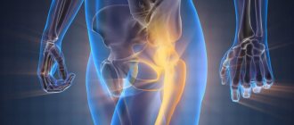
Sarcomas are tumors that develop from precursor cells of connective tissue - mesoderm. Like cancer, they are malignant neoplasms, but with the difference that the source of the cancerous tumor is the epithelium lining the internal organs and the human body. Therefore, these tumors are tied to a specific organ, for example, prostate cancer, breast cancer, etc. Sarcomas develop from connective tissue and can arise in any organ where such cells are present, that is, virtually everywhere.
- Types of soft tissue sarcomas
- Stages of development
- Risk factors
- Symptoms of soft tissue sarcoma
- Diagnosis of soft tissue sarcoma
- Treatment of soft tissue sarcoma
- Survival prognosis for soft tissue sarcoma
Types of soft tissue sarcomas
Soft tissue sarcoma is a collective term that unites a large number of malignant tumors, among which the most common are the following:
- Leiomyosarcoma is a tumor of smooth muscle cells.
- Rhabdomyosarcoma is a tumor of striated muscle.
- Liposarcoma is a malignant tumor of adipose tissue.
- Angiosarcoma is a malignant neoplasm that grows from the tissue of blood vessels.
- Fibrosarcoma is a tumor of fibrous tissue.
Also, soft tissue sarcomas are usually divided according to the degree of malignancy. Low-grade neoplasms rarely metastasize, but are prone to persistent recurrence. Highly malignant tumors tend to metastasize hematogenously (through the bloodstream), lymph nodes are affected less frequently.
Stages of development
The stage of soft tissue sarcoma is determined by the degree of malignancy of the tumor, its size and the presence of metastases.
- Stage 1 A includes neoplasms of low malignancy, less than 5 cm in size, without metastases in regional lymph nodes and distant organs.
- Stage 1 B - low-grade tumors, more than 5 cm without metastases.
- Stage 2A - neoplasms of moderate malignancy, less than 5 cm without metastases.
- Stage 2B - neoplasms of moderate malignancy, more than 5 cm in greatest dimension, without metastases.
- Stage 3A - sarcomas of a high degree of malignancy, less than 5 cm without damage to the lymph nodes and internal organs.
- Stage 3B - high-grade sarcomas, more than 5 cm without metastases.
- Stage 4 - sarcoma of any degree of malignancy, of any size in the presence of damage to the lymph nodes or metastases in the internal organs.
Risk factors
In most cases, soft tissue sarcomas develop spontaneously, that is, for no apparent reason. However, there are certain circumstances that increase the likelihood of developing this disease:
- Exposure to chemical carcinogens, in particular herbicides, chlorophenols.
- Genetic predisposition - neurofibromatosis, Li-Fraumeni syndrome, Gardner syndrome.
- Exposure to ionizing radiation, such as undergoing radiation therapy.
- Immunodeficiency states - immunosuppression after organ transplantation, chemotherapy, AIDS.
- Some types of soft tissue sarcomas can develop against the background of lymphostasis of the limb.
Symptoms of soft tissue sarcoma
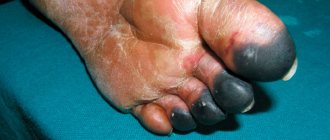
In the initial stages, soft tissue sarcoma appears as a painless tumor that slowly increases in size. It may have a capsule, which, however, does not interfere with metastasis. Also, the tumor may have infiltrative growth, in which case it is problematic to determine its edges.
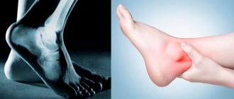
With further growth, symptoms will depend on the location of the tumor. For example, with uterine sarcomas there will be bleeding; if the tumor is superficial, it causes swelling and pain, and the function of the affected organ, for example, an arm or leg, is impaired.
Resources
The Kristen Ann Carr Fund www.sarcoma.com The Kristen Ann Carr Fund is a resource for people diagnosed with soft tissue sarcoma. Every year the foundation prepares a newsletter “Up-to-date information on the problem of sarcoma.” You can find more information about soft tissue sarcoma on the foundation's website.
Cancer.Net www.cancer.net/cancer-types/sarcoma-soft-tissue/introduction Cancer.Net provides information about soft tissue sarcoma, including videos and blog posts.
This website also has a Spanish version. Visit the website for more information. to come back to the beginning
Diagnosis of soft tissue sarcoma
As part of the diagnosis of soft tissue sarcoma, a physical examination is performed, medical imaging methods are used, followed by a biopsy and morphological examination of the obtained material.
A CT or MRI of the affected area is required. Firstly, the study helps to detect a tumor, and secondly, to monitor the radicality of the cure.
To make a diagnosis, a morphological examination of a tumor fragment is required. To obtain it, a trephine biopsy is performed using a special thick needle under ultrasound control, or an incisional biopsy, which involves surgical excision of a fragment of the tumor. In some cases, to clarify the type of sarcoma, a more detailed study is necessary, then immunohistochemical and molecular genetic analyzes are performed.
In order to determine the extent of tumor spread, a CT scan of the chest, abdominal cavity, and ultrasound examination of the lymph nodes is performed.
Book a consultation 24 hours a day
+7+7+78
Long Term Care
Your health care experience at Memorial Sloan Kettering (MSK) is a long process. You will see your doctor every 4 to 6 months for the first 3 years after treatment ends. At each of these follow-up visits, you will be examined by a doctor and have an X-ray or CT scan.
After the early follow-up period, you will see your doctor every 6 to 12 months for the next 5 years. After this, you will only see your doctor once a year or once every two years.
Your doctors and nurses are committed to your long-term health care. Please contact them at any time during the period of such service if you have any questions or concerns.
to come back to the beginning
Treatment of soft tissue sarcoma
The choice of treatment for sarcoma is determined by the degree of its malignancy, location and presence of metastases. For the treatment of low-grade tumors, treatment is predominantly surgical; in other cases, combined methods are used, including radiation and chemotherapy.
Surgery
The following types of operations are used in the surgical treatment of sarcomas:
- Wide local resection. In this way, low-grade small tumors located in the skin and subcutaneous tissue are removed.
- Wide excision. With this intervention, the sarcoma is removed within the anatomical zone, retreating from the edge of the tumor by at least 4-6 cm.
- Radical organ-preserving surgery. With this volume of intervention, the tumor is removed along with the muscle fascia and intact muscles, which are cut off at the site of their attachment. If blood vessels, nerve trunks and bones are involved in the process, they are resected with one-stage plastic surgery using a skin or musculocutaneous flap.
- Amputation or disarticulation is the removal of a limb or its disarticulation along the line of the joint space. Such interventions are carried out in cases of extensive, locally advanced, highly malignant processes involving the main blood and nerve trunks over a long distance, as well as bone structures and muscles.
During surgery, an urgent intraoperative histological examination is required. If there are malignant cells at the cutting edges and with the potential for radical removal of the tumor, reoperation is performed.
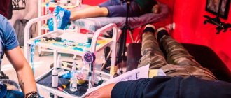
Radiation therapy
Radiation therapy is used as part of combined treatment in the preoperative and/or postoperative stages. In the first case, the goal is to reduce the size of the sarcoma and create conditions for radical intervention. In the second - the destruction of the remaining malignant cells. To ensure a uniform effect on the tumor, multifield irradiation techniques are used.
Preoperative radiotherapy
During preoperative RT, the boundaries of the irradiation fields (FI) should extend beyond the size of the tumor tissue by at least 3-4 cm. If the tumor is large, irradiation is performed within 10 cm from its edge until a total focal dose of 45-50 Gy is achieved. Then the software is reduced to the size of the tumor.
If the tumor is located on a limb, there is a possibility of developing osteoradiation necrosis, muscle contractures and edema. To avoid this, it is recommended to irradiate no more than 2/3 of the circumference of the limb, if possible. The minimum thickness of non-irradiated tissue in the forearm area is 2 cm, on the thigh 4 cm, and on the lower leg - 3 cm.
Postoperative radiotherapy
Postoperative radiation therapy is indicated in the following cases:
- High degree of malignancy of sarcoma according to histological examination.
- A non-radical operation is the presence of malignant cells at the cutting edges when reoperation is impossible.
- Opening the sarcoma capsule during surgery.
Postoperative RT begins no later than 4 weeks after the end of surgical treatment.
If preoperative RT was not performed, the radiation fields include the following areas:
- Tumor bed.
- Tissues that are located at a distance of 2 cm from the removed tumor.
- Postoperative scar.
Irradiation is carried out at a total focal dose of 60 Gy. If non-radical surgery was performed, the dose is increased to 70 Gy. Radiation therapy is carried out in a similar mode if surgical treatment is not possible.
Chemotherapy
Chemotherapy is widely used to treat high-grade soft tissue sarcomas. As with radiation therapy, it can be prescribed preoperatively and/or postoperatively. Neoadjuvant (preoperative) chemotherapy is designed to affect the tumor site, destroy micrometastases and create conditions for organ-preserving surgical treatment. For this purpose, 2-3 courses of chemotherapy are carried out with a break of 3-4 weeks.
Postoperative chemotherapy can be performed to destroy micrometastases or already visualized metastases. For this purpose, 3-4 courses of treatment are performed. In the chemotherapy treatment of sarcomas, various combinations of the following drugs are used:
- Doxorubicin.
- Etoposide.
- Vincristine.
- Ifosfamide.
- Dacarbazine.
- Cisplatin, etc.
The chemotherapy regimen will be determined by the morphological variant of the tumor and its sensitivity to previous treatment.
Diagnosis of pathology
Unfortunately, a standard scheme for diagnosing this oncopathology has not been developed to this day. The clinical picture may be similar to a regular bruise or swelling resulting from sprains. For this reason, it is impossible to accurately detect sarcomatous soft tissue lesions through external examination. But an external examination of the patient and palpation of problem areas is also extremely important, because this helps to raise at least rough suspicions. Thanks to palpation, the doctor can find out the approximate dimensions of the tumor and the depth of its localization. In addition, this method assesses the degree of involvement of nearby tissue structures.
Further diagnosis involves undergoing the following procedures:
• histological analysis of the biopsy; • x-ray examination; • CT; • MRI; • angiography.
In the presence of cancer lesions localized in the extremities and with a diameter greater than 5 cm, a biopsy is a mandatory procedure. As a rule, a subcutaneous method is used - fine needle aspiration. The needle is inserted into the affected area so that no difficulties arise during further treatment (surgeries, radiation treatment). An open biopsy is performed less frequently (if the tumor lesion is located in the deep tissue layers). This procedure is dangerous in its own way, because it can provoke the spread of cancer cells through the bloodstream. The extracted material is examined in laboratory conditions. Thanks to this analysis, it is possible to find out the histological appearance of the cancer lesion and prescribe effective treatment. For imaging purposes, an MRI is usually prescribed. With its help, you can clearly determine the contrast between the lesion, muscles and blood vessels.