Sarcomas are the general name for malignant tumors that are formed from different types of connective tissue. They are often characterized by progressive, very rapid growth and frequent relapses, especially in children. This behavior of sarcomas is explained by the accelerated development of connective and muscle tissue cells at a young age.
- Causes of sarcomas and risk factors
- Development of sarcoma
- Prevalence of sarcoma
- Symptoms
- Diagnostics
- Treatment of sarcoma
Sarcomas are divided into two large groups: soft tissue and bone.
Unlike cancer, which is a malignant neoplasm of the epithelium, sarcomas grow from connective tissue and are not associated with any specific organ, but can occur anywhere in the body. In everyday life, all malignant neoplasms are usually called cancer, but this is incorrect.
Bone sarcomas can develop both directly from bone tissue (paraosteal sarcoma, chondrosarcoma) and from tissues of non-bone origin (Ewing's sarcoma - from mesenchymal stem cells, angiosarcoma - from blood vessel cells, etc.). They can affect internal organs, skin, lymphoid tissue, and the central and peripheral nervous system. The classification, based on the type of tissue from which the tumor was formed, includes the following types:
- osteosarcomas grow from bone tissue;
- mesenchymomas - from embryonic ones;
- liposarcoma - from adipose tissue;
- myosarcoma - from muscle tissue;
- angiosarcomas - from blood and lymphatic vessels, etc.
In total, about 100 different types of sarcomas are known, each of which has its own characteristics of development, treatment and, accordingly, its own prognosis.
According to the degree of tissue maturity, they are divided into low, medium and highly differentiated. The degree of malignancy of the tumor, as well as the tactics of its treatment, depend on this feature: the less differentiated the tumor cells, the more aggressive the tumor and the more serious the prognosis.
Book a consultation 24 hours a day
+7+7+78
Etiology and pathogenesis
The causes of soft tissue sarcomas have not been identified. Factors that increase the risk of developing this pathology:
- Paget's disease (ostosis deformans);
- Recklenhausen's disease (neurofibromatosis);
- Gardner's syndrome (diffuse polyposis of the colon);
- Werner's syndrome (multiple endocrine adenomtaosis);
- Taking anabolic steroids.
The clinical course of malignant soft tissue tumors is characterized by variability.
Common signs of soft tissue sarcomas include frequent local relapses. Why do sarcomas often recur? This is explained by the following reasons:
- Lack of true capsule;
- Tendency to infiltrative growth;
- Multicentric growth.
Symptoms
Manifestations of sarcoma can be very different depending on the place of its origin, size and direction of their growth. In most cases, a neoplasm is first detected, which gradually increases in size. As it grows, neighboring tissues are involved, growing into which, the sarcoma damages the nerves and blood vessels in them. This is manifested by a pain syndrome that is poorly relieved by analgesics.
Characteristic symptoms:
- with Ewing's sarcoma - pain in the legs at night;
- with intestinal leiomyosarcoma - progressive signs of intestinal obstruction;
- with uterine sarcoma - intermenstrual bleeding;
- with extraperitoneal sarcoma - lymphostasis and elephantiasis of the lower extremities;
- with mediastinal sarcoma - swelling of the neck, expansion of the saphenous veins in the chest, shortness of breath;
- for sarcomas of the face and neck - asymmetry, head deformation, disturbances in the functioning of the masticatory and facial muscles, etc.
As sarcoma of the extremities develops, skin changes appear above it in the form of redness, dilated veins, thrombophlebitis and a local increase in temperature. The mobility of the arms and legs becomes limited, and dysfunction is accompanied by constant pain.
Diagnosis of soft tissue sarcomas
Diagnostic tests include:
— Examination by an experienced specialist, oncologist; In the photo you can see what soft tissue sarcoma looks like:
- X-ray examination. Allows you to visualize the shadow of the tumor, deformation of the fascial bridges adjacent to the tumor, and identify changes in the bones;
— Ultrasound of soft tissue tumors. This is a method for diagnosing both the primary focus and damage to regional zones. Allows you to determine the boundaries of the tumor, connections with surrounding organs, and tumor structure;
Figure No. 1. Soft tissue sarcoma of the thigh. Ultrasound image in gray scale and vascular imaging modes
— CT scan of the primary tumor, chest and abdominal organs;
— MRI of the primary tumor
Figure No. 2. Soft tissue sarcoma of the upper third of the left thigh
— Morphological verification (puncture biopsy, trephine biopsy) is the most important diagnostic method.
After receiving the results of the examination, laboratory and instrumental diagnostics, the oncologist determines the tactics for managing the patient, according to modern recommendations.
General principles of treatment of soft tissue sarcomas
Treatment of soft tissue sarcomas should be multicomponent. A council of doctors gathers to determine treatment tactics for the patient.
Basic method:
- Surgical
Additional methods:
- Radiation therapy
- Chemotherapy
Preoperative radiation therapy in combination with surgical treatment can reduce the number of relapses by reducing the malignant potential of the tumor and reducing its volume.
Intraoperative radiation therapy is used, i.e. irradiation of the tumor during surgery in order to suppress subclinical lesions and increase the radiation dose.
Chemotherapy is also used in combination with surgical treatment. There has been a trend towards improved survival rates, but this method is not considered the standard of care for the treatment of soft tissue sarcomas.
A general name for a large group of malignant tumors growing from connective tissue cells. Since connective tissue is found almost everywhere in the body, a tumor can begin to grow almost anywhere. Lesions are relatively rare in adults and in Russia they account for 0.7% of all cancers. In childhood, the frequency reaches 6.5% (and this is the 5th place among malignant neoplasms in children). Sarcomas grow very quickly, begin to metastasize early and tend to recur (even after successful operations), especially in childhood.
Causes and risk factors
The exact causes of tumors are not known. Some risk factors for the development of sarcomas have been established:
- Exposure to ionizing radiation, ultraviolet irradiation
- Previous chemotherapy or radiotherapy
- Contact with carcinogens (work in chemical plants)
- Immunity disorders (including HIV)
- Some types of benign tumors
- Heredity (cases of sarcoma in blood relatives)
- Previous operations involving removal of lymph nodes
Types and localization of sarcomas
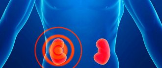
Unlike other types of cancer that affect a specific organ, sarcomas occur in any part of the body and organs: extremities (50% of all tumors), torso (40%), head and neck (10%), and less commonly, the gastrointestinal tract. Soft tissue sarcomas include:
Angiosarcomas are formed from cells of blood and lymphatic vessels.
Mesenchymomas are made from embryonic cells.
Leiomyosarcoma - from smooth muscle cells of internal organs (uterus, stomach, small intestine).
Rhabdomyosarcoma - from skeletal muscle cells.
Liposarcoma - from adipose tissue cells.
Malignant fibrous histiocytoma - from cells of fibrous tissue (tendons and ligaments).
In total, there are more than 50 types of soft tissue sarcomas. In adults, malignant fibrous histiocytoma (up to 40% of all sarcomas) and liposarcoma (25%) are more common. In children - rhabdomyosarcoma.
Stages of sarcomas
To establish the stage of the tumor process, specialists evaluate the size of the tumor, the presence or absence of distant metastases, damage to regional lymph nodes, and the degree of malignancy of the tumor. The stage of the disease largely determines the treatment plan and the methods and/or chemotherapy regimens used.
The size of the primary tumor is determined clinically and according to ultrasound, X-ray computed tomography or magnetic resonance imaging. The malignancy of the tumor is determined by examining tissue obtained from a biopsy. The extent of damage to the lymph nodes and the presence of distant metastases is difficult to identify; for this, different research methods are used, depending on the location of the primary tumor.
Classification by stages
(WHO, with recommendations of the American Joint Committee on Cancer - UICC and AJCC - 1994) use the 2011 classification:
- Stage IA - low grade, tumor less than 5 cm, no lymph node involvement and no distant metastases
- Stage IB - low grade, tumor larger than 5 cm, no lymph node involvement and no distant metastases
- Stage IIA - moderate malignancy, tumor less than 5 cm, no lymph node involvement and no distant metastases
- Stage IIIA - high grade, tumor less than 5 cm, no lymph node involvement and no distant metastases
- Stage IIIB - high grade, tumor larger than 5 cm, no lymph node involvement and no distant metastases
- Stage IV - tumor of any size and grade, but there is involvement of lymph nodes and/or distant metastases
What happens during the development of sarcoma
Under the influence of provoking factors, a mutation occurs in connective tissue cells. The changed cells begin to divide uncontrollably. The tumor grows and penetrates neighboring tissues, which are destroyed in the process. A special feature of sarcomas is the presence in many cases of a so-called pseudocapsule, which, however, most often does not limit the tumor, and malignant cells penetrate beyond its boundaries. Some types of tumor (rhabdomyosarcoma) have several growth centers.
Sarcomas spread throughout the body through the bloodstream (hematogenous metastasis), metastases most often form in the lungs. Damage to nearby (regional) lymph nodes is less common (5-10%).
Most soft tissue sarcomas tend to reappear after seemingly successful operations.
Symptoms
In most cases, the first manifestation of soft tissue sarcoma is the appearance of a painless tumor that gradually grows, often forming a pseudocapsule. In some cases, the patient associates the appearance of such a tumor with injury. Other symptoms of soft tissue sarcoma of the extremities:
- pain (their presence depends on the location and size of the tumor).
- The shape of the tumor can be very different: from round to spindle-shaped. With infiltrative growth, the boundaries of the tumor are unclear, as a rule, there is no pseudocapsule.
- The tumor is most often densely elastic to the touch, in advanced cases with areas of softening (signs of tumor disintegration).
- Ulcers on areas of the skin located above large tumors.
- Usually the tumor is inactive or completely immobile (when fused with the bone)
- Depending on the location and size of the tumor, limb function is impaired.
If sarcoma grows in the connective tissue of internal organs, the retroperitoneal space, then other symptoms appear. Their nature depends on the location of the tumor and its size, as well as on penetration into healthy surrounding tissue. For example, with leiomyosarcoma of the uterus, there may be bleeding or long and painful menstruation, and if the sarcoma develops in the intestines, the first sign may be intestinal obstruction (first partial, then complete).
About 87% of patients are admitted to specialized oncology institutions with an advanced tumor process. Therefore, timely diagnosis of sarcomas is extremely important.
Diagnosis of sarcoma
If a tumor is detected, further examinations are necessary to accurately determine its location and size. Typically, a thorough ultrasound examination, X-ray computed tomography and MRI (magnetic resonance imaging) are performed, with the latter method being the most informative.
Angiography may be necessary to determine the blood supply to the tumor and its connections to the vessels.
A tumor biopsy is then required. It is necessary to determine the type of tumor cells and, very importantly, the degree of malignancy. This makes it possible to establish the stage of sarcoma and develop treatment tactics.
For a biopsy to be informative, it is necessary to obtain a sufficient amount of material; electron microscopy is often required.