Otitis externa
is an inflammation of the outer ear.
Otitis externa
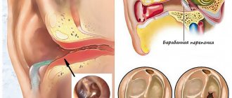
Our ear is a complex organ consisting of three parts: the outer ear, which includes the pinna and the ear canal, which rests on the eardrum, the middle ear (the cavity behind the eardrum) and the inner ear. Inflammation of the ear is called otitis media; it can develop in each of these three parts, and the causes and symptoms of the disease will vary significantly.
Inflammation of the outer ear is caused by local infection. In most cases, the development of inflammation is preceded by damage to the skin of the ear canal. A small wound resulting from a minor injury disrupts the integrity of the skin, opening the way for infection into the depths of the tissue.
The skin lining the ear canal is highly vulnerable. This is due to the fact that it is very thin, and closer to the eardrum it is rigidly fixed to the bone base. As a result, pressure on the skin does not lead to tissue displacement, as usually happens elsewhere first, but immediately to injury.
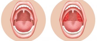
There are limited and diffuse external otitis. Limited external otitis
called inflammation that occurs in a localized area.
This form of the disease develops as a result of inflammation of the hair follicle. If the inflammation spreads to large areas of the ear canal, involving both the cartilaginous (the beginning of the ear canal) and the bone part, they speak of diffuse external otitis
. With diffuse external otitis, inflammation can cover the skin tissue, subcutaneous fatty tissue, and spread to the eardrum.
What is otitis externa?
This is an inflammation of the tissues of the external auditory canal, eardrum and auricle. Otitis externa is widespread. It is considered acute if it lasts less than 4 weeks, chronic if it lasts longer and/or occurs more than 4 times a year.
IMPORTANT! Information from the article cannot be used for self-diagnosis and self-medication! Only a doctor can prescribe the necessary examinations, establish a diagnosis and draw up a treatment plan during a consultation!
Dermatitis of the ear
It can be separated into a separate form, since inflammation of the skin of the auricle often occurs, without involving the NSP. The causes of the disease are a bacterial infection plus a factor of sensitization of the body to bacterial toxins. With this type of inflammation, redness and thickening of the skin occurs, with the formation of small serous blisters, similar to those in eczema. When the bubbles open, yellow crusts form in their place. Diagnosis is made based on examination, and consultation with a dermatologist may be required. Treatment is also antibacterial and anti-inflammatory, and antihistamines may be prescribed.
Symptoms of external otitis
There are three degrees of severity of otitis externa.
- Mild external otitis: itching in the ear canal, redness inside the ear, discomfort in the ear, which intensifies when pressing on the auricle or tragus (small protrusion in the center of the auricle). A small amount of clear, odorless fluid may come out of the ear.
- Moderate external otitis: the itching becomes stronger, the ear hurts, there is obvious redness inside, the discharge intensifies, and pus may appear. The auditory canal feels full as a result of swelling and retention of discharge.
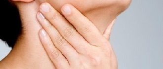
- Diffuse external otitis: severe pain in the ear, radiating to the neck, temple, cheek; the ear canal is completely blocked; the auricle becomes red and swollen, as do the lymph nodes in the neck. The temperature is elevated, sometimes significantly.
Chondroperichondritis of the auricle
This disease is an inflammation of the perichondrium and cartilage of the auricle caused by bacterial flora. Chondroperichondritis can be caused by trauma to the auricle with damage to the skin, suppuration of an auricular hematoma, deep burns of the auricle, a boil or suppurating atheroma of the ear, and erysipelas. There are infiltrative and purulent forms. A few days after the described traumatic factor, infiltration and hyperemia of the skin of the auricle develops, accompanied by pain, with or without fever. An important diagnostic criterion is that the earlobe remains unaffected, since there is no cartilage in its thickness. Treatment: antibacterial and anti-inflammatory therapy. If there are purulent cavities, an opening with removal of pus and gentle necrotomy of the destroyed cartilage is performed. The disease is difficult to treat and can last for weeks or months. Often the consequence of chondroperichondritis is cicatricial deformation of the auricle; sometimes there may be complete destruction of the cartilage.
Possible complications of otitis externa
As a rule, otitis externa does not cause complications and is easily treated. However, if complications do occur, they may include the following:
- temporary hearing loss in the affected ear. Goes away after recovery from otitis media;
- chronic external otitis. It usually occurs when there are difficulties in treating external otitis, for example, in fungal and mixed bacterial-fungal forms;
- spread of infection to deep tissues - cellulitis of the neck, lymphadenitis, osteomyelitis. Similar complications (malignant otitis media) can occur in patients with immunodeficiency conditions, diabetes, and those receiving chemotherapy. Such complications can be life-threatening.
Erysipelas of the auricle
Acute erysipelas is an infectious disease and is caused by streptococcus pyogenes; later, other flora may join. With such inflammation there are symptoms of skin damage and a general inflammatory syndrome - weakness, fever, chills. Factors provoking the disease: decreased immunity, abrasions and burns of the skin, swimming in polluted waters, contact with a carrier of pyogenic streptococcus or a sick person. Depending on the depth of damage to the skin, three forms are distinguished:
- Erythematous, when pain occurs, redness of the skin (it has a “varnished” appearance) slightly rises above the level of healthy skin and has a clear border with it.
- The bullous form, more severe, is characterized by the same skin lesions as in the erythematous form, but with the formation of large blisters above the surface of the skin with transparent or cloudy contents.
- Necrotic form. With this form, deep damage to the skin occurs, right down to the reticular and papillary layers, and deep ulcers with purulent plaques form. Due to the addition of various microflora, it is more severe than other forms. Typically, patients with this form require surgical debridement of areas of necrosis.
How to prevent otitis externa?
- Gently blot the ear canal after bathing, but do not wipe the ear canal itself with anything.
- If water gets into your ear, you can shake it out by jumping on one leg and tilting your head to the same side. You can also dry the water in your ear with a hairdryer, setting it to the lowest setting and holding it about 30-40 cm from the ear.
- If you know your eardrum has been damaged or punctured, you can use ear drops that will prevent bacteria from growing in your ear once water enters the eardrum.
- Don't swim in dirty waters.
- Do not reach into the ear canal with a finger, a stick, a cotton swab or a swab - in a word, nothing.
- Protect your ears when using hairspray if you know it may irritate the skin inside your ear canal;
- If you have had ear surgery or ear infections, see an ENT before swimming.
Prevention
Prevention of ONO is carried out in case of frequently recurring episodes of the disease, concomitant dermatological diseases, as well as with regular contact with water - in athletes.
When engaging in water sports or frequently visiting the pool, it is recommended to use swimming caps or special earplugs, and dry your ears after swimming. The outer ear can be gently wiped with a towel. To remove residual water, shake your head vigorously in different directions.
Author:
Chekaldina Elena Vladimirovna otorhinolaryngologist, Ph.D.
Diagnosis of external otitis
Usually it is not difficult. Otitis externa is easily identified by its symptoms and the appearance of the ear and ear canal. The doctor may examine your ear with an otoscope. If he wants to make sure that the eardrum is intact, he can use a curette to clean the ear and take a deeper look.
If the otitis is diffuse, the doctor may need additional diagnostics of the condition of the middle ear, determining the nature of the otitis (bacterial or other), etc.
Causes of the disease
If the cause of the disease in adults is bacteria and viruses, we are talking about the infectious nature of the inflammation. The causative agents of otitis media can be Pseudomonas aeruginosa, staphylococcus, fungi, etc. External factors can also cause inflammation. What provokes the development of the disease?
- failure to comply with basic hygiene rules, when ears are washed rarely and poorly;
- in contrast to the first point - excessive hygiene, when an adult cleans wax from the ear canal almost every day and with great diligence. This is a big mistake - the sulfur released has an antibacterial effect and is a natural defense against infection. It is enough to clean your ears twice a week;
- damage to the tissues of the ear canal in the process of improper cleaning of the ears - many adult patients go to the doctor because they choose objects not intended for ear hygiene: knitting needles, toothpicks, matches. The skin of the ear canal is very delicate, and picking at it with sharp objects can easily injure it. Pathogenic microflora penetrates through these microtraumas;
- very deep cleaning of the ear is also fraught with damage to it - do not insert the cotton swab deep into the cleaning process; for children's hygiene, use special cotton swabs with limiters;
- insufficient sulfur production;
- excessive sulfur production, leading to sulfur plugs;
- foreign objects getting into the ear - this problem is especially relevant for children. While playing, children put everything into their ears out of curiosity: beads, buttons, small parts from toys, which damage the delicate tissues of the ear canal;
- water often getting into the ear - the water washes away the natural defense - wax, and accordingly, the infection easily settles on the skin; and if swimming takes place in dirty stagnant water, for example, in a polluted pond, this is an additional chance of getting sick;
- reduced immunity especially in adult patients at risk (HIV infection, tuberculosis, oncology, diabetes, etc.);
- infectious diseases currently occurring in the body (tonsillitis, sinusitis, otitis media, etc.);
- improper use of medications;
- allergic reactions;
- skin diseases.
Make an appointment right now!
Call us by phone or use the feedback form
Sign up
As we can see, the list is extensive. In order to start treatment on time, you must be able to recognize the symptoms of acute inflammation.
Treatment of external otitis
The goal of treatment is to stop the spread of infection and allow the ear canal to clean itself as it normally does.
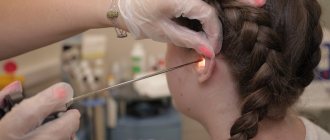
- Cleaning and rinsing the ear canal. As a rule, the doctor cleans it with a curette to free it from skin particles, stuck earwax and dried discharge (serous or purulent). This is necessary so that the ear drops can spill to the entire depth of the ear canal.
- Ear drops prescribed by an ENT or general practitioner (GP). Usually these are drops with antibiotics and/or corticosteroids. For severe pain, analgesics can be used.
- If there is severe swelling of the external auditory canal, the doctor may first replace the drops with turunda soaked in medicine. When the swelling subsides, the turunda can be easily removed from the ear, and you can continue to be treated with drops.
- When instilling cold drops, hold them in your palm for a while to reduce discomfort. After the drops are in your ear, lie down on your healthy side for a few minutes so that the drops are better absorbed. You can ask someone to instill drops for you - this is more convenient.
- If the infection is widespread, the doctor may prescribe oral antibiotics in addition to drops.
Sources
- Kesser BW Assessment and management of chronic otitis externa. Current Opinion in Otolaryngology & Head and Neck Surgery. 2011, 19: 341–347.
- Roland PS, Stroman DW Microbiology of acute otitis externa // Laryngoscope. - 2002. - Vol. 112, No. 7, Pt. 1. - P. 1166-1177.
- Rosenfeld RM, Brown L, Cannon CR et al. Clinical practice guideline: acute otitis externa // Otolaryngology. Journal of Otolaryngology - Head & Neck Surgery. - 2006. - Vol. 134 (4 Suppl). - P. 4-23.
- Schaefer P., Baugh RF Acute otitis externa: an update. American Family Physician. 2012, Dec; 1-86 (11): 1055-61.
- Wiegand S., Berner R., Schneider A., Lundershausen E., Dietz A., Otitis Externa: Investigation and Evidence-Based Treatment. Archive of "Deutsches Ärzteblatt International". 2021, Mar; 116 (13): 224–234.
Gurov A.V., Yushkina M.A. Features of the clinical course and etiotropic therapy of external otitis. Russian medical journal. 2016. – T. 24. – No. 21. – P. 1426-1431.