28.06.2017
Diseases of the chest and abdominal cavity | Spleen
There are no extra organs. Despite the fact that surgery to remove the spleen is by no means uncommon, and people who have undergone it are able to live full lives, this organ has an important role in the body. And not alone.
- Firstly, the spleen is an organ of immune defense - a large filter that retains those whose presence in the body is undesirable. These can be either foreign substances and pathogens, or one’s own potentially dangerous elements (damaged and altered cells).
- But this is by no means a passive filter. It analyzes the “detainees”, destroys them (if they are dangerous or useless for the body) and activates special reactions that work for the long term (for example, the production of antibodies that search for and predetermine the death of “enemies” far from the spleen itself).
- “Own” cells retained by the spleen are red blood cells. Not just any, but those who are old or sick. The selection of viable and full-fledged red blood cells occurs due to narrow gaps in the path of blood through the splenic filter. Changed by age or disease, red blood cells get stuck and go straight to be eaten by phagocytes. And useful elements from destroyed cells (for example, iron) are again included in the creation of new blood components. It is not for nothing that the spleen is also called the “graveyard of red blood cells.” Here, outdated blood platelets - platelets - are also disposed of.
- The spleen does not simply destroy cellular elements that are unusable for work. She also keeps in touch with the main place of their birth - the red bone marrow. In order to promptly warn a “colleague” in the event of mass cell death about the need to quickly replace the loss.
- In addition to the above, the spleen is the place of cultivation of the most important soldiers of the “salvation army” (lymphocytes); platelet storage; a significant link in metabolic processes in the body.
How then can you survive without a spleen? By redistributing its responsibilities among the remaining bodies. The main saviors of the situation are the liver and lymph nodes, of which there are more than 600 in our body. The completeness and duration of compensation is determined by the state of the body as a whole (the susceptibility of people with a removed spleen to bacterial infections, especially meningitis and pneumonia, remains statistically significant). And yet, now, if the spleen is damaged, they try to save at least a fragment of its tissue (for example, the organ is removed, and its fragment is transplanted into the “pocket” of the greater omentum). (There are also pathologies of the spleen when its complete removal is necessary).
General information
The spleen is an unpaired organ located in the abdominal cavity.
Its size increases with age. In adults, the weight of the spleen is 150-170 g. Normally, the spleen is hidden by the lower ribs and is not palpable. It can be detected by palpation with a magnification of 1.5-3 times. It is a well-supplied lymphoid organ that performs various functions. Functions of the spleen in the body:
- Participation in the formation of an immune response to foreign antigens. This organ is involved in the production of antibodies (synthesis of IgG , properdin and tuftsin ).
- Filtration of blood and elimination (removal) of microorganisms and blood elements loaded with antibodies. This organ is a large accumulation of reticuloendothelial tissue, the main function of which is phagocytosis performed by macrophage cells. Normal and abnormal blood cells are also eliminated.
- Participation in lymphopoiesis. The white pulp of the spleen, as a lymphatic organ, produces lymphocytes .
- Depositing blood. This function is performed by the red pulp of the spleen. Normally, one third of all platelets and a large number of neutrophils, which are produced during bleeding or infection, are deposited in it.
- Participation in hematopoiesis in certain diseases (in adults, the organ of hematopoiesis is the bone marrow).
- Regulation of portal blood flow due to the common vascular network with the liver.
- Participation in metabolic processes.
In many conditions and diseases, changes in the spleen in the form of splenomegaly are observed. Splenomegaly, what is it? This term is used to refer to the enlargement of a given organ to one degree or another. A synonym is the term megasplenia. Changes in size and structure occur in response to infectious, immune and hemodynamic factors. Changes in the spleen always indicate an acute or chronic process in the body - this is an indicator of pathological conditions.
Splenomegaly is an early and sometimes leading symptom of a number of diseases, without being an independent nosological entity. You need to know that an increase in this organ is always the body’s reaction to hematological, non-hematological diseases or a consequence of portal hemodynamic disorders. Therefore, the classification always takes into account the underlying disease against which this symptom developed. The splenomegaly code according to ICD-10 R16.1 is established if the enlargement of this organ is not classified in other categories.
Splenomegaly is often, but not always, accompanied by hypersplenism . Hypersplenism is a pathological condition that is characterized by a decrease in the number of formed elements (erythrocytes, leukocytes, platelets) in the circulating blood due to increased spleen function. pancytopenia develops , which is life-threatening.
Hypersplenism is associated with increased blood-destructive function of the spleen. This may be due to a number of reasons: difficulty in the outflow of blood from this organ, increased contact of formed elements with spleen macrophages, or increased activity of macrophages. Changes appear only in the peripheral blood, and the bone marrow remains normal. In many cases, the spleen is enlarged without symptoms of hypersplenism , and the latter can also occur with normal spleen sizes.
In most patients, hypersplenism requires removal of the spleen, after which the blood picture and the patient’s condition return to normal.
How to treat
“In the presence of pathology, the most common conservative treatment method is observation of the patient. When severe abdominal pain appears, as well as when the spleen is enlarged, a decision is made towards splenectomy - surgical removal of the organ,” notes the surgeon.
But, of course, removing an organ is not the best or most favorable outcome. After all, there are no empty and unnecessary organs in the human body. After removal of the spleen, the risks of more frequent acute respiratory viral infections and other infections increase. The composition of the blood may also change, and the changes will persist for life. In addition, after the operation you will need to follow a diet for at least six months in order to normalize and adjust the body’s functioning without the spleen.
Article on the topic
I look at myself as if in a mirror. What to do if the heart is on the right?
Pathogenesis
With splenomegaly of various etiologies, different changes occur; therefore, several mechanisms of this symptom are considered:
- In response to antigenic stimulation during bacterial and viral infections, with a large number of abnormal cells during hemolysis hyperplasia of its reticuloendothelial tissue develops Hyperplasia of lymphoid tissue also occurs as antibody synthesis increases. All these mechanisms are associated with an increase in the protective function - “working splenomegaly”.
- An increase in size against the background of abnormal blood flow in the organ is observed with blood stagnation due to increased pressure in the portal vein or thrombosis of the splenic vein. In both cases, the outflow of venous blood from the spleen becomes difficult. Overflow of blood causes hyperplasia of the connective tissue, and it increases. The spleen compensates for the increased pressure in the portal vein by taking on the increased load. Splenomegaly during congestion occurs with hyperplasia of the reticular tissue and proliferation of connective tissue. The degree of organ enlargement depends on the level of blockage - extrahepatic (or subhepatic, which is associated with pathology of the portal vein vessels), intrahepatic (associated with cirrhosis ) or suprahepatic ( hepatic vein abnormalities ). subhepatic portal hypertension occurs , in which there is a significant increase in the organ in question. To a lesser extent, it increases with intrahepatic and suprahepatic portal hypertension . Ascites develops with suprahepatic and intrahepatic portal hypertension.
- With increased intrasplenic breakdown of erythrocytes, the pulp strands expand and the number of phagocytic cells increases, and hyperplasia of sinus cells occurs.
- In malignant and benign neoplasms, spleen tissue is affected ( lymphomas , lymphoblastic leukemia , congenital cysts, metastases ).
- Increase due to abnormal accumulation of metabolic products and lipids (storage diseases).
- Due to extramedullary hematopoiesis in thalassemia .
Treatment of inguinal hernia - types of surgery
Doctors operate on hernial protrusions in the groin area using the most modern techniques, choosing the type of intervention that is optimal for the patient. And just two or three decades ago, tension repair was considered the only and main surgical method (the opening of the hernial orifice was “covered” with flaps formed from the tissue of the patient’s abdominal wall). Now they are moving away from this method: it involves a significant number of side effects and cases of recurrent hernias.
Thanks to progress, surgery is shifting towards gentle, innovative operations. The technologies for removing indirect and direct inguinal hernias used in hospitals and clinics are much more effective, less painful, and less long-term in terms of rehabilitation. The use of artificially synthesized materials minimizes rejection reactions. What hernioplastic measures are there to eliminate the pathology quickly and harmlessly:
1. Tension-free method according to Liechtenstein . They use “tension-free” plastic surgery, one in which the material involved – a polymer synthetic mesh – does not require tension and is not rejected by the body. There are already 3D varieties of mesh. Through an incision in the groin, the resulting hernia is reduced or removed, after which an onlay is made instead of the hernia orifice. The surgeon installs a mesh implant in their place with a hole for the spermatic cord and attaches it to the abdominal cavity. The patient can go home a few hours after the operation! Over time, the mesh becomes part of the body. The material under the skin is intertwined with natural tissues and acts as a frame that supports the organs. The downside of the technique is that a small scar remains visible at the incision site.
2. Laparoscopic suturing . They practice the most effective and low-traumatic method of hernia excision - laparoscopic inguinal hernia repair. There is a limitation - it cannot be used in cases where the hernia has become very large.
This operation requires general anesthesia. The technique involves making three neat punctures in the anterior abdominal wall. An instrument is inserted laparoscopically through them and an auxiliary substance, carbon dioxide, begins to be pumped. First, under the influence of gas, the wall of the peritoneum is stretched, and then it is possible to move the hernial sac into the abdominal cavity. Using instruments introduced into the patient’s tissue, the surgeon closes the hernial orifice from the inside with a mesh polymer “patch,” and the excess internal tissue is sutured.
3. Endoscopic surgery . It is recognized in the medical world as the safest and most effective, as it is performed with local anesthesia (rarely with general anesthesia). Tissue damage is minimal, there will be no scar marks on the skin. It can be argued that this is rather a masterly reduction of the protrusion under local anesthesia. Feature - mesh retaining material is implanted between the layers of the abdominal wall. A self-supporting effect is created due to natural pressure in the peritoneum. The progress of the process is monitored by an endoscope, and the surgeon’s actions are broadcast by a video camera on a screen with multiple magnification.
Surgery and rehabilitation for inguinal hernia
Manipulations to eliminate an inguinal hernia in men on the left or right are considered easy for both patients and surgeons - a small surgical field is involved, large incisions are not required. The maximum is done to remove the hernia and prevent its recurrence. As a result of the intervention, doctors surgically remove the protrusion (hernial sac), returning the displaced viscera, vessels and lobes of human tissue to the desired “correct” position. After plastic surgery of the hernial orifice, a polypropylene endoprosthesis-mesh made of synthetic threads serves as support for the inguinal canal and the peritoneum as a whole, and in the future does not interfere with leading a normal, familiar life.
In order to eliminate postoperative complications, it is necessary to remember and carefully follow the doctor’s recommendations:
- take only medications prescribed by the doctor orally and externally;
- bed rest in the first days (up to a week),
- stop smoking and do not drink alcohol;
- adhere to dietary nutrition,
- carefully begin physical activity;
- use the breathing technique “abdominal breathing”;
- keep the incision/puncture site clean and change bandages;
- after removing stitches and bandages, wear and often change underwear made of natural material, ironed with a preheated iron,
- avoid rooms where the temperature, concentration of dust, and humidity are high;
- do not allow any intimate contacts earlier than 14-20 days;
- The first sexual intercourse after surgery should be performed in a gentle manner, without squeezing the hernia area.
The operation in commercial clinics is estimated in the range of 25-90 thousand rubles. The cost consists of the type of manipulation chosen (laparoscopy is the most expensive), medical supplies, anesthesia used (local, epidural or general), the size and location of the hernia, and the qualifications of the operating specialist.
Classification
Degrees of severity of spleen enlargement according to Siegenthaler W:
- Mild degree - moderate splenomegaly. Protrudes from under the edge of the rib by 2 cm. A moderate degree is noted with infection (acute or chronic), cirrhosis , acute leukemia , hemolytic anemia and portal hypertension .
- A significant degree. Increases to 1/3 of the distance to the navel. Such an increase, in addition to the diseases listed above, may occur with Hodgkin lymphoma .
- High degree. Increases to 1/2 the distance to the navel. It is found in Gaucher disease , splenic cysts , chronic myelogenous leukemia , and hemolytic anemia .
- Very high degree. Reaches the level of the navel. Characteristic of chronic myeloid leukemia , huge cysts and Gaucher disease .
Differential diagnostic value of the degree of enlargement of the spleen
Megasplenia in acute illness is regarded as a short-term macrophage reaction and a “physiological” response to inflammation. As a rule, such an increase does not cause further chronic pathology of the organ. With constant and prolonged increase, matrix proliferation occurs, which leads to disruption of the structure and function of the organ.
Symptoms of recurrent inguinal hernia
In the vast majority of cases, the prognosis for men after surgery is favorable. After undergoing treatment, he will soon be able to return to his usual rhythm of life. An exception is if the progress of the operation/healing was disrupted. Then there is a danger that the protrusion will appear again within a year (such hernias are called recurrent).
It is worth noting that the risk of recurrent hernia rarely exceeds 5-10%. Most often, the recurrence of pathology occurs due to ignoring or negligent implementation of medical recommendations. It is extremely rare that it is provoked by technical errors in the operation or untreated chronic diseases of the patient.
Symptoms of recurrent inguinal hernia:
- hematomas that do not resolve;
- suppuration of the suture or wound area;
- recurrence of constipation, diarrhea with blood discharge;
- persistent cough;
- protrusion of the peritoneum and internal organs (looks like a spherical swelling).
Causes of splenomegaly
The causes of splenomegaly in adults can be grouped into several categories:
- Inflammatory splenomegaly, which occurs in response to infectious and inflammatory processes. Among the causes include acute infections: infectious mononucleosis , typhoid and paratyphoid infections, brucellosis , sepsis (microbial and fungal), endocarditis , viral hepatitis . Chronic infections: malaria , leishmaniasis , tuberculosis , syphilis , trypanosomiasis , sarcoidosis , bronchiectasis , toxoplasmosis , visceral mycoses and helminthiases ( echinococcosis , schistosomiasis ).
- Congestive, developing with hemodynamic disorders: cirrhosis with portal hypertension, thrombosis of the splenic vein and in the portal vein system, congestive heart failure .
- Tumor. An enlarged spleen in adults occurs due to blood diseases: hemolytic anemia , pernicious and sideroblastic anemia , leukemia (acute and chronic), lymphomas (Hodgkin's and non-Hodgkin's), thrombocytopenic purpura , myelofibrosis , myeloma , cyclic agranulocytosis . Its excessive increase is due to chronic myeloproliferative diseases.
- Infiltrative, developing with the accumulation of inclusions in the macrophages of the spleen (storage disease) and the presence of metastases . Gaucher disease is a lysosomal storage disease (lipids accumulate in macrophages of the spleen and liver).
- Infectious organ enlargement. an abscess is formed , which causes an increase in size with mild symptoms. Rarely seen.
- Focal organ lesions: tumors (benign and malignant), abscesses , cysts .
- Autoimmune diseases: systemic lupus erythematosus , Felty's syndrome , rheumatoid arthritis , periarteritis nodosa , Still's disease .
If we consider the causes of splenomegaly in children from 1 year to 18 years, the main ones are:
- Epstein-Barr infection (EBV).
- Cytomegalovirus infection.
- Chronic hepatitis B.
- Chronic hepatitis C.
- Parasitic infestation.
- An enlarged spleen in a child under one year of age is most often caused by TORCH infection ( toxoplasmosis , rubella , cytomegalovirus , herpes ), EBV infection and chronic hepatitis C (transmission in utero from the mother).
Who's at risk
Despite the fact that the pathology is not so common, people still worry about who may be more susceptible to such a disorder. After all, from this you can predict your examination plan. “Both adults and children are susceptible to this disease. In children, it can be detected with acute chronic abdominal pain resulting from twisting of the ligamentous apparatus that holds the spleen. If there is a developmental defect, the child may be born with this pathology. In an adult, a “wandering spleen” can occur as a result of injury, an accident, or connective tissue diseases,” says Tatyana Galich.
Symptoms of an enlarged spleen
An enlarged spleen occurs in various diseases, but symptoms of organ damage rarely come to the fore. The most common complaint of patients is slight pain and heaviness in the upper left quadrant of the abdomen. With massive splenomegaly, the patient notes an enlarged abdomen and a feeling of fullness when the stomach is compressed. The main thing in the clinical picture is the symptoms of the underlying disease.
Thus, any inflammatory enlargement of the spleen is always accompanied by fever . Typically, the spleen in acute conditions is detected in the first days of fever. This happens with acute bacterial infections ( paratyphoid fever , typhoid fever , brucellosis , miliary tuberculosis , sepsis ), as well as viral infections in which the number of lymphocytes increases: acute viral hepatitis , measles , infectious mononucleosis , viral pneumonia , rubella . Each of these infectious diseases has a characteristic clinical picture.
abscess is a rare condition. Its development is facilitated by injuries, transfer of infection from nearby organs, purulent-inflammatory processes in other organs ( pleural empyema , osteomyelitis ). Characterized by fever and symptoms of intoxication, pain in the left hypochondrium, aggravated by exercise.
Sepsis is a complication of an inflammatory or wound process. Occurs acutely or subacutely against the background of heart disease , meningococcal infection , purulent infection ( abscess , cellulitis , carbuncle ), after abortion or surgery. It occurs with intermittent fever (intermittent - high numbers and normal temperatures constantly change) or remitting fever (decrease during the day by 1-2 degrees, but not to normal levels) type. Chills and heavy sweating are also common. Severe megasplenia is observed in chronic malaria . With this disease, it reaches enormous sizes and descends into the pelvis. Patients usually experience weight loss ( malarial cachexia ) and severe anemia .
If the patient does not have a fever with an enlarged spleen, then the causes of non-inflammatory splenomegaly are considered: blood diseases, amyloidosis , storage diseases, portal hypertension , neoplastic processes , cysts, lymphomas. Hemolytic anemias are accompanied by accelerated destruction of mature red blood cells, so the clinical manifestations will be anemia and jaundice , and an enlarged spleen occurs due to increased workload. The disease occurs with hemolytic crises - a rapid (over several days) increase in anemia and hemolytic jaundice. In this case, the patient experiences nausea, vomiting, shortness of breath, abdominal pain, tachycardia , pale skin , followed by jaundice , which rapidly increases. In childhood, crises can cause the death of a child. With a long course of the disease, stones appear in the gall bladder, and trophic ulcers on the legs are also observed.
Splenomegaly in adults is observed in chronic myeloid leukemia , chronic lymphocytic leukemia and hairy cell leukemia , and in this disease it is pronounced - the enlarged organ occupies almost the entire abdominal cavity.
Its significant size is observed in 90% of patients. Patients also complain of weakness (due to anemia), hemorrhages and nosebleeds, as well as frequent infectious complications.
Myeloid leukemia is characterized by the appearance of immature precursor cells of myelopoiesis, enlargement of the lymph nodes and liver. With lymphocytic leukemia, immature lymphoid cells appear, and the lymph nodes and liver also enlarge. Mature and elderly men are affected. Patients note weakness, fatigue, sweating , and low-grade fever. In chronic lymphocytic leukemia, the first symptom is enlargement of the subcutaneous lymph nodes. With myeloid leukemia, the lymph nodes are not very enlarged, but there is pain in the bones. The spleen is significantly enlarged, reaching gigantic sizes and compressing the organs of the chest and abdominal cavity.
Lymphomas of the spleen (benign and malignant) always occur with early enlargement, which is determined during the initial examination of the patient. Its sizes vary - it can occupy most of the abdomen. There are primary lymphomas of the spleen, and later the lymph nodes and bone marrow are affected. In secondary cases, the spleen is affected secondarily after the primary tumor in the lymph nodes.
Marginal zone lymphoma is asymptomatic and is detected incidentally. As with all cancers, the patient is concerned about weakness, fatigue, and excessive sweating. Due to the enlarged spleen, heaviness appears in the left hypochondrium and symptoms of compression of the stomach (early satiety) and intestines ( constipation ). The temperature may rise in the evenings and rapid weight loss may occur.
With Hodgkin's lymphoma, organ enlargement occurs in 10-45% of cases. The tumor process affects the cervical, retroperitoneal and inguinal lymph nodes, mediastinal nodes and also organs rich in lymphoid tissue, except the spleen - the liver and bone marrow. Patients experience symptoms of intoxication (fever to febrile levels, night sweats). The progression of Hodgkin's lymphoma is manifested by severe weakness, weight loss, decreased ability to work, and itchy skin. perisplenitis and splenic infarctions often occur , which is manifested by intense pain in the left hypochondrium and increased temperature. The tumor process can only affect the spleen, in which case the disease is benign, and treatment consists of its removal.
With hemolytic anemia , in addition to the symptom considered today, jaundice appears, not without itching, weakness and fatigue due to anemia. When red blood cells are destroyed in the bloodstream, patients develop black/dark brown urine. The enlargement of the spleen with this type of hemolysis is insignificant or the spleen remains of normal size.
Chronic hepatitis of alcoholic and viral origin at the onset of the disease is accompanied by megasplenia in 25% of patients, and in the last stages - in the majority. The spleen enlarges during exacerbation, and during remission it decreases significantly. With hepatitis, complaints of nausea, a feeling of heaviness in the right hypochondrium and icteric discoloration of the skin and sclera come to the fore. The outcome of hepatitis is cirrhosis of the liver - necrosis of the liver parenchyma, with a diffuse predominance of connective tissue and restructuring of the lobular structure of the liver.
The difference between cirrhosis and hepatitis is portal hypertension , which leads to dilation of the veins of the esophagus, stomach, hemorrhoidal veins and splenic vein. In this regard, bleeding from dilated veins (esophagus and stomach) is observed. Hemorrhoidal veins can prolapse and become pinched and cause bleeding. In patients, the saphenous veins near the navel dilate, which gives a characteristic picture of the manifestations of cirrhosis of the “head of the jellyfish”. Ascites also develops .
In adults with rheumatoid arthritis and a special form of rheumatoid arthritis, Felty syndrome , with pronounced activity of the process, megasplenia is also observed, but the leading symptoms are characteristic joint lesions. Felty syndrome is observed in women over 50 years of age and develops in parallel with seropositive rheumatoid arthritis , 10 years after the onset of the disease.
Systemic manifestations are noted: fever , weight loss , lymphadenopathy , myocarditis , polyserositis , Sjögren's sicca syndrome and severe joint deformity. There is an increased susceptibility to infections.
Hernia in the groin in men: treatment
There is no option that an inguinal hernia disintegrates or disappears on its own. In the absence of proper treatment, there is a high risk of a chain development of serious complications - necrotic changes in organs and tissues, even death. The only effective method of curing a male inguinal hernia is surgery to remove it in a hospital setting (also called hernioplasty). In modern equipped clinics, an intervention aimed at radically eliminating the problem of hernia filling takes from 30-40 minutes to an hour and a half.
Conservative methods of control are just a list of preventive measures. Folk remedies: compresses, ointments, infusions from plants, as well as special physical exercises will temporarily ease some symptoms, but will not be able to achieve the main thing - to eliminate the reason why inguinal hernias appear and progress: atrophy of the abdominal wall and inguinal canal.
It is better to discuss the use of any non-surgical or alternative therapy with your doctor so as not to cause additional harm to yourself. With a diagnosis such as an inguinal hernia, we do not recommend self-medication - you need qualified help from doctors.
Tests and diagnostics
Before examining the patient, a history of the disease is collected, identifying the presence of febrile fever several weeks before treatment, the current presence of elevated temperature and increased sweating. The dynamics of weight over the past months are clarified and attention is drawn to weight loss, if any. It is important to find out the patient’s attitude towards alcohol and family history (congenital anemia, thalassemia , whether there have been cases of enlarged spleen in relatives).
Objective information about the size of an organ is obtained by palpation, but more accurate dimensions, the condition of the splenic vein and parenchyma are obtained using instrumental research methods, which include:
- Ultrasound of the abdominal organs, which reveals not only an enlarged spleen, but also the condition of other organs of the liver, gallbladder, pancreas and kidneys.
- Computed tomography of the abdominal cavity. Assess the condition of the organ in more detail, identifying difficult-to-diagnose tumors of the spleen, enlarged lymph nodes and other formations in the abdominal cavity.
- Plain radiography of the abdominal cavity. Radiologically, megasplenium is determined by the displacement of organs (stomach, intestinal loops).
- Spleen scintigraphy is performed if necessary. After intravenous administration of the radiopharmaceutical, a scan is performed in a gamma camera. Focal changes in the parenchyma (neoplasms, cysts, abscesses, infarctions) are detected.
- Splenoportography. Injection of a contrast agent (20-30 ml) into the spleen tissue during puncture and taking x-rays at certain intervals. The contrast agent quickly enters the splenic vein and other veins of the portal system. If the blood circulation in the portal system is not changed, the splenic and portal veins are visible on the images at the 3-4th second. As portal pressure increases, the portal and splenic veins become elongated and dilated, and the intrahepatic branches become poorly filled. In case of portal/splenic vein thrombosis, they are not filled with contrast. This is an unsafe intervention - there is a risk of significant damage to the spleen and the development of severe bleeding.
- Sternal puncture. Bone marrow sampling by sternal puncture is performed according to the decision of a hematologist if malignant blood diseases are suspected.
Clinical, biochemical and immunological blood tests:
- For blood diseases: anemia , altered forms of red blood cells, the appearance of blast forms of reticulocytosis/reticulopenia, signs of hemolysis .
- Hemolytic anemia. An important sign of intravascular hemolysis is an increase in lactate dehydrogenase and an increased level of bilirubin .
- Biochemical markers for systemic diseases. Rheumatoid arthritis and Felty's syndrome are characterized by hypergammaglobulinemia , high levels of rheumatoid factor, and circulating immune complexes.
- Genetic studies for suspected hereditary diseases accompanied by megalosplenia.
- With sarcoma and tuberculoma - lymphocytosis , anemia (not always), high ESR.
- In rheumatoid arthritis and Felty's syndrome, leukopenia and neutropenia are found .
Hypersplenism syndrome is characterized by:
- normocytic anemia , after bleeding - hypochromic microcytic anemia ;
- leukopenia with neutropenia ;
- thrombocytopenia , when platelets decrease to 30-40 per 10 in 9/l, hemorrhagic syndrome ;
- compensatory hyperplasia of the bone marrow (immature precursors of erythrocytes and platelets predominate in it).
results
After the screening is completed, the patient receives a research protocol, which will indicate the location of the spleen, its shape and size, tissue structure, condition of the capsule, diameter of the vessels, and the calculated area of the maximum oblique cut. The doctor's report will contain a list of all identified pathologies. With the ultrasound results obtained, the patient should be referred to the attending physician for a final diagnosis and treatment.
Previous NextSplenomegaly in children
In the prenatal period (from 22 to 38 weeks), the spleen in the fetus performs the function of hematopoiesis. At nine months of age, the bone marrow begins to produce red blood cells and white blood cells of the granulocyte series, and the spleen produces monocytes and lymphocytes . In this regard, young children may experience lability of the hematopoietic system and, under unfavorable factors, a return to embryonic hematopoiesis (splenic) is possible, which affects the size of this organ. In 5-15% of cases, an enlarged spleen occurs in a child without any disease and is considered with increased immunological reactivity at this age.
The most common diseases in children associated with megalosplenia are immune disorders and infections. Megalosplenia often develops in children in response to cytomegalovirus , toxoplasmosis , herpesvirus infection (infectious mononucleosis), congenital rubella , and with age - to the hepatitis B virus . In the first year of life, the cause of enlargement of this organ is abdominal tumors and hemolytic anemia, after 7 years - Wilson's disease and oncohematological diseases ( leukemia ), and even at an older age - systemic diseases ( rheumatoid arthritis , lupus erythematosus , ulcerative colitis ).
In children, infectious mononucleosis comes to the fore (in 90% caused by the Epstein Barr virus , in 10% by cytomegalovirus ), which is accompanied by prolonged fever (from 1 to 4 weeks) and lymphadenopathy (enlargement of different groups of lymph nodes). Tonsillitis (catarrhal or ulcerative-necrotic) often develops Enlargement of the spleen and liver is observed from the 4-5th day of the disease and lasts up to a month. In some cases, there is significant megasplenia with unexpressed other manifestations. Since during this period the spleen is loose, any rough examinations, injuries, or sudden movements are contraindicated, since rupture of the spleen is possible (sometimes it can be spontaneous). Some babies develop jaundice. Changed lymphocytes (they are called mononuclear cells ), which have a wide cytoplasm with a purple tint and a different shape (round and irregular). The nuclei in them are also of different shapes (polymorphic). Mononuclear cells are also called “virocytes,” which emphasizes the viral cause of the disease.
Acute inflammatory megasplenia occurs with typhoid fever , paratyphoid fever , miliary tuberculosis , as well as with viral infections - hepatitis , rubella , viral pneumonia . The organ in these diseases increases in size from the first days of fever. It should be remembered that it often does not increase with bacterial pneumonia , influenza , pyelitis , fever , cholera , measles , neurotropic viral infections , shigellosis and diphtheria . leukopenia , lymphopenia or lymphocytosis is detected in the analysis, infectious mononucleosis , typhoid fever and brucellosis are excluded Each of these diseases has characteristic clinical manifestations. Prolonged fever in a child requires the exclusion of lymphogranulomatosis (Hodgkin's disease) and collagenoses . If prolonged fever is combined with a chronic bacterial infection, a septic process , and in case of heart defects, infective endocarditis .
When megasplenia and hepatomegaly are combined, you need to think about portal hypertension in children, which has features in this age group. In childhood, the most common extrahepatic form of hypertension is associated in 80% of cases with anomalies of the portal vein - cavernous transformation . Also important in the development of portal hypertension is thrombosis of the portal vein system, which develops with thrombophlebitis of the umbilical vein , if it is catheterized after the birth of the child. Intrahepatic portal hypertension in children is associated with congenital liver diseases: fetal hepatitis , viral hepatitis , cholangiopathies with damage to the bile ducts of varying severity (from hypoplasia to complete failure of function).
In these diseases, severe fibrosis , which contributes to the rapid progression of portal hypertension. A sign of the extrahepatic form of portal hypertension is severe megalosplenia and symptoms of hypersplenism . , varicose veins of the esophagus often occur , which leads to periodic bleeding. Between bleedings the child's condition remains satisfactory. Liver enlargement is not typical in this form; it appears with portal vein thrombosis due to the development of umbilical sepsis . Ascites is rare in children Decompensation of portal hypertension in cirrhosis occurs in older children, but bleeding rarely develops.
A radical method of treatment is the application of vascular anastomoses. Due to the small diameter of the vessels in children, surgical intervention is fraught with difficulties, and there are also no full-fledged anatomical formations on which anastomoses can be applied. Therefore, operations are carried out after 7-8 years. The prognosis after anastomosis on the vessels is favorable. Young children are treated conservatively.
If we consider rare diseases that occur with megasplenia, then we should mention Lettepera-Siwe disease , which is characterized by high mortality and is observed up to the age of 3 years. Characterized by fever , hepatomegaly and megalosplenia , hypersplenism , lung damage (a “honeycomb lung” is formed), enlarged lymph nodes , erythrematous ulcerative rashes and bone destruction .
Hand-Schüller-Christian disease is a slowly progressive systemic disease in which granulomatous lesions of the lymph nodes, bones and internal organs are found. In the later stages, the disease occurs with the classic triad: diabetes insipidus , exophthalmos and granulomas in the bones of the skull. In 1/3 of cases, skin lesions are found - infiltrates in the form of large papules on the chest, in the groin and axillary areas.
Treatment of spleen cancer
Treatment approaches depend on the type and stage of the malignant tumor. For example, with lymphomas, if there are no symptoms, it is enough to go to the doctor for examinations and take blood tests every six months. If, for example, a metastasis of a solid tumor is detected in the spleen, active treatment is required. Below we will consider the basic principles of treatment of lymphomas as the most common tumors of the spleen.
Surgical interventions
The main type of treatment for splenic lymphoma is organ removal, splenectomy. Statistics show that most patients who undergo such surgery do not require any other types of treatment over the next five years.
Chemotherapy
In some cases, chemotherapy is used - drugs that destroy tumor cells. Some are administered intravenously, others are taken in tablet form. The type, dosage and schedule of administration of chemotherapy drugs are selected depending on the type and stage of the tumor.
Studies have shown that rituximab, a drug from the group of monoclonal antibodies, can be an effective alternative to splenectomy for lymphomas. It helps to get rid of symptoms and avoid complex surgery.
Radiation therapy
In some cases, radiation therapy is used to combat lymphomas. X-rays and proton therapy are used.
Radiation therapy is used as an independent type of treatment to combat tumors in the early stages. In later stages and with aggressive tumors, it is combined with chemotherapy. In addition, radiation helps combat symptoms caused by metastases.
Stem cell transplant
Stem cell transplantation (red bone marrow transplantation) is indicated in cases where the tumor cannot be treated with chemotherapy and radiation therapy. High doses of chemotherapy or radiation are given to suppress the patient's own red bone marrow, and then red bone marrow stem cells, either their own or a donor's, are injected into the patient's blood.
Consequences and complications
- Hypersplenism.
- Splenic rupture.
Due to the possibility of organ rupture, patients should refrain from physical activity and sports that pose a risk of abdominal injury, since rupture causes uncontrolled bleeding. Persons with megasplenia should not engage in heavy physical labor and work with increased trauma; moderate physical activity is indicated for them. Rupture is more often observed in older patients, given the thinning of the capsule and its significant increase.
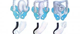
CAUSES OF PROPRESSION OF INTERNAL ORGANS (GIT)
This pathology, like many other diseases, can be congenital or acquired . The cause of chronic organ prolapse may be the already mentioned weakness of the ligamentous apparatus and muscles.
ᅠᅠ The reasons related to acquired omission include:
- Hard physical labor;
- Regular physical or strength exercises for which the body is not prepared;
- Metabolic disorders, which can result in various diseases;
- Overweight;
- Age (especially after forty);
- Consequences after childbirth.
This disease was once considered the province of the elderly, but over time, prolapse of one or more internal organs is increasingly found in middle-aged and even young people.
Forecast
The prognosis is determined by the timeliness of diagnosis and treatment of the underlying disease, as well as its severity. With correct diagnosis, acute infectious diseases are cured and at the same time megasplenia disappears. In chronic myeloid leukemia , in which the organ reaches enormous size, the prognosis depends on age, the number of blast elements and the response to treatment. In general, medications ( Imatinib ) can increase the life expectancy of patients by many years while significantly improving its quality. Five-year survival in chronic myeloid leukemia with the use of imatinib is observed in more than 90% of patients.
With thrombocytopenic purpura, with modern treatment, the prognosis for life in most cases is favorable. Outcomes can be: recovery, remission or chronic relapsing course. In rare cases, death can occur if there is bleeding in the brain.
In classic Hodgkin's lymphoma, no signs of the disease are considered cured for five years after the end of treatment. When chemotherapy with or without radiation therapy, Relapse after 5 years is rare. Risk factors for relapse include: male gender, advanced stage, age over 45 years, as well as low albumin , anemia and leukocytosis . Patients who do not achieve remission after treatment or have a relapse in the first year have a poor prognosis.
Prevention
It is now known that there is a connection between the development of malignant neoplasms of the spleen and infections such as hepatitis C, HIV infection, and infection caused by the T-cell lymphoma virus. It is recommended to follow recommendations that help prevent infection:
- Use condoms.
- If you decide to get a piercing or tattoo, choose a reliable salon.
- Do not inject with used needles.
A high fat diet and excess weight are two other risk factors for developing non-Hodgkin lymphoma. You need to eat a healthy diet and maintain a normal weight.
| More information about treatment at Euroonco: | |
| Oncologist-hematologist | 10500 rub. |
| Chemotherapy appointment | 6900 rub. |
| Emergency oncology care | from 11000 rub. |
| Palliative care in Moscow | from 40200 per day |
| Radiologist consultation | 10500 rub. |
Book a consultation 24 hours a day
+7+7+78
List of sources
- V.V. Voitsekhovsky, N.D. Goborov. Splenomegaly in clinical practice / Amur Medical Journal. — No. 2 (26) 2021, p. 62-77.
- Russian clinical guidelines for the diagnosis and treatment of lymphoproliferative diseases / ed. ed. I.V. Poddubnoy, V.G. Savchenko. M. 2021. 419 p. Fainshtein F.E., Kozinets G.I.,
- Vorobiev A.I. Guide to Hematology. 3rd ed., revised, additional. M.: Newdiamed, 2005. T.3. 409 p.
- L.M. Pasieshvili, L.N. Beaver. Splenomegaly syndrome in the practice of a family doctor / Ukrainian Therapeutic Journal. - No. 2, 2007, pp. 112-119.
- S.P. Krivopustov. Splenomegaly in pediatric practice / Children's doctor. — No. 5 (26), 2013. pp. 5-8.


