- home
- Neurology
- Intracranial hypertension
—
—
Intracranial hypertension is increased pressure in the cranial cavity. It is observed when the outflow of cerebrospinal fluid (CSF) is impaired. Severe intracranial hypertension can be caused by head trauma, stroke, or a tumor of the central nervous system. In addition, benign intracranial hypertension is common, in which the pressure of the cerebrospinal fluid is constantly increased, but not so much as to provoke a sharp deterioration in the patient’s condition.
Hospitalization of patients. Daily. Around the clock
9,500 patients trust us annually.
Call your doctor now!
Causes
The main category of patients with benign intracranial hypertension are women aged 30-50 years who are obese. In them, pathology is detected 12 times more often compared to the average in the population.
The disease can also develop at any other age, including men. Its causes and risk factors:
- endocrine pathologies;
- systemic connective tissue diseases;
- infectious lesions of the membranes of the brain;
- previous head injuries;
- obesity;
- thrombosis of the venous sinuses of the dura mater;
- chronic kidney disease;
- heavy metal poisoning;
- taking antibiotics, glucocorticoids.
If a cause can be found, intracranial hypertension is considered secondary. If the etiological factor is not identified during the examination, intracranial hypertension is called idiopathic.
Our expert in this field:
Shakhnovich Viktor Alexandrovich
Neurologist, MD
Call the doctor
Call the doctor
Reasons for the formation
If we elaborate on what was said earlier about the disease, we can name the most common provoking factors:
- Brain tumors. Malignant ones, due to rapid expansive growth, sooner lead to a disorder such as intracranial hypertension. When removed, doctors can achieve high-quality results. Benign neoplasia develops slowly. But with significant sizes, the result is the same. Both conditions are treated with surgery.
- Other volumetric formations. First of all, cysts. Special bags filled with liquid. Formally, they are not considered tumors, because the cells do not divide, and therefore rapid growth cannot be expected.
- Hemorrhagic stroke. Acute circulatory disorder in the brain with a parallel violation of the anatomical integrity of blood vessels. Leads to the development of large hematomas. Often this condition becomes fatal.
- Hydrocephalus. Increased amount of fluid, cerebrospinal fluid. Usually a congenital disorder. Most often acquired as a result of injury.
- Cardiovascular diseases. Congestive failure, recent heart attack and others.
- Lung pathologies. COPD, asthma, chronic bronchitis, history of pneumonia. All of them cause impaired blood flow and insufficient nutrition of the brain.
- Pregnancy. Not a pathological condition, but the increased load on the expectant mother’s body creates risks of negative consequences. During pregnancy, it is recommended to regularly visit a neurologist
In children, congenital defects and hypoxia most often occur. Premature patients are at high risk. You also need to carefully monitor the child’s condition if gestation occurs with complications.
Symptoms
Headache is the main symptom observed in 90% of patients with intracranial hypertension. It may have a pulsating character. Some patients complain of pain without a clear localization, others suffer from pain in the orbital area. It mainly appears or intensifies in the morning.
Other symptoms:
- 58% of patients develop tinnitus that resembles a whistle;
- Every third patient has an inability to coordinate a sideways gaze due to impaired function of the abducens nerves;
- nausea is observed in 20% of patients;
- dizziness occurs in 25% of patients.
High cerebrospinal fluid pressure leads to stagnant changes in the fundus. They appear especially often in patients whose intracranial hypertension is caused by thrombosis of the venous sinuses. In 50% of them, hemorrhages in the fundus are detected. They are also found in 20% of patients with endocrine disorders. Over time, visual acuity and visual fields may decrease.
Send documents by email The possibility of treatment will be considered by the head of the neurology department, neurologist Roman Viktorovich Shakhnovich
Intracranial hypertension: treatment, drugs
Increased intracranial pressure can lead to a decrease in the patient’s intellectual abilities and disruptions in the normal functioning of internal organs. Therefore, this pathology requires immediate initiation of treatment aimed at reducing intracranial pressure.
Treatment can only be carried out if the causes of the pathology are correctly diagnosed. For example, if intracranial hypertension occurs due to the development of a tumor or hematoma of the brain, then surgical intervention is required. Removal of a hematoma or neoplasm leads to normalization of intracranial pressure.
When increased intracranial pressure is a consequence of inflammatory processes in the body (meningitis, encephalitis, etc.), then the only effective method of treatment is massive antibiotic therapy. In this case, antibacterial drugs can be injected into the subarachnoid space in combination with the extraction of part of the cerebrospinal fluid.
Therapy is aimed at reducing the release of cerebrospinal fluid while simultaneously increasing its absorption. For this purpose, patients are prescribed diuretics.
Quite often, treatment does not require taking any medications. A set of gymnastic exercises is developed for the patient, the implementation of which leads to a decrease in intracranial pressure. Adjustments are also made to the diet and a drinking regime is developed individually. Light manual therapy, acupuncture and physiotherapy have a beneficial effect. The effectiveness of non-drug treatment is observed within the first week from the start of therapy.
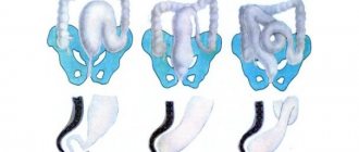
In case of postoperative, congenital cerebrospinal fluid block or other severe cases, surgical treatment is indicated. The most common type of surgical intervention is bypass surgery, that is, the insertion of a special tube with one end into the abdominal cavity or heart cavity, and the other into the cerebrospinal fluid space of the brain. Thus, excess cerebrospinal fluid is constantly removed from the skull, leading to a decrease in pressure.
When intracranial pressure increases at a very high rate and the patient’s life is threatened, urgent measures are required to save the patient. In this case, the patient is injected intravenously with a hyperosmolar solution, artificial ventilation is performed, the patient is put into a medically induced coma, and excess cerebrospinal fluid is removed by puncture.
The most aggressive treatment measure, which is resorted to in the most difficult cases, is decompressive craniotomy. At the time of the operation, a skull defect is created on one or both sides so that the brain does not rest against the bones of the skull.
Intracranial hypertension can be completely eliminated if the causes that caused it (tumor, poor blood flow, etc.) are eliminated.
Diagnostics
To examine patients with suspected intracranial hypertension, the following methods are used:
- ophthalmological examination, including assessment of the condition of the fundus;
- MRI of the brain;
- cerebrospinal fluid puncture - if signs of increased intracranial pressure are detected according to MRI results.
Instrumental studies, such as MRI or ultrasound, can help suspect intracranial hypertension, as well as exclude other causes of the patient’s symptoms. But the final diagnosis can only be established by measuring the pressure of the cerebrospinal fluid based on the results of the puncture. It exceeds the normal value and is 200-400 mm aq in most patients. Art., and in a third of patients it exceeds 400 mm aq. Art., including in 10% of patients the pressure reaches 500 mm aq. Art. and more.
Intracranial hypertension (intracranial hypertension, increased intracranial pressure)
Conservative treatment of cerebrospinal fluid hypertension is carried out when it is residual or chronic in nature without pronounced progression, in acute cases - with a slow increase in ICP, there is no evidence of dislocation syndrome and serious disorders of consciousness. The basis of treatment is diuretic pharmaceuticals. The choice of drug is dictated by the level of ICP. In acute and severe cases, mannitol and other osmodiuretics are used; in other situations, the drugs of choice are furosemide, spironolactone, acetazolamide, hydrochlorothiazide. Most diuretics should be used in conjunction with the administration of potassium preparations (potassium aspartate, potassium chloride).
At the same time, the causative pathology is treated. For infectious-inflammatory lesions of the brain, etiotropic therapy is prescribed (antiviral drugs, antibiotics), for toxic ones - detoxification, for vascular ones - vasoactive therapy (aminophylline, vinpocetine, nifedipine), for venous stagnation - venotonics (dihydroergocristine, horse chestnut extract, diosmin + hesperidin) etc. To maintain the functioning of nerve cells in conditions of intracranial hypertension, neurometabolic agents (gamma-aminobutyric acid, piracetam, glycine, pig brain hydrolysate, etc.) are used in complex therapy. Cranial manual therapy can be used to improve venous outflow. In the acute period, the patient should avoid emotional overload, avoid working at the computer and listening to audio recordings with headphones, sharply limit watching movies and reading books, as well as other activities that put strain on the eyesight.
Surgical treatment of intracranial hypertension is used urgently and planned. In the first case, the goal is to urgently reduce ICP to avoid the development of dislocation syndrome. In such situations, neurosurgeons often perform decompressive craniotomy and, if indicated, external ventricular drainage. Planned intervention aims to eliminate the cause of increased ICP. It may involve removal of an intracranial space-occupying lesion, correction of a congenital anomaly, elimination of hydrocephalus using cerebral shunting (cystoperitoneal, ventriculoperitoneal).
Forecast and prevention of intracranial hypertension
The outcome of liquor-hypertensive syndrome depends on the underlying pathology, the rate of increase in ICP, the timeliness of therapy, and the compensatory abilities of the brain. With the development of dislocation syndrome, death is possible. Idiopathic intracranial hypertension has a benign course and usually responds well to treatment. Long-term liquor hypertension in children can lead to delayed neuropsychic development with the formation of debility or imbecility.
The development of intracranial hypertension can be prevented by the prevention of intracranial pathology, timely treatment of neuroinfections, discirculatory and liquorodynamic disorders. Preventive measures include maintaining a normal daily routine, rationing work; avoiding mental overload; adequate management of pregnancy and childbirth.
Book your room today
6 seats
1 local ward
- 4 meals a day
- Bathroom in the room
- Anti-decubitus mattresses
8200 rub.
Book
13 places
2 local ward
- 4 meals a day
- Bathroom in the room
- Anti-decubitus mattresses
5700 rub.
Book
2 seats
VIP chamber
- a guest room
- 4 meals a day
- Bathroom in the room
- Anti-decubitus mattresses
17700 rub.
Book
Treatment
Therapy is usually medication. The use of a group of pharmaceuticals is indicated.
- Diuretics. Diuretics are prescribed to quickly relieve attacks. They are also used systematically as part of therapy. The most actively prescribed thiazide names (Hypothiazide and others). Loops are indicated in acute cases (Furosemide and analogues).
Caution:
Concomitant use of potassium-sparing medications is recommended to negate the removal of electrolytes.
- Cerebrovascular. To normalize brain nutrition. Piracetam as the main one. It is possible to prescribe Actovegin. If there is no intolerance to the drug.
- Nootropics. Accelerate metabolic processes in cerebral structures. Glycine, Phenibut. You cannot take them without permission, especially if there are brain tumors.
- Venotonics. For blood stagnation (medicines based on diosmin and hesperidin).
Massage also worked well. It is carried out by a qualified specialist and forms part of a comprehensive treatment.
In urgent cases, surgery for direct drainage and removal of excess fluid is possible. This is a last resort. This procedure is carried out in a neurosurgical department.
Reviews
30.09.2021
Feedback on the treatment of ovarian cancer at the NACFF clinic
07.09.2021
NACFF patient about treatment, quick examinations and attentive attitude
03.09.2021
I have healed again, I have been born again
10.08.2021
Treatment of multiple fractures and traumatic brain injury
View all reviews
Hospital, hospitalization, emergency medical care - 24/7, 7 days a week
+7 (495) 259-44-44
What needs to be examined
Diagnosis is carried out on an outpatient or inpatient basis. The task is to identify not only the violation itself, but also its culprit.
The problem is that there are no specific assessment measures, so doctors have to be insightful and draw conclusions in the face of limited information.
Possible activities include:
- Oral questioning of the patient or his parents. All health complaints are identified. The symptomatic complex is assembled in parts. Further, it is already possible to draw intermediate conclusions.
- Anamnesis collection. Past illnesses, current diagnoses, habits. Lifestyle, professional activity and other points. As part of identifying a possible cause, the etiology of the process.
- EEG. Encephalography indicators provide indirect signs of intracranial hypertension: a decrease in the activity of certain areas of the brain or, conversely, an increase in it. But the information is sketchy; the methodology is only suitable for a comprehensive assessment.
- X-ray. Provides data only at advanced stages of the disease.
- MRI diagnostics. It also does not allow us to say anything concrete until the condition is sufficiently “ripe”.
- The doctor must conduct a routine neurological examination. Evaluates reflexes.
- In particularly controversial cases, an invasive procedure is possible: puncture. It is used extremely rarely.
Attention:
Making a diagnosis of intracranial hypertension creates a lot of difficulties. The best solution, especially if the discomfort is severe, is hospitalization.
Symptoms of high intracranial pressure
Based on the severity of neurological signs and intracranial pressure indicators, the following degrees of increase in ICP are distinguished:
- weak (from 16 to 20 mm Hg);
- moderate (from 21 to 30 mm Hg);
- pronounced (from 31 to 40 mm Hg);
- extremely pronounced (over 41 mm Hg).
The leading symptom of intracranial hypertension is headache. With an acute increase in intracranial pressure, the pain is intense and increases quickly; in the case of a chronic increase, it periodically intensifies or is constant. Unpleasant sensations are localized in the fronto-parietal areas, characterized by symmetry of pain and an accompanying feeling of pressure on the eyeballs. Some patients speak of a bursting feeling pressing on the eyes from the inside. Often headaches are accompanied by nausea and pain when moving the eyes. If intracranial pressure has increased significantly, vomiting occurs at the height of the headache, which does not bring relief.
In the case of acute liquor hypertension, convulsions appear, disturbances of consciousness quickly increase until the person falls into a coma. With chronic intracranial hypertension, the patient’s general condition worsens gradually: irritability appears, sleep is disturbed (insomnia at night, drowsiness during the day), mental and physical fatigue is noted, and weather sensitivity develops (deterioration of well-being with changes in atmospheric pressure).
There are also disorders of the functioning of the autonomic nervous system, manifested by fluctuations in blood pressure, increased heart rate, and increased sweating. Chronic intracranial hypertension can be accompanied by cerebrospinal fluid-hypertensive crises, which are provoked by a sharp rise in intracranial pressure.
Clinically, a hypertensive crisis is manifested by intense headache with nausea and vomiting, and in some cases short-term fainting. Also with intracranial hypertension, symptoms of visual impairment are observed:
- blurred vision,
- diplopia (double vision),
- decreased visual acuity.
The severity of clinical manifestations is determined by the nature of the underlying disease and the rate of rise in intracranial pressure. In chronic liquor hypertension, the symptoms increase gradually (headache at first only bothers you in the morning, slow deterioration of vision); in the case of acute intracranial hypertension, cerebral edema develops very quickly, followed by disruption of vital functions. Secondary pathology is accompanied by signs of the primary disease.
Symptoms of intracranial hypertension due to liquorodynamic disorders
The presence of liquor hypertension may be indicated by indirect signs:
- excessive activity, agitation;
- deterioration in work ability;
- nosebleeds;
- pale skin, bruises under the eyes;
- decreased libido;
- chin trembling.
Complications
Chronic intracranial hypertension leads to atrophy of the brain matter, which affects the neuroregulation of internal organs and mental abilities (memory deterioration, decreased ability to concentrate, weakened intelligence). In addition, constant increased intracranial pressure negatively affects vision (deterioration up to blindness) due to atrophy of the optic nerve.
The most serious complication of the pathology with an acute increase in intracranial pressure is considered to be the development of dislocation syndrome (herniation of the brain stem into the foramen magnum), which leads to respiratory arrest and impaired circulation and ends in death.