Hospitalization and treatment under the compulsory medical insurance quota. More details after viewing the pictures.
- Aneurysm clipping
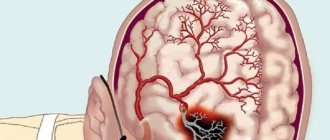
A cerebral aneurysm is a local protrusion of the arterial walls. The disease often occurs without symptoms. As the brain aneurysm grows, a significant thinning of the walls of the formation occurs, which can lead to its rupture and the development of hemorrhagic stroke.
Depending on the shape, a cerebral aneurysm can be spindle-shaped or saccular. The exact type of disease can only be determined during a comprehensive diagnosis. Saccular aneurysm is much more common.
Causes of the disease
An aneurysm is a dangerous condition that primarily requires surgical treatment. Otherwise, complications will not be avoided. Experts believe that the disease is a consequence of developmental abnormalities that disrupt the structure of the vascular wall. Often, a cerebral aneurysm is combined with connective tissue dysplasia, polycystic kidney disease, and vascular disorders. The acquired form of the disease can be a consequence of traumatic brain injury. Also, a cerebral aneurysm occurs against the background of atherosclerosis and hypertension.
The vascular wall defect itself occurs gradually. Against the background of degenerative processes, tissue damage or underdevelopment, the area loses its former elasticity. Under the pressure of blood flow, the vascular wall begins to bulge. This is how a brain aneurysm forms. Most often, the formation occurs in the place where the arteries branch and there is the strongest pressure on the vessel wall.
Hemorrhagic stroke and other consequences
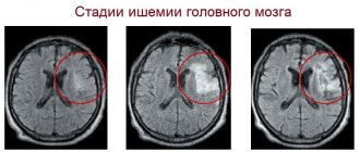
An aneurysm is a dangerous rupture of the vessel wall, which is subject to constant stress due to blood flow. When a rupture occurs, a hemorrhagic stroke occurs. It is a very serious medical and social problem. Due to hemorrhage, a hematoma is formed, which compresses the brain tissue and puts pressure on neighboring vessels. In the absence of treatment, irreversible changes occur in the affected structures within a few hours.
A hemorrhagic stroke can be suspected by severe headaches, sudden problems with speech and coordination of movements. The person feels disoriented and goes into a state of shock. In this case, you need to urgently contact a medical facility. The pathology requires emergency surgery and long-term treatment. Patients who have suffered a hemorrhagic stroke require long-term rehabilitation.
A complicated aneurysm, which is accompanied by thrombosis, is extremely dangerous. Due to a blood clot, blood circulation in the vessel slows down or stops completely. Another common complication is aneurysm dissection. In this case, a lumen is formed in the inner lining of the vessel through which blood passes.
The presence of an aneurysm significantly impairs the quality of life, especially if the pathology affects an important part of the brain. The larger the protrusion, the stronger the disease manifests itself. Due to neurological symptoms, people are forced to give up their usual activities and constantly take medications for headaches. The patient’s well-being worsens in the presence of cardiovascular diseases, increased stress, and bad habits.
Symptoms of an aneurysm
Two variants of the course of the disease can be distinguished: apoplexy and tumor-like. The characteristics of the clinical picture depend on the types of aneurysm. The tumor-like form of the disease is accompanied by an active course. In a short period of time, the aneurysm increases in size and compresses important anatomical structures of the brain.
A tumor-like aneurysm is most often localized in the area of the cavernous sinus or chiasm. When the chiasmal region is damaged, visual acuity decreases. Over a long period of time, optic nerve atrophy occurs. If a cerebral aneurysm is located in the cavernous sinus, paresis occurs and damage to the branches of the trigeminal nerve occurs. As the pathology develops, oculomotor disturbances occur and strabismus may appear.
Signs of a ruptured aneurysm
A sharp headache is the first symptom of a ruptured aneurysm. The pain syndrome is initially local in nature. But very quickly the headache spreads and becomes diffuse. In this case, patients note the appearance of nausea and repeated vomiting.
In the future, characteristic meningeal symptoms arise:
- stiffness of the neck muscles;
- Kernig's and Brudzinski's symptoms;
- loss of consciousness.
When a cerebral aneurysm ruptures, subarachnoid hemorrhage occurs. It may be accompanied by focal symptoms, which depend on the location of the formation. The most severe is hemorrhage into the ventricles, which often leads to the death of the patient.
With an aneurysm of the anterior cerebral artery, leg paresis develops, which is often combined with mental disorders. Damage to the middle cerebral artery leads to speech impairment and the development of hemiparesis.
If the pathology is localized in the area of the vertebrobasilar system, during the rupture of the aneurysm, swallowing disorders occur, nystagmus develops in combination with central paresis of the face and trigeminal nerve.
At the first sign of a possible aneurysm, you should immediately seek emergency medical help. It is important to maintain physical rest and avoid overexertion. Specialists will conduct an examination, diagnose, make an accurate diagnosis, and prescribe the necessary medications to reduce the risk of life-threatening complications.
Medicines
Photo: grud.guru
If a strange and sharp headache occurs, a person must immediately contact the nearest medical institution for qualified help. The disease is not treated with medication, but there is prevention and rehabilitation after surgery.
Surgical intervention is currently the only and most promising method of treating aneurysm. Treatment with special drugs is used only to stabilize the patient or in a situation where surgery is contraindicated or not possible at all.
Chemical drugs cannot eliminate an aneurysm; they only reduce the likelihood of vessel rupture by eliminating critical factors. Some medications are included in a complex of general therapy, which is aimed primarily at alleviating the symptoms of the initial pathology in patients. What vitamins and medications are taken for cerebral aneurysm?
Calcium channel blockers
The main representative of the nimodipine group. The chemical drug reliably blocks calcium channels in the muscle cells of the vascular walls. The vessels dilate. Blood circulation in the cerebral arteries is significantly improved. These drugs are simply irreplaceable in the prevention of dangerous arterial spasms.
Antacids
The principle of action is based on blocking H2 histamine receptors in the stomach. As a result, its acidity decreases and the secretion of gastric juice is significantly reduced. This group includes Ranitidine.
Anticonvulsants
Today, Fosphenytoin is the main representative of this group. The drugs cause reliable stabilization of membranes in nerve cells. Pathological nerve impulses noticeably slow down and do not spread.
Antiemetic drugs
Prochlorperazine is mainly used. The gag reflex is reduced by blocking postsynaptic dopamine receptors in the mesolimbic region of the brain.
Painkillers
Morphine is very effective for pain relief. The level of pain is reduced as a result of the effect on specific opioid receptors.
Antihypertensive drugs
Recently, three main drugs have been used: labetalol, captopril, hydralazine. Due to the effect on enzymes and receptors, the overall tone of the arteries is reduced and rupture is prevented.
How is it diagnosed?
Since an arterial aneurysm can occur without significant symptoms, it is necessary to pay special attention to the first signs in women and men. A neurologist must diagnose the disease based on the examination, the diagnostic results obtained and the study of cerebrospinal fluid.
During a neurological examination, focal and meningeal symptoms are detected. Based on the location of complaints, it is possible to determine presumably where the pathological focus is located.
The neurologist obtains accurate information about the patient’s condition using the following studies:
- X-ray of the skull. A cerebral aneurysm is characterized by signs of bone tissue destruction.
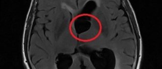
- Computed tomography, magnetic resonance imaging of the brain.
- Angiography. Basic information about the location, shape and size of the brain aneurysm can be obtained.
- Lumbar puncture. The study reveals blood in the cerebrospinal fluid, which indicates the presence of hemorrhage.
Specialists carry out differential diagnosis of cerebral aneurysm. It is important to make an accurate diagnosis in a short time. This will avoid the adverse effects of a brain aneurysm. It is necessary to differentiate an aneurysm from tumor-like processes, cysts and abscesses.
Surgical removal
After diagnosing and assessing the patient’s condition, the doctor selects a treatment regimen. If the aneurysm is large and there is an increased risk of rupture, surgery to remove the protrusion is recommended. The procedure is performed by a neurosurgeon. It can be planned or emergency if a rupture occurs.
During surgery, the aneurysm is coagulated. It can also be excised or bandaged. The method of eliminating the pathology is selected based on the diagnostic results, depending on the characteristics of the clinical case. The purpose of the operation is to exclude the damaged area of the vessel from the bloodstream. Minimally invasive techniques are used during the procedure.
Principles of treatment
Aneurysm treatment is carried out by a neurologist. The operation is performed only by a neurosurgeon. Surgical methods are aimed primarily at preventing aneurysm rupture, which can result in the death of the patient. Conservative therapy for this pathology is used to slow the progression of the disease.
Surgical treatment of a cerebral aneurysm involves two types of main operations: clipping and endovascular occlusion. Surgery prevents the aneurysm from rupturing. A radical method of treatment is the removal of arteriovenous malformation during a microsurgical operation.
Aneurysm clipping
Aneurysm clipping is an open operation during which the neurosurgeon blocks blood flow in a certain area of the vessel and applies a clip. At the stage of preparation for surgery, specialists prescribe a procedure that allows them to stabilize the patient’s condition and minimize the risk of possible complications. Preventive examination makes it possible to identify possible disorders and diseases that can lead to adverse consequences of surgical intervention.
The operation is performed using modern microsurgical techniques through a small trepanation hole. During surgery, the neurosurgeon prevents rupture of the wall of the malformation.
After isolating the neck of the aneurysm, the specialist places a clip on it. Additional control with Doppler ultrasound allows you to assess the state of blood flow and ensure the effectiveness of the operation.
Treatment of an aneurysm using the clipping method is considered quite complex and should only be performed by an experienced neurosurgeon. A qualified specialist will do everything possible to ensure accurate and technically correct application of the clip. Otherwise, dangerous complications and a long rehabilitation period cannot be avoided.
The clip is applied to the neck of the aneurysm vessel. The accuracy of the neurosurgeon’s actions is confirmed by conducting high-quality diagnostics (Dopplerography) during the procedure.
Endovascular occlusion
During endovascular occlusion of an aneurysm, the neurosurgeon blocks the lumen of the dilated vessel with a special implant. The operation is used when clipping is not possible, for example, when the aneurysm is spindle-shaped. During surgery, a specialist inserts a catheter balloon through the femoral artery using angiographic control. It closes the lumen of the aneurysm. You can also use a microspiral to perform thrombosis. The choice remains with the attending physician. The microspiral in the cavity of the affected vascular area forms blood clots, which clog the lumen of the vessel and shut off the aneurysms from the blood circulation.
Rehabilitation after surgical treatment of an aneurysm
The rehabilitation period takes place under the supervision of specialists. Professionals do everything necessary to prevent the occurrence of postoperative complications. The patient remains in the intensive care unit for several days under the constant supervision of specialists. Afterwards, the patient is transferred to a general ward, where strict bed rest must be observed for some time. The return to a normal lifestyle occurs gradually.
If the patient's well-being worsens after surgery, transcranial Doppler ultrasound may be prescribed. Rehabilitation is aimed primarily at preventing vasospasm and increased blood pressure. Specialists also monitor the general condition of the cardiovascular system and, if necessary, prescribe osmodiuretics to eliminate swelling of the brain tissue. Additionally, anti-inflammatory therapy is carried out.
Rehabilitation after surgical treatment of an aneurysm necessarily includes a program to restore impaired body functions and further socialize the patient. The recovery period lasts at least 6 months. With the help of high-quality rehabilitation, it is possible, among other things, to eliminate the adverse consequences of surgical treatment.
Rehabilitation measures include physiotherapy, massage, and the implementation of an individually selected rehabilitation program. After endoscopic clipping, the patient can return to normal life within a few weeks.
Forecast for life
The prognosis of the disease depends on the location of the vascular protrusion and its size. The initial condition of the patient also has a huge impact. The mortality rate in case of rupture of a cerebral aneurysm exceeds 30-50%. Surviving patients sometimes retain the consequences of the condition in the form of movement restrictions, cognitive impairment and a significant decrease in quality of life. Mortality after recurrent hemorrhage exceeds 70%.
Therefore, it is so important to carry out surgical treatment in time, turning to a qualified neurosurgeon. The operation is postponed only if there are certain medical indications.
Successful neurosurgical operations are carried out at the Burdenko Research Institute. Patients with signs of an aneurysm or suspected aneurysm are admitted here. At the Institute of Neurosurgery, it is possible to carry out comprehensive diagnostics using the latest technology and further treatment of identified diseases.
Pregnancy and aneurysm
Women who are planning to conceive need to be aware of the threat aneurysms pose. During pregnancy, the risks of hemorrhagic stroke for physiological reasons increase. In the second and third trimester, the volume of circulating blood increases and blood pressure rises. The situation may worsen if the patient has concomitant diseases: hypertension, diabetes.
According to statistics, the frequency of hemorrhagic strokes in pregnant women with an aneurysm is 2-5 cases per 10,000. The disease is accompanied by depression of consciousness, profound motor disorders, and respiratory failure. Neurological disorders pose a threat to the life of the expectant mother and child. Due to respiratory failure, the supply of oxygen to organs and tissues is disrupted, and fetal hypoxia develops. Lack of oxygen is a dangerous condition that leads to pathologies of intrauterine development.
A pregnant woman with a ruptured aneurysm requires urgent neurosurgical treatment and oxygen support.
Women of reproductive age who have been diagnosed with a bulging artery wall should consult a neurologist in advance and undergo treatment.