Intracranial neoplasms of a benign or malignant nature are called brain tumors. They can affect not only tissues, but also nerves, blood vessels, membranes, and endocrine structures. Depending on the location of the lesion, different symptoms appear. To make a correct diagnosis, it is necessary to undergo an examination by a neurologist and an ophthalmologist, as well as undergo a number of tests, including MRI of the brain and MR angiography. For treatment, surgery is most often used, which is supplemented with radiation or radiotherapy.
Symptoms of brain cancer
Brain cancer is characterized by the following symptoms:
Pain syndrome in the head, a headache that can appear periodically or be present constantly. As the brain tumor enlarges, the pain intensifies, and in some cases the patient may lose consciousness.
- Visual impairment, double objects.
- The appearance of attacks of dizziness.
- Constantly rising temperature.
- Fatigue, tiredness
- Numbness, impaired movement.
- The appearance of hallucinations.
- Sharp weight loss.
- Impaired consciousness.
- The appearance of vomiting.
- Hearing loss.
All symptoms of brain cancer occur due to the fact that the tumor grows and compression of the brain occurs. Its intensity depends on the location, size of the tumor and which parts of the brain are located nearby.
We strongly recommend that you undergo an annual routine brain examination, which does not take much time and is necessary for every person. Since brain cancer can develop asymptomatically!
Clinical manifestations
Symptoms are usually divided into general and focal. In the latter case, the symptoms depend on where the tumor forms.
General symptoms
The clinical picture is caused by an increase in pressure inside the skull and the toxic effect of metabolites of pathogenic cells on healthy tissue. Common signs include:
- headache not relieved by conventional analgesics;
- various visual impairments, for example, fog, doubling, glare, decreased visual acuity and others;
- loss of appetite, poor health, nausea and vomiting that are not caused by food poisoning;
- changes in the usual emotional state: mood swings, depression or aggressiveness, and others;
- symptoms of an epileptic nature;
- weakening of the brain: it is difficult for a person to concentrate, mental activity is disrupted, there is a loss of concentration, memory problems, etc.
Important. Early signs of cancer are often misunderstood by sick people. They seek help when clinical signs significantly impair quality of life. In this case, the brain tumor is diagnosed late, which significantly complicates treatment and worsens the prognosis.
Focal symptoms
Signs of this type arise due to changes in the functioning of cerebral structures, so the clinic depends on the location of the neoplasm:
- when the hemispheres are damaged, tactile sensitivity decreases, muscles in the opposite part of the body weaken (in the presence of pathogenesis in the right hemisphere, problems arise on the left side);
- disease in the temporal lobe leads to a deterioration in the ability to speak, memory suffers, and the perception of smells changes;
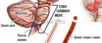
- pathological changes in the cerebellum lead to disturbances in gait and coordination of movements;
- damage to the frontal lobe causes mental and cognitive changes;
- neoplasia in the parietal lobe changes tactile sensations and fine motor skills;
- damage to the occipital zone negatively affects vision, and hallucinations are possible.
Important. The presence of these signs (of any type and intensity) is a good reason to seek medical advice. Early detection of pathologies improves the prognosis of treatment.
CAUSES OF BRAIN CANCER
There are risk factors for brain cancer; it is quite difficult to name the exact cause of the disease:
- Radiation therapy performed for other tumor diseases.
- Accommodation near high-voltage lines within a radius of 500 meters.
- It most often affects older people, although brain cancer also occurs at a young age.
- Hereditary factors increase the risk of the disease.
- Constant contact with toxic substances.
- Alcohol and smoking abuse.
- Infectious and viral pathogens.
- Immune system disorder.
- Traumatic brain injuries.
- Hormonal disorders.
Until now, science has not come to the exact results of studying the causes of the disease; these are only factors that provoke the development of brain cancer.
List of sources
- Pavlenko, A.Yu. Cerebral edema: conceptual approaches to diagnosis and treatment / A.Yu. Pavlenko // Emergency Medicine. - 2007. - No. 2 (9). — P. 11-15.
- Principles and methods of diagnosis and intensive therapy of cerebral edema and swelling: method. recommendations / V.I. Cherny [and others]. - Donetsk, 2003. - 49 p.
- Slynko E.I. Traumatic injuries of the spine and spinal cord / E.I. Slynko, A.N. Honda. - K.: PP Gamma-Print, 2010. - 288 p.
- Martynov V.A., Zhdanovich L.G., Karaseva E.A., Ageeva K.A., Khasanova L.A. Edema-swelling of the brain: tactics of patient management // Infectious diseases: news, opinions, training. 2018. – T. 7. – No. 1. – S. 124-13.
- Zadvornov A.A., Golomidov A.V., Grigoriev E.V. Clinical pathophysiology of cerebral edema. Part 2 // Bulletin of anesthesiology and resuscitation. – 2021. – T. 14. – No. 4. – S. 52-60.
DIAGNOSIS OF BRAIN CANCER
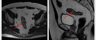
If you experience the above symptoms or are at risk, it is recommended to consult a neurologist, therapist, or ophthalmologist. The specialist will take a history and examine the patient. Magnetic resonance imaging of the brain is prescribed.
MRI with contrast
This study helps to assess brain activity, blood flow in the brain, metabolic processes, detect metastases, and distinguish benign from malignant tumors.
- Computed tomography, which determines the location and size.
- Positron emission tomography
- Single photon emission computed tomography
- Magnetoencephalography
- A spinal tap is performed to collect cerebrospinal fluid material and test it for cancer cells.
- Biopsy. The procedure allows you to determine the type of cancer cells.
If you suspect brain cancer, your doctor will recommend that you undergo the necessary examination, depending on your symptoms.
Early diagnosis of the disease allows for guaranteed rehabilitation treatment in a short time!
Pathogenesis
With any tumor, the defining feature is cell proliferation that gets out of control. The cause of this reproduction is an oncogene. Cell proliferation, invasive growth and the immune response of cells are interrelated, but are independent processes associated with genetic changes. Thus, the TSC-22 gene has been studied, which is important in various malignant brain tumors. In brain gliomas, the metabolism of non-heme iron is important.
Changing the cell genome activates antitumor immunity. However, the immune system also contributes to tumor progression by producing an immunogenic tumor phenotype.
As the tumor grows, the following occurs:
- its invasion into surrounding tissues and their destruction;
- compression of underlying tissues;
- increased intracranial pressure;
- cerebral edema;
- difficulty in the outflow of cerebrospinal fluid;
- the growth of new vessels in the tumor, becoming a source of bleeding;
- disturbance of venous and arterial blood flow.
TYPES OF BRAIN CANCER
Brain tumors are classified depending on location:
Neuroma is characterized by damage to the cranial nerves.
Sarcoma develops in connective tissue cells
Glioma develops in nerve tissues.
Meningioma affects the meninges
Pituitary adenoma is localized in the gland.
Brain tumors can be primary or secondary.
Primary tumors are formed from brain tissue, membranes, cranial nerves, and secondary tumors are metastases to the brain from other organs.
Foci in the nervous system are detected in lymphomas.
Secondary brain tumors are more common than primary ones.
Consequences and complications
The consequences of cerebral edema, even in cases of rapid relief, can manifest as long-term consequences (absent-mindedness, disturbances in sleep/motor activity, communication abilities, depression). In severe cases, the consequences of cerebral edema can develop in the form of:
- decerebral syndrome (characterized by persistent extensor muscle rigidity, strabismus, severe mental defect);
- decortication syndrome (characterized by impairment/disappearance of speech, motor and mental skills;
- posthypoxic encephalopathy (characterized by dysfunction of the cortex with the development of a cerebral intellectual-mnestic defect).
The most severe complication is death from cerebral edema.
STAGES OF BRAIN CANCER
A cancerous tumor goes through several stages in its development.
1. First stage. At this stage, a person develops a small number of cancer cells; the malignant neoplasm is characterized by slow growth.
2. Second stage. At this stage, the tumor grows and grows into other brain tissues.
3. Third stage. Here, accelerated proliferation of cancer cells is observed. They spread to large areas of healthy tissue.
4. Fourth stage. It is the most dangerous; oncological tumors are difficult to treat.
Cost of treatment
| Name of service | price, rub. | Unit measurements |
| Consultation with an oncologist and radiotherapist | 1 500 | PC. |
| Consultation with a pediatric oncologist | 0 | PC. |
| Repeated consultation with specialists | 500 | PC. |
| Primary topometry on a specialized computed tomograph | 15 000 | procedure |
| Repeated topometry on a specialized computed tomograph | 7 000 | procedure |
| Primary dosimetric planning of radiation therapy (tomotherapy) | 20 000 | PC. |
| Repeated dosimetric planning of radiation therapy (tomotherapy) | 7 000 | PC. |
| Radiation therapy (tomotherapy), including IMGRT (*) | 315 000 | well |
| Radiation therapy (tomotherapy) stereotactic radiofrequency surgery (*) | 315 000 | well |
| Accompanying drug therapy: intravenous administration in the treatment room (excluding the cost of medications) | 1 000 | procedure |
| Accompanying drug therapy: intramuscular administration in the treatment room (excluding the cost of medications) | 200 | procedure |
| Topometric marking | 750 | procedure |
| Conducting a study of the radiation therapy plan using absolute dosimetry on a specialized phantom “Cheese Phantom Accuray” | 15 000 | procedure |
| Carrying out the quality assurance (QA) procedure for the radiation therapy plan on the specialized phantom “PTW Octavius” | 30 000 | procedure |
The type of radiation therapy and the number of sessions of the course are determined by the medical commission individually for each patient based on the location, nosology of the tumor and taking into account the medical history.
Free online consultation
Sign up
Treatment of brain cancer
Treatment of brain cancer is a set of measures aimed at getting rid of the disease, improving the patient’s well-being and returning him to a full life.
There are protocol methods for treating brain cancer, such as radiotherapy, surgery, and chemotherapy.
The Russian-Japanese Oncology Center uses only proven and well-proven methods.
The maximum effectiveness of treatment for brain cancer is achieved only if there is a strong desire of the patient for recovery, the support of loved ones, and the trust of the doctor and the patient. Treatment is carried out in accordance with high international standards, using the scientific clinical base and experience of scientists from the Russian Federation and Japan.
Statistics from the Oncology Center show stable results in the treatment and rehabilitation of brain cancer.
Patients who applied at stage I.
Within a year after the discovery of brain cancer, after rehabilitation treatment, 100% of those who applied survive and live a full life for more than 20 years - almost 90% of patients
Patients who applied at stage II.
Within a year after the discovery of brain cancer, after rehabilitation treatment, 100% of those who applied survive and live a full life for more than 15 years - almost 72% of patients
Patients who applied at stage III.
Within a year after the discovery of brain cancer, after rehabilitation treatment, 100% of those who applied survive and live a full life for more than 10 years - almost 48% of patients
Patients who applied at stage IV.
Within a year after the discovery of brain cancer, after rehabilitation treatment, 100% of those who applied survive and live a full life for more than 5 years - almost 28% of patients
Procedures and operations
Brain cancer is a serious disease and tumor removal is important to perform accurately, so a neuronavigation system is used during the operation. Before surgery, the patient’s research data is entered into the navigation system program, and a 3D computer model of the brain with tumors and blood vessels is obtained, which helps virtually plan the operation. Surgical treatment is based on the principle: extracerebral tumors are removed completely, and intracerebral tumors are removed as much as possible, so that brain function does not suffer. Any operation should involve minimal trauma, preservation of brain structures, blood vessels and veins. Some areas of the brain are considered inoperable - these are the motor and speech areas.
Minimally invasive surgery to remove a tumor is performed using laser, stereotactic and endoscopic techniques. Minimally invasive operations are performed through endonasal access (through the nostril), supraorbital (through an incision above the eyebrows), retrosigmoid (incision behind the ear), as well as through any point convenient for removing deep-lying tumors. Minimally invasive approaches are as effective as invasive ones. If the operation is performed on functionally significant areas, then laser technologies are used. This could be laser thermal destruction. For deep-lying tumors, brachytherapy and photodynamic therapy are used to destroy them.
Highly effective technologies allow treatment without surgery. These include radiosurgery methods: linear accelerator, gamma knife. However, they can be used for small lesions.
Gamma Knife is a form of radiation therapy. This ultra-precise method destroys even several formations per session. The method is applicable for multiple lesions (tumors of metastatic origin). The CyberKnife system is equipment based on a linear accelerator that delivers high doses to the tumor and destroys it. The operation does not require open intervention. If the tumor is large, the bulk of the tumor is first surgically removed, and then irradiation with the CyberKnife system is used.
Boron-neutron capture therapy method. With this method of exposure, the destruction of a tumor that has accumulated the boron-10 isotope is carried out by neutron irradiation. Boron accumulated in cells increases sensitivity to radiation. Boron isotopes absorb neutrons and a “nuclear” reaction occurs, during which energy is released that destroys the new formation. Brachytherapy has not become widespread. It is carried out by implanting radioactive sources (iridium-192, iodine-125, palladium-103) into the tumor tissue.
Before the operation, the possibility of its implementation and safety are taken into account. Without these predictions, surgical treatment is considered inappropriate. With any type of intervention, consequences are possible in the form of disturbances in movement, speech, hearing, vision, reading, and mental changes. It all depends on the location of the tumor and the degree of trauma to surrounding tissues. When a pituitary tumor is removed, hormonal function is disrupted. Motor disturbances that occur immediately after surgery are associated with cerebral edema. But with the use of decongestants, patients recover quickly.
REHABILITATION OF PATIENTS
THE RUSSIAN - JAPANESE ONCOLOGICAL CENTER HAS DEVELOPED REHABILITATION PROGRAMS FOR PATIENTS AFTER TREATMENT, CHEMOTHERAPY AND RADIATION THERAPY.
Rehabilitation is aimed at areas of the brain that are responsible for important body functions: speech, movement, sensory organs, thinking, memory.
With rehabilitation treatment, the risk of complications after chemotherapy is reduced to a minimum. The patient does not experience severe symptoms after the procedures and does not lose quality of life.
Feel the quality of life after rehabilitation!
The patient should never forget that brain cancer is an aggressive type of cancer and develops quite quickly. Relapses are possible, so we make sure to monitor our patients at least 2 times a year and repeat the supporting set of rehabilitation measures after a year or a year and a half.
Our experience shows that treatment of brain cancer is always possible, even regardless of its stage and location.
The developed and tested methods of our Oncology Center are aimed at curing this disease, reducing its symptoms, preventing the progression of the disease and improving the quality of life!
Prevention
There is no specific prevention of cerebral edema, since it develops secondary. Among the general recommendations, we can only cite the need to follow some rules:
- Minimize the risk of head injury: use protective devices when moving (protective helmet when rollerblading/biking/skating, skiing; seat belts when driving in a car, do not jump your head into a body of water, etc.).
- Treat infectious/somatic diseases in a timely manner.
- Monitor your blood pressure .
- Lead a healthy lifestyle.
- When climbing to altitude (mountains), do not forget about the need to acclimatize to the altitude.
How are brain tumors treated?
Most often, three methods are used to treat brain tumors:
- surgery;
- radiation therapy;
- chemotherapy.
The most effective treatment is surgery. The other two methods have many disadvantages, so they are used as additional techniques or in patients with contraindications to surgery.
The problem with drug treatment is that most drugs do not penetrate the blood-brain barrier well. In addition, the success of treatment is far from the most outstanding. For glioblastoma, a standard chemotherapy drug increases a person's life expectancy by an average of 10 weeks.
Radiation therapy methods are often positioned as an alternative to surgery. Patients are also impressed by the names of the procedures: CyberKnife, Gamma Knife. One gets the impression that the tumor is “cut out” from the head by radiation, quickly and painlessly. But the truth is this:
- programs of simultaneous irradiation with the entire dose of radiation are not used for tumors with unclear boundaries (that is, for malignant neoplasms);
- CyberKnife and Gamma Knife can only be used for tumors of minimal size;
- they do not destroy the tumor, but at best reduce it in size, and sometimes it only stabilizes in size;
- rays pass through healthy tissue, so post-radiation complications often develop - their risk increases sharply when the tumor depth is 3 cm or more from the surface of the head;
- if the tumor decreases, the effect develops gradually, so the technique does not eliminate the symptoms of the disease immediately, and sometimes does not eliminate them at all.
For these reasons, radiation therapy is still not considered the main radical method of treating malignant brain tumors and cannot be used as an alternative to surgery. Radiation should complement, and not replace, neurosurgical treatment. If radiation therapy is used instead of, and not in addition to, surgery, then such cases are associated exclusively with a deep tumor, which cannot be reached due to the threat of severe neurological deficit, or the presence of medical contraindications to surgery.
Advantages of surgery:
- the tumor is physically removed from the body;
- it is possible to remove tumors of almost any size;
- immediately after surgery, even the most severe cerebral symptoms regress;
- the best patient survival rates among all types of treatment.


