Medium-sized AVM of the lower cerebellum
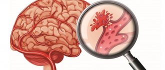
Alteriovenous malformations (AVMs) include congenital, non-hereditary defects of the vascular system, in which there are no normal capillary networks between the artery and vein, and arterial blood is discharged directly into the venous system. Over time, aneurysms form arteriovenous malformations .
AVM most often occurs in the brain, but it can also occur in the spinal cord. One or more aneurysms may be found in association with an AVM. AVM begins to develop during the fetal stage. It grows unpredictably with age.
What is AVM?
Normally it is transported from the heart through large arteries leading to the main parts of the body. Gradually, the arteries branch, become smaller and transform into capillaries with a diameter of only one cell (approximately 5 microns). The network of capillaries is called the vascular bed; here the blood gives oxygen and nutrients to the tissues and, in turn, receives metabolic products. The blood then returns through the veins to the heart. With an AVM, the arteries intertwine with the veins, forming a pathological tangle rather than a vascular bed. In this case, a vascular shunt with high internal pressure, or fistula, is formed. Veins are not able to withstand the pressure of blood coming from the arteries. As a result, the vein dilates, the venous wall stretches and, in many cases, forms a “sac”, later called an aneurysm. The blood inside the aneurysm accumulates and does not circulate, so the surrounding tissues lack blood supply. If blood pressure continues to be high, the vein or aneurysm itself will eventually rupture, causing bleeding.
There are four types of AVM
- Arteriovenous malformation is an abnormal plexus of blood vessels in which blood from the arteries enters directly into the veins, bypassing the capillaries, which is characterized by the absence of a capillary bed and high pressure.
- Cavernous hemangioma is an abnormal collection of dilated capillaries; as a rule, veins and feeding arteries are absent; characterized by low blood pressure.
- Venous malformation is an abnormal collection of dilated veins in the shape of wheel spokes; characterized by the absence of feeding arteries and low pressure; usually does not require treatment because it rarely bleeds.
- Capillary telangiectasia - abnormal capillaries with increased surface area (similar to cavernous hemangioma); pressure is very low.
AVMs can form in any part of the body; symptoms correspond to location. In the brain, arteriovenous malformations can be localized on the surface of the brain (cortical), inside brain tissue (hypothalamus, basal ganglia or brainstem), and also inside the dura mater - the dense protective layer of the brain. AVMs located within the membrane are called arteriovenous fistulas (AVFs). Normally, blood moves through the venous vessels of the brain and is transported to the venous sinuses located in the dura mater of the brain, after which it moves to the heart muscle. In AVF, the arteries are connected directly to veins or sinuses. In addition to the arteriovenous fistula of the lining of the brain, the most common are carotid-cavernous fistulas (shunts between the carotid artery and the venous sinus). Spinal AVMs form either on the surface of the spinal cord (extramedullary) or inside it (intramedullary). There are four types of such AVMs:
- Type 1 – (most common) arteriovenous tunica fistula. This AVF usually has a single feeding artery, and symptoms are caused by increased venous pressure.
- Type 2 - (glomus) is localized intramedullary, consists of a very dense tangle of vessels in a small area of the spinal cord.
- Type 3 – (juvenile) extensive AVM, consisting of anomalous vessels and localized intra- and extramedullary.
- Type 4 – (located within the sheath) extramedullary AVF on the surface of the spinal cord
Why is arteriovenous malformation dangerous?
It is a very dangerous and insidious disease, since its complications are always sudden and with a high probability can cause death or disability of a person. There are:
- intracerebral, intraventricular and subarachnoid (in the cavity between the arachnoid and soft membrane) hemorrhages. Occurs in 55–65% of cases. Symptoms are similar to those of a stroke: numbness of the limbs, difficulty speaking, impaired vision and coordination, sudden onset of severe headache;
- epileptic seizures in about 25–40% of cases;
- disruption of adequate blood supply to the brain leads to the death of a significant part of the neurons and, as a consequence, this leads to significant impairments in speech, intellectual abilities, and in some cases to paresis or paralysis of various parts of the body.
Arteriovenous malformations of internal organs are rarely complicated; an asymptomatic course of the disease is more typical for them.
Symptoms
AVMs have different symptoms depending on the type and location. Common symptoms are migraine-like headaches and seizures. However, most AVMs do not cause symptoms until they rupture and begin bleeding.
Common signs of cerebral AVMs:
- sudden attack of headache, vomiting, neck stiffness (patients often complain of “the worst pain of my life”)
- convulsions
- migraine-like headaches
- unusual tinnitus (whistle or ringing) caused by pulsating blood
General signs of spinal AVMs:
- sudden severe back pain
- weakness in the legs or arms
- paralysis
Where can the malformation be located and how does it manifest itself?
Arteriovenous malformations can occur in:
- brain;
- spinal cord;
- internal organs.
Vascular malformation of the brain manifests itself depending on the location of the lesion:
- Damage to the frontal lobe is manifested by slurred speech, decreased intellectual abilities, headaches, decreased ability to work, less commonly, stretching of the lips in a “tube,” and seizures.
- When the cerebellum is damaged, there is a lack of coordination of movements, muscle hypotonia (weakness), horizontal nystagmus (involuntary eye movements), falling during normal walking, instability in the Romberg position (in a standing position, stretch your arms forward and close your eyes).
- When the temporal lobe is involved in the pathological process, severe headaches, deterioration in speech perception occur (patients hear well, but do not understand the essence of what they say), pulsation in the temporal region, convulsive seizures, and a decrease in visual fields.
- If arteriovenous malformation occurs at the base of the brain, it manifests itself as: strabismus, one- or two-sided blindness, paralysis (complete loss of movement) of the lower or upper extremities. The movements of the eyeballs are impaired.
The largest arteriovenous malformations occur in the posterior hemispheres of the brain.
When the spinal cord is damaged, the following symptoms occur:
- with a superficial location, a pronounced pulsating formation is noted;
- pain in the spinal column, especially in the thoracic or lumbar region, can radiate (give) to the thoracic region, upper and lower extremities;
- decreased muscle tone;
- convulsive twitching of the arms or legs;
- it is possible to disrupt the functional activity of organs located in the pelvis. This manifests itself in severe cases as fecal or urinary incontinence;
- increased fatigue, especially when walking or exercising.
From internal organs, arteriovenous malformations can occur between the aorta and the pulmonary trunk (two large vessels emerging from the heart muscle) - patent ductus arteriosus (white heart defect). This condition can be provoked by chromosomal diseases, for example, Down syndrome; rubella suffered by the mother while carrying a child; prematurity of the baby. Normally, it overgrows no more than eight weeks, in rare cases up to fifteen. This condition manifests itself:
- an increase in the size of the heart muscle;
- shortness of breath;
- “machine noise”, which can be heard in the second or third intercostal spaces;
- increased heart rate (tachycardia);
- slow growth and development of the child;
- cyanosis of the skin;
- in severe cases, swelling of the upper or lower extremities, hemoptysis, impaired functioning of the heart, difficulty breathing. Spontaneous cardiac arrest may occur.
Arteriovenous malformation arising in the kidneys manifests itself:
- pain in the lumbar region on one or both sides;
- hematuria (the appearance of blood in the urine);
- an increase in blood pressure that is difficult to correct with antihypertensive drugs.
With liver damage, which occurs very rarely, specific symptoms cannot be detected. They occur only with concomitant bleeding. Pallor of the skin, weakness, fainting (syncope), dizziness, stool may turn black (melena), and in severe cases, blood pressure drops.
If a vascular malformation affects the lungs, it manifests itself:
- headaches;
- pulmonary hemorrhages in 10 - 15% of cases;
- increased amount of carbon dioxide in the blood;
- increased susceptibility to thrombosis.
AVMs cause the following injuries to the brain or spinal cord:
- When an AV malformation ruptures, there is bleeding in the brain or in the space between the brain and the skull (subarachnoid hemorrhage). Small AVMs (less than 3 cm) are more likely to rupture than large ones. Hemorrhage can lead to a stroke.
- If the AV malformation grows, it increases pressure on the brain tissue, causing seizures and hydrocephalus. Such complications are usually typical for large AVMs.
- AV malformations can cause oxygen starvation of surrounding brain tissue. Since the AVM vessels lack capillaries, brain cells do not receive enough oxygen from the blood and begin to die.
The risk of bleeding from an AV malformation is estimated to be 2–3% per year. Mortality due to first bleeding is approximately 10-30%. If a hemorrhage occurs, the likelihood of a repeat incident during the first year increases 9 times. Before deciding to have surgery, patients often want to know what the long-term likelihood of bleeding is. Here's a simple formula for this assessment: % bleeding risk = 105 - age For example, a 25-year-old man with a diagnosed AVM has an 8 out of 10 chance (i.e., an 80% chance) of having at least one bleed at some point. A number of factors influence these indicators, in particular the location and type of AVM. It is best to consult your doctor to discuss these risks as they apply to your specific case.
Why does it occur?
The issue of genetic predisposition to this pathology has been discussed for a long time, but no reliable evidence has been found. It is a congenital disease. The processes of vascular disruption that lead to this pathology occur in the first or second month of intrauterine fetal formation. Currently, predisposing factors are considered:
- uncontrolled use of medications, especially those affecting the fetus (with a teratogenic effect);
- past illnesses that occur in the first trimester of pregnancy, of a viral or bacterial nature;
- rubella disease during pregnancy;
- smoking and drinking alcohol during pregnancy. Not all girls, even when “in position”, give up these habits;
- ionizing radiation;
- pathology of the uterus;
- severe poisoning with chemicals or other toxic agents;
- intrauterine fetal injuries;
- exacerbation of a chronic pathology in a pregnant woman (diabetes mellitus, glomerulonephritis, bronchial asthma, etc.)
As a result, the vascular bundles connect incorrectly, become intertwined, and arteriovenous malformation occurs. When it forms to a significant size, cardiac output increases, the walls of the veins hypertrophy (compensatory enlargement), and the formation takes on the image of a large pulsating “tumor.”
How is the diagnosis made in Israel?
Seeing a doctor about an AVM is usually associated with either its rupture or the intention to begin treatment for an AVM that was previously discovered. In any case, the doctor reviews symptoms and other medical problems, family history, a list of medications taken on an ongoing basis, and also conducts a physical examination. Next, the patient will be referred for additional tests to determine the type and size of the AVM, its location and interaction with other structures.
- Computed tomography (CT) is a non-invasive technique that takes detailed X-ray images of the brain to detect the presence of blood inside or outside the brain tissue. A relatively new modification of CT is called CT angiography. In this case, contrast is injected into the general bloodstream to visualize the cerebral arteries. The combination of standard CT and angiography makes it possible to assess the condition of brain tissue and blood vessels.
- Magnetic resonance imaging (MRI) is a non-invasive test that uses a magnetic field and radio frequency waves. Its type, magnetic resonance angiography (MRA), allows you to obtain detailed images of the vascular bed in a non-invasive way.
- Angiography is also used as an invasive procedure, when a catheter is inserted into a peripheral artery and passed into the vessels of the brain. After this, contrast is injected into the blood and a series of x-rays are taken.
Arteriovenous malformations
Arteriovenous malformations (AVMs) are a congenital vascular pathology in which a plexus (knot) of pathological arteries and veins is formed in the substance of the brain, in the absence of a capillary network. Thus, AVMs do not participate in the blood supply to brain tissue. The walls of these vessels lack full muscle and elastic layers, as a result of which they have reduced resistance to blood pressure. In this case, arterial blood from the arteries enters (shunted) directly into the veins, as a result of which the veins experience high pressure uncharacteristic of their structure. The incidence of AVM is 2 people per 100,000 population.
AVMs may have one or more feeding vessels (arteries) from one or more blood supply basins.
AVMs can be located both on the surface of the brain and in its deep parts, in functionally “silent” or functionally important areas
Drainage (diversion) of blood is most often carried out through one, rarely through several veins, which due to high pressure greatly increase in diameter (hypertrophy). Draining veins can drain into both the superficial and deep veins of the brain
The sizes of AVMs vary from small (several millimeters) to giant ones, occupying entire lobes of the brain.
What is the pathological effect of AVM on humans?
- Due to increased pressure and low vascular resistance, AVMs can manifest themselves as spontaneous (random) hemorrhages into the substance of the brain, into its ventricular system and under the membranes of the brain. In this case, both direct damage to the brain substance occurs with focal disruption of its functions (for example, the development of motor or sensory disorders in the opposite half of the body), and indirectly, due to a reflex spasm of healthy cerebral vessels, which can lead to cerebral ischemia with depression of consciousness, and also with impaired breathing regulation. The annual probability of developing hemorrhage in patients with AVM is on average 3-5% and is essentially a “lottery” among such patients. Once experienced hemorrhage increases the risk of repeated hemorrhages several times. The mortality rate from AVM hemorrhage ranges from 10 to 30%. Patients who have survived hemorrhage may experience persistent neurological deficits in the form of impairment of certain brain functions, with varying degrees of disability
- The presence of a pulsating AVM node can lead to the development of epileptic syndrome in the form of seizures of various types: convulsive and non-convulsive, primary or secondary generalized (with loss of consciousness), in the form of absence seizures, in the form of a complex structure, etc. The presence of seizures requires constant and regular use of antiepileptic drugs
- Large AVMs (occupying large parts of the brain) cause the development of the so-called. “steal” syndrome, when large volumes of blood are directly shunted through the AVM into the venous bed (path of least resistance), and nearby healthy brain tissue experiences chronic insufficiency of blood flow. This leads to the early and significant development of first hypoxic and later ischemic disorders in the brain, also with disruption of its function
How to diagnose an AVM?
AVMs are often diagnosed only after a hemorrhage or during examination for seizures. But there are also accidental findings during routine CT or MRI.
Computed tomography (CT) can detect only fairly large AVMs, and only with the introduction of a contrast agent. CT without contrast is not informative in diagnosing AVM. Spiral CT with bolus injection of contrast (SCT angiography) not only has higher sensitivity compared to conventional CT, but also allows you to build three-dimensional computer models of the malformation, helping to better understand the features of its structure and blood supply
Magnetic resonance imaging (MRI) is quite sensitive to detecting AVMs in T2 and T1 modes with contrast. MRI angiography allows you to see relatively large (5-10 mm) formations, but is inferior in sensitivity to SCT angiography
Direct selective cerebral angiography is the most sensitive method for diagnosing AVMs. Its essence is to install a catheter in the femoral (sometimes ulnar) artery and carry it to the mouth of the internal carotid and vertebral arteries (the main vessels supplying the brain), followed by the introduction of contrast and dynamic registration of its passage through the vessels, starting from the intracranial arteries through the arterioles, capillaries, venules and veins with further outflow into the venous sinuses. At the same time, in the AVM zone there is a pathological vascular network and an abnormally early, in comparison with healthy areas, discharge of blood into the venous bed. Direct angiography makes it possible to detect AVMs with a diameter of up to several mm, and digital angiography makes it possible to computer process the resulting images with the construction of three-dimensional models of the malformation
Treatment
The choice of treatment method for AVM and the associated risks primarily depend on the size of the malformation, its location in relation to the functional areas of the brain, and the nature of the venous outflow. Additional factors are the caliber of the feeding arteries and the presence of residual atrophic changes (posthemorrhagic cysts) from previous hemorrhages.
Microsurgical resection (removal) of AVM allows the patient to be cured if the malformation is completely removed. Most often, this is possible with small and/or superficially located nodes, outside functionally important areas. Incomplete removal of the AVM node increases the risk of recurrent hemorrhage due to hemodynamic restructuring in the residual part of the malformation.
Endovascular embolization is performed using superselective angiography, when a catheter is installed directly in the small artery feeding the AVM. In this case, after carrying out special functional tests confirming the safety of treatment, a special adhesive, quickly polymerizing composition is injected through the catheter, filling the AVM node and turning off the blood flow in it. Complete exclusion of the AVM from the bloodstream is usually achieved in approximately half of the patients.
Both microsurgery and endovascular surgery, provided that blood flow into the AVM is completely stopped, can immediately relieve the patient from subsequent hemorrhages.
If surgical treatment methods are impossible or ineffective, radiation techniques are used.
Stereotactic irradiation (radiosurgery, radiotherapy) is a method of targeted irradiation of the AVM in one or several sessions. At the same time, as a result of irradiation, a post-radiation inflammatory reaction develops in the walls of the pathological vessels of the AVM node, ultimately leading (like any inflammation) to the development of connective tissue (scar) restructuring of the vascular wall, called hyalinosis. Due to hyalinosis, the vascular wall thickens, becomes more rigid, the vascular lumen decreases and completely disappears. This process takes some time and, on average, is 2-3 years. There is a direct relationship between the dose applied to the vascular malformation and the rate of development of hyalinosis
Fig 1. Post-radiation hyalinosis of the AVM walls with complete obliteration of the vascular lumen (microscopy)
Radiosurgery involves a single dose of radiation, which, on the one hand, is necessary to obtain the desired result, and on the other hand, is sufficiently safe for the surrounding brain matter. Therefore, radiosurgery is most effective in treating AVMs. Up to 80% of AVMs “close” within 2-3 years after Gamma Knife application. Repeated radiosurgery of residual functioning AVM fragments allows achieving the desired result in 95% of cases. However, the size (volume) of AVMs does not always allow the safe use of the desired radiation doses, and a decrease in the dose leads to a decrease in the effectiveness of the exposure.
Fig 2. Result of radiosurgery for arteriovenous malformation. Above: AVM in the territory of the left anterior and posterior cerebral arteries at the time of radiosurgery. Bottom - absence of pathological blood flow 2 years after radiosurgery
Hypofractionated radiotherapy is used for AVM sizes of 3-6 cm. For this purpose, linear accelerators of various designs are used. The meaning of the technique is that the desired dose is delivered not in one, but in several (3-5) sessions, which reduces the risks of post-radiation pathological reactions. However, a reduction in a single dose also leads to a decrease in the effectiveness of exposure to 60-70%
Stereotactic radiotherapy in the conventional fractionation mode (small doses over a large number of sessions - up to 25) is rarely used, mainly for giant AVMs and has an effectiveness of less than 20%.
Trained radiosurgery is an alternative to hypofractionated and standard radiation therapy. This treatment method is used mainly with Gamma Knife and involves irradiating large AVMs (up to 6-8 cm) with large doses, but in small portions, with an interval of at least 6 months. This approach has proven to be quite effective and safe. In some cases, the first stage is sufficient to obtain the effect, due to the post-radiation “closure” of the entrance part of the AVM. However, it may usually take several stages of radiosurgery and, accordingly, several years to complete the treatment and achieve the desired result.
Radiosurgical treatment can be used both for newly diagnosed AVMs and for residual malformations after any surgical treatment.
The fundamental difference between surgical and radiation treatment methods for AVM is that after surgery the effect occurs immediately, and after radiation it takes several years until the blood flow completely stops.
Does radiation increase the risk of hemorrhage?
As long as blood flow through the AVM remains, the risk of hemorrhage from it also remains. However, according to numerous studies conducted abroad and in our country, radiation does not increase this risk. Moreover, as obliteration of the AVM node progresses and the associated slowing of blood flow, the likelihood of AVM rupture gradually decreases.
Why is angiography used when planning radiosurgery?
Angiography (AG) - gives an idea of the dynamics of blood flow in the AVM, allowing one to identify the most functionally important part of the AVM node. But AG presents an X-ray image only in two planes - direct and lateral, not allowing a full assessment of the volume and shape of the AVM. On the contrary, MRI gives a static picture of blood flow, but allows you to build a three-dimensional model of a malformation of the most complex shape. Thus, the combination of two methods makes it possible to most reasonably identify the volume of AVM that needs to be irradiated.
Rice. 3 The principle of combining AG and MRI images
How to choose the optimal treatment method?
First of all, you need to contact specialists who are professionally involved in the treatment of AVMs. Not all neurosurgeons, especially radiotherapists, have sufficient experience in “dealing” with such pathology. From the opinions of several specialists, you will form your idea of the method of influence you personally need, its pros and cons, effectiveness and possible complications.
Rice. 4 Algorithms for the treatment of intracranial arteriovenous malformations
BIBLIOGRAPHY
- B. Karlsson: Gamma Knife Surgery of cerebral arteriovenous malformations, Stockholm 1996 (Thesis)
- Hartmann, A., H. Mast, et al. //Treatment of AVMs of the brain// Curr Neurol Neurosci Rep 2007 7(1): 28-34
- Lunsford LD, Kondziolka D //Stereotactic radiosurgery for arteriovenous malformations of the brain.// J Neurosurg 75:512–524, 1991
- Pollock BE, Flickinger JC // A proposed radiosurgery-based grading system for arteriovenous malformations // J Neurosurg 96:79–85, 2002
Radiosurgery
During this procedure, precisely focused radiation is directed at the abnormal vessels. Accurate focusing requires several hours of preparation; The irradiation itself takes another hour. The patient is discharged on the same day. Over the next 6 to 24 months, the vessels gradually close and are replaced by connective tissue. The advantages of this method are that the skull is not opened and the procedure is painless. The disadvantages of the method are that the best results are obtained only with small AVMs, and the therapeutic effect is achieved after a long time. During this time, the risk of hemorrhage remains. In a recent long-term experiment conducted in one of the large clinics in America, 73% of patients showed excellent or good results after radiosurgery. In these patients, the risk of future bleeding was reduced to almost zero.
Endovascular therapy
Treatment in Israeli clinics is carried out in the angiographic unit of the X-ray department by an experienced physician specialist in the field of neurovascular procedures. During this procedure, a catheter is used to inject an adhesive or other blocking material directly into the arteriovenous malformation to stop the flow of blood through the area. The patient is under general anesthesia. A catheter is inserted through a small incision in the groin and passed through a series of blood vessels directly to the artery that supplies the AVM. A blocking material (coil or medical adhesive) is then injected to narrow this artery and stop the flow of blood through the AVM. The procedure time may vary and the patient will remain in the hospital for several days for observation. This method is less invasive than surgery and can be used to treat deep and inoperable AVMs.
Surgery
Under general anesthesia, part of the skull is opened, called a craniotomy. Arteriovenous malformation is carefully detected. Using a laser and other instruments, abnormal vessels are cauterized. Once the AVM has emptied and shrunk, it is removed from the brain tissue. The hospital stay usually does not exceed 5-7 days, followed by short rehabilitation. The type of craniotomy depends on the size and location of the AVM. In general, the decision about surgical treatment is made depending on the general health of the patient. The advantage of surgery is that recovery is immediate if the AVM is removed. Disadvantages include the risk of bleeding, damage to surrounding brain structures, and stroke.