Causes of concussion
Concussion is a fairly common injury in children. The frequency of this injury is explained by the mobility and hyperactivity of children. This injury may occur if the child:
- stumbled;
- fell;
- hit;
There are also more serious causes of concussions - car accidents, criminal acts, etc.
A concussion is considered a minor brain injury, but medical intervention, examination and treatment should not be neglected. When diagnosing a concussion, the first step is to determine whether the child needs hospitalization or whether this is not necessary. It is worth noting that mild concussions can be successfully treated at home: it will be more convenient and enjoyable for both the baby and his parents. Doctors note that an examination by a neurologist is mandatory even for mild forms of concussion, when treatment occurs at home.
What is traumatic brain injury
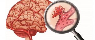
Traumatic brain injury (TBI) is mechanical damage to the skull and intracranial structures (brain, blood vessels, nerves, meninges).
Manifestations of traumatic brain injury in children differ significantly from symptoms characteristic of adults, and they are due to the characteristics of the child’s body, namely:
- the process of ossification of the baby’s skull is not yet complete, the bones of the skull are plastic, flexible, their connection with each other is loose;
- the brain tissue is immature, saturated with water, differentiation of the structures of the nerve centers and the cerebral circulatory system is not complete. Thus, on the one hand, brain tissue has greater compensatory capabilities and a so-called safety margin (soft bones of the skull and a larger amount of fluid in the brain than in adults can absorb shock). On the other hand, since it is the immature brain tissue that is exposed to trauma, which can lead to disruption of the development of its structures and provoke further limitation of mental development, emotional disturbances, etc.
Signs of a concussion in a child
The most common symptoms of a concussion in children are:
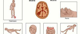
- brief loss of consciousness (usually less than a minute);
- temporary loss of orientation in space;
- weakness:
- headache;
- reading difficulties;
- It may also be difficult for a child to focus his or her gaze on something.
When diagnosing a concussion, it is very important to know whether the parents saw the fall or blow of the child. If parents saw their child fall or hit, it will be easier for doctors to find out exactly how it happened, whether the child lost consciousness after the blow or not, where he fell from or from what height, what he hit, how strong the blow was. In addition, it is important for doctors to understand whether the child remembers what happened to him. In medical practice, a condition when a person does not remember the events that happened to him is called retrograde amnesia.
Possible injuries
Several possible injuries can result from a fall, including:
Shake
A physician should evaluate head injuries for concussions.
A concussion is a type of head injury that usually occurs when a blow to the head causes the brain to receive a jolt inside the skull. It is difficult to detect a concussion in a toddler because they cannot describe their symptoms.
Signs of a concussion in an infant include:
- loss of consciousness
- inconsolable crying
- vomit
- excessive sleepiness
- long periods of silence
- refusal to eat
- temporary loss of recently acquired skills
- irritability
Damage to soft tissues.
The scalp is the skin that covers the head and contains many small blood vessels. Even a minor cut or injury can bleed profusely, making the injury appear more serious than it is.
Sometimes, an internal hematoma under the scalp can cause swelling on the baby's head, which can last for several days.
Skull fracture
The skull is the bone that surrounds the brain. Possible skull fracture if falling from a high place.
Infants with a skull fracture may have:
- depressed area on the head
- clear fluid coming from the eyes or ears
- bruises around the eyes or ears
Take your child to the emergency room immediately if he has any of these signs.
The brain is a delicate structure that contains many blood vessels, nerves and other internal tissues. Falls can damage these structures, sometimes severely.
Parents have powerful intuition. If something seems wrong with your child, it is important to understand it so you can contact a doctor. It's always better to be safe and make sure there are no serious injuries.
Treatment
Severe injuries (skull fractures, brain contusions, soft tissue injuries) will be excluded with proper examination and diagnosis. MRI and head x-rays can help diagnose internal brain damage. A neurologist examines the symptoms and makes a final conclusion.
You can treat your child at home after doctors have ruled out all possible complications. When treating at home, parents should ensure that the child maintains bed rest. The duration of bed rest directly depends on the symptoms. Doctors recommend keeping your child in bed for a while even after visible symptoms disappear. On average, complete recovery takes from 3 days to two weeks; in some cases, treatment can be longer.
Incorrect treatment can lead to serious complications in the future, so do not forget to constantly consult with your doctor during the treatment process and tell him about any changes in your baby’s condition.
Diagnostics
After the examination, the doctor may prescribe additional tests, namely: x-rays, MRI, neurosonography, computed tomography or electroencephalography.
X-rays help reveal the integrity of the skull bones.
Neurosonography is prescribed for children under 2 years of age. Using this procedure, the doctor will be able to identify this or that brain pathology.
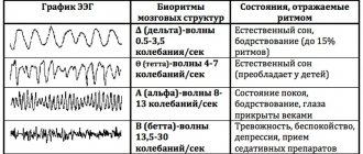
Electroencephalography will allow you to study the bioelectrical activity of the brain and assess the severity of the injury.
Computed tomography is an effective method for detecting brain damage.
What to do first if a child falls
We strongly advise parents whose children have suffered a head injury: even if, in your opinion, nothing bothers the baby, he fell from a small height, stopped crying, etc., immediately seek help from the following doctors: a pediatric neurologist, a traumatologist, a neurosurgeon. To do this, you need to call an ambulance at home and you and your child will be taken to a specialized hospital. Or, go to the emergency surgery department of any large children's hospital yourself, where the child will be consulted by the specified specialists. If they do not confirm the pathology, you can safely return home.
Failure to consult a doctor is dangerous due to late diagnosis of the injury, worsening of the child’s condition, and the possibility of coma. All this requires treatment in intensive care, and in some cases, surgical intervention. Delayed consultation with a doctor increases the risk of death and complications, lengthens the recovery period and worsens its outcome, to the point that the child may become disabled.
Short-term complications of concussion
Some patients who have suffered a concussion may experience so-called post-stress disorders:
- persistent headaches lasting 7-14 days, the intensity of which decreases when taking analgesics or painkillers of other groups;
- attacks of dizziness, difficulty concentrating, difficulty performing any normal activities (reading, writing, etc.);
- periodic vomiting for no apparent reason, feeling of nausea.
Most often, the side effects of a concussion disappear after some time without treatment; if they bother the patient for several months, you should visit a doctor and get an appointment for a consultation with a neurologist or a brain tomography (magnetic resonance, computer) to clarify the diagnosis.
Methods for diagnosing TBI
An important examination for head trauma in infants is neurosonography - a study of the structure of the brain using an ultrasound machine through the child’s large fontanel (such a study is possible until the large fontanelle closes, up to 1 - 1.5 years). This method is easy to use, does not have a negative effect on the body, and provides enough information to determine treatment tactics for the patient. With its help, you can first of all exclude or determine the presence of intracranial hemorrhages (the most life-threatening). The only limitation to its use may be the absence in the hospital of an ultrasound machine or a specialist who knows how to operate it (for example, not all hospitals in the country that have ultrasound machines can conduct emergency neurosonography at night, since the specialist works during the day).
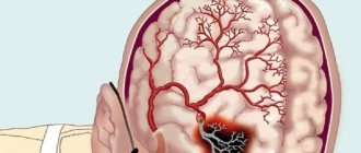
If intracranial hemorrhage is suspected (especially if for various reasons it is not possible to do neurosonography), a lumbar puncture is performed - a therapeutic and diagnostic procedure in which a hollow needle connected to a syringe is used to puncture the area of the second to fourth lumbar vertebrae of one of the spaces of the spinal cord (subarachnoid space) and taking a portion of cerebrospinal fluid for examination under a microscope. The presence of intracranial hemorrhage is determined by the presence of blood cells in the cerebrospinal fluid.
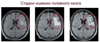
In addition, there are more complex methods for examining a child’s head: computed tomography (CT) and magnetic resonance imaging (MRI).
Computed tomography (CT) (from the Greek tomos - segment, layer + Greek grapho - to write, depict) is a research method in which images of a certain layer (slice) of the human body (for example, the head) are obtained using X-rays. With CT, the rays hit a special device that transmits information to a computer, which processes the received data on the absorption of X-rays by the human body and displays the image on the monitor screen. In this way, the smallest changes in the absorption of rays are recorded, which in turn allows you to see what is not visible on a regular x-ray. It should be noted that radiation exposure with CT is significantly lower than with conventional X-ray examination.
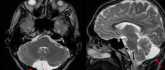
Magnetic resonance imaging (MRI) is a diagnostic method (not associated with x-rays) that allows you to obtain layer-by-layer images of organs in various planes and construct a three-dimensional reconstruction of the area under study. It is based on the ability of some atomic nuclei, when placed in a magnetic field, to absorb energy in the radio frequency range and emit it after the cessation of exposure to the radio frequency pulse. For MRI, various pulse sequences have been developed to image the structures under study to obtain optimal contrast between normal and altered tissues. This is one of the most informative and harmless diagnostic methods.
But the widespread use of CT and MRI in early childhood is difficult due to the need to conduct this examination in children in a state of immobility (under anesthesia), since an important condition for the successful implementation of the technique is the immobility of the patient, which cannot be achieved from an infant.
Typical cases of traumatic brain injury in infants
- The baby lies on the changing table or on the sofa, the mother turns away for a few moments, and the baby falls on his butt.
- The baby is left unattended in a high chair. He pushes off the table with his feet and falls on his back along with the chair.
- The baby is trying to get up in the crib. Something on the floor interested him, and he hangs over the side and falls.
- The little one was left sitting in the stroller, not expecting that he would try to stand up in it and, not finding support, would fall down.

